
Method and device for magnetic resonance imaging with improved sensitivity by noise reduction

a magnetic resonance imaging and noise reduction technology, applied in the field of image processing of magnetic resonance images, can solve the problems of inherently low sensitivity, low signal-to-noise ratio, high cost of strategies regarding the necessary hardware, etc., and achieve the effect of improving image quality, avoiding loss, and reducing nois

Active Publication Date: 2018-03-29
MAX PLANCK GESELLSCHAFT ZUR FOERDERUNG DER WISSENSCHAFTEN EV
View PDF0 Cites 17 Cited by
  • Summary
  • Abstract
  • Description
  • Claims
  • Application Information

AI Technical Summary

Benefits of technology

The invention is a method for improving the quality of magnetic resonance imaging (MRI) by denoising the coefficient data sets of wavelets in the wavelet frequency domain. The method involves statistical amplitude processing and scaling factors to remove noise from the data. The method also includes an adaptive averaging over image data sets to recover details hidden by noise. The method preserves signal phase coherence and avoids loss of signal contributions. The method can be applied to multiple complex MR images and provides improved image quality without the need for repeated acquisitions of images under identical conditions.

Problems solved by technology

Thus, despite its success, MRI is often limited by a low signal-to-noise ratio (SNR).
The problem of an inherently low sensitivity is becoming increasingly important when applying MRI techniques, in which a certain preparation of the spin system and spatial encoding by the imaging pulse sequence is used to achieve a quantitative parametric characterization of the object under investigation (e.g., a tissue or an organ or a whole organism).
However, such strategies are demanding and expensive regarding the necessary hardware.
Furthermore, the gain achieved by a higher magnetic field is only moderate (approximately linear increase with B0) whereas the applicability of hyperpolarization is rather limited to specific applications.
Appropriate choice of the size of the array elements reduces the effective size of that part of the sample that acts as a noise source, which is, however, limited by the overall size of the object to be imaged.
This method requires an identical contrast for all images that are supposed to be averaged and is time consuming (e.g., to double the SNR requires four times the number of scans).
In particular, while averaging of all data, e. g. from an image series acquired with variation of specific acquisition parameters to extract a quantitative property of the tissue after application of a fitting procedure, would lead to a final dataset with high SNR, it would—at the same time—destroy all underlying information about the parameter to be extracted or the time-dependence of the dynamic experiment, and hence, traditional averaging has substantial limitations.
Similarly, smoothing of the images unavoidable leads to a smearing of contrast and thus effectively reduces the image resolution.
A substantial disadvantage of these approaches is the suppression of information that result from weak and fine details of the imaged object that are hidden by overlaying noise contributions and, hence, misclassified as noise.
Thresholding techniques typically aim at smooth images with intact sharp edges characterizing the object, and maximum SNR, which may come at the cost of a partial loss of finer details.

Method used


Image

  • Method and device for magnetic resonance imaging with improved sensitivity by noise reduction
  • Method and device for magnetic resonance imaging with improved sensitivity by noise reduction
  • Method and device for magnetic resonance imaging with improved sensitivity by noise reduction

Examples


examples of application

[0124]In a first example of applying the invention, the gain in SNR by application of ‘standard AWESOME’ to a series of 2D spin-echo MR images is demonstrated. In particular, the images of the series are assumed to be recorded as a function of the echo time (TE), whereby TE increases progressively over the image series in order to quantitatively map the transverse relaxation time, T2, after a voxel-by-voxel fit if the image intensity to an exponential, S(TE)=S(0) exp(−TE / T2). To assess the performance of the method of the invention in a a situation, where the underlying ground truth is known, the noisy images were simulated from a standard brain image by adding Gaussian complex noise to the real-valued data. For simplicity, a single exponential decay was assumed for each pixel. A total of 16 echo times (M=16) were used. The resulting images, which are used as input data to the method of the invention are shown in FIG. 7, which shows a series of simulated 2D spin-echo MR images demon...


PUM


Abstract

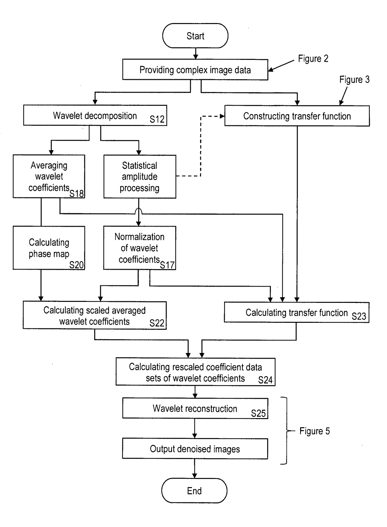
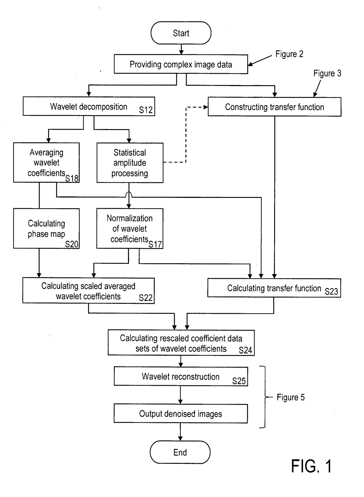
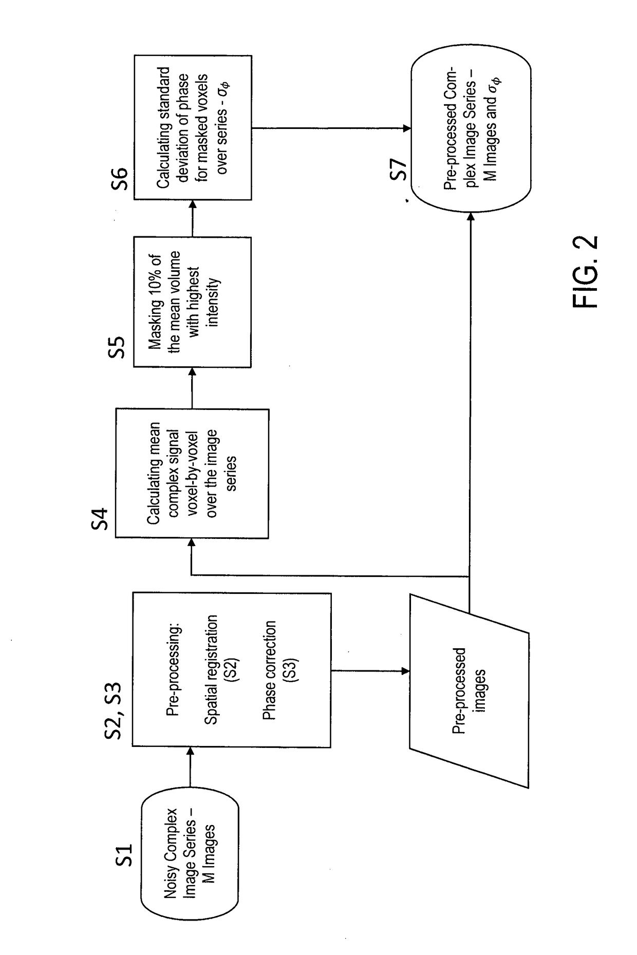
A method of image processing of magnetic resonance (MR) images for creating de-noised MR images, comprises the steps of providing image data sets including multiple complex MR images (S7), subjecting the MR images to a wavelet decomposition (S12) for creating coefficient data sets of wavelet coefficients (Sn,m) representing the MR images in a wavelet frequency domain, calculating normalized coefficient data sets of wavelet coefficients Formula (I) (S17), wherein the coefficient data sets are normalized with a quantitative amount of variation, in particular standard deviation Formula (II), of noise contributions included in the coefficient data sets (Sn,m), averaging the wavelet coefficients of each coefficient data set (S18) for providing averaged wavelet coefficients Formula (III) of the coefficient data sets, calculating phase difference maps (Δφn,m) for all coefficient data sets (S20), wherein the phase difference maps provide phase differences between the phase of each wavelet coefficient and the phase of the averaged wavelet coefficients Formula (III), calculating scaled averaged coefficient data sets of wavelet coefficients by scaling the averaged wavelet coefficients Formula (III) with scaling factors (Cn,m), which are obtained by comparing parts of the normalized wavelet coefficients of the normalized coefficient data sets Formula (I) that are in phase with the averaged wavelet coefficients Formula (III) (S22), calculating rescaled coefficient data sets of wavelet coefficients Formula (IV) (S24) by applying a transfer function Formula (V) on the coefficient data sets (Sn,m) and on the scaled averaged coefficient data sets, wherein the transfer function includes combined amplitude and phase filters, each depending on the normalized coefficient data sets Formula (I) and me phase difference maps (Δφn,m), resp., and subjecting the rescaled coefficient data sets to a wavelet reconstruction Formula (IV) (S25) for providing the denoised MR images.

Description

TECHNICAL FIELD OF INVENTION[0001]The invention relates to a method of image processing of magnetic resonance (MR) images for creating denoised MR images, in particular to a method of denoising image data in wavelet domain. A magnetic resonance imaging device for implementing the method is also disclosed. Applications of the invention are available in all fields of magnetic resonance imaging, in particular functional and / or quantitative medical imaging.TECHNICAL BACKGROUND[0002]For describing the background of the invention, particular reference is made to the following publications:[0003][1] V. R. Fuchs et al. in “Health Aff.” 20: 30-42 (2001);[0004][2] D. I. Hoult. Sensitivity of the NMR experiment. In: D. M. Grant, R. K. Harris (eds.); Encyclopedia of Nuclear Magnetic Resonance, Vol. 7, pp. 4256-4266, Wiley, Chichester (1996);[0005][3] S. Zaroubi et al. in “Magn. Reson. Imaging” 18: 59-68 (2000);[0006][4] J. B. Weaver et al. in “Magn. Reson. Med.” 21: 288-295 (1991);[0007][5] U.S...

Claims


Application Information

Patent Timeline
IPC IPC(8): G06T11/00G01R33/56G01R33/565G06T5/00G06T5/10A61B5/055A61B5/00
CPCG06T11/005G01R33/5608G01R33/56545G06T5/002G06T5/10A61B5/055A61B5/7203G06T2207/10088G06T3/0068G06T2207/30016G06T2207/20064G06T5/70G06T3/14
Inventor MARSCHNER, HENRIKPAMPEL, ANDREMOELLER, HARALD
Owner MAX PLANCK GESELLSCHAFT ZUR FOERDERUNG DER WISSENSCHAFTEN EV
Learn More