

Orthopaedic medical device
a medical device and orthopaedic technology, applied in medical science, surgery, vaccination/ovulation diagnostics, etc., can solve the problems of limited repair propensity of cartilage tissue, limited propensity for cartilage repair, and “wear and tear” of osteoarthritis (oa), so as to reduce the likelihood of tissue damage, less constricted areas, and more control
- Summary
- Abstract
- Description
- Claims
- Application Information
AI Technical Summary
Benefits of technology
Problems solved by technology
Method used
Image

Examples
example 1
[0114]Synovial compartments of cadaveric knees were washed out using 50 mL phosphate buffered saline (PBS) to determine number of MSCs within the synovial fluid prior to release from synovium. After this a further 50 mL PBS was injected into the knee cavity. Through a small incision a brush with a number of projecting elements attached to a motorised drill for increased abrasion was inserted and used to agitate the superficial synovium. The 50 ml of PBS containing released cells were aspirated from the joint and cells within this and the initial washout were grown for 14 days to produce colonies (the so called colony forming unit-fibroblastic, CFU-F assay, FIG. 9). Results show that FIG. 9A had substantially less colonies (186) as compared to FIG. 9B which had 448 colonies from the stimulated synovial tissue and released MSCs. This was a significant difference (p=0.002) and showed that contacting the synovium with an abrasive surface and agitation the abrasive surface of the device ...
example 2
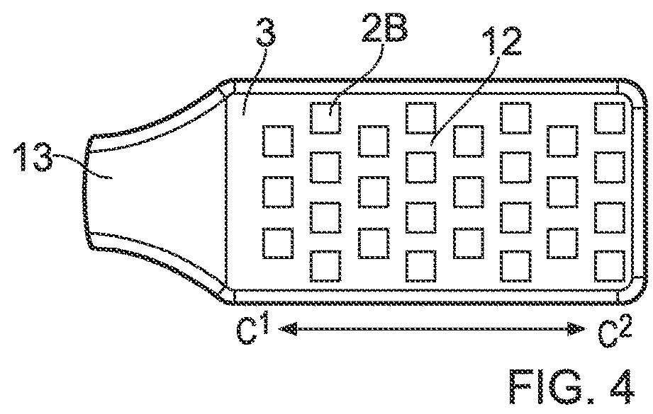
[0115]Synovial membrane (synovium) was taken from patients undergoing total knee replacement surgery. After washing of the membrane to remove loosely bound cells, the synovium was held in place using forceps. The devices (C1 with circular cross-sectional projecting elements; C2 with square cross-sectional projecting elements; C3 with continuous wave projections and C4 with “V” shaped or chevron cross-sectional projecting elements) were applied to the surface of the synovium using downward pressure and vertical stokes to detach superficial cells (including MSCs). The synovium was then washed to remove any remaining detached cells. These cells were then grown for 14 days in a CFU-F assay. The colonies were fixed in formalin, stained and counted (FIG. 10A). The comparative results (FIG. 10B device C1 versus C2 and FIG. 10C devices C3 and C4) showed that in order of performance, based on the number of colonies, that of the projecting elements the circular cross-sectional projecting elem...
example 3
[0116]MSCs are thought to be key cellular mediators for the repair bone and cartilage by their ability to differentiate into mesenchymal tissue such as bone, cartilage and fat. The ability of the device of the present invention to release MSCs from the synovium is shown in FIG. 9 by the formation of colonies in the CFU-F assay. To further demonstrate these cells are MSCs and to show their ability to form tissue of mesenchymal origin, tri-lineage differentiation was performed on culture expanded cells released from human synovium by the intended device. FIG. 11A shows the ability of the released cells to form cartilage (chondrogenesis), bone, (osteogenesis) and fat (adipogenesis), further confirming the cells as MSCs.
[0117]The present invention comprises an orthopaedic device capable of detaching and releasing superficial cellular material from the synovium membrane of articulating joints. This cellular material contains viable MSCs as characterised by colony forming ability (FIG. 9)...
PUM
 Abstract
Description
Claims
Application Information