Antibodies for treatment and diagnosis
a technology for diagnosis and antibodies, applied in the field of diagnosis and treatment of diseases, can solve problems such as unsuitable targeting applications, and achieve the effect of facilitating production of clinical-grade materials and being easier to produce and purify
- Summary
- Abstract
- Description
- Claims
- Application Information
AI Technical Summary
Benefits of technology
Problems solved by technology
Method used
Image
Examples
example 1
on and Characterisation of Two New Antibodies Against the IIICS Isoform of Fibronectin
[0093]Antibodies SW01 and SW02 were isolated in single-chain Fv (scFv) configuration from phage display libraries which include the libraries described in PCT / EP2009 / 006487, in Weber et al. (PLoS One, 2014, 9 (6) doi: 10 / 1361) and in Silacci et al. (Protein Engineering Design & Selection, 2006, 19, 471-478) according to the screening technique described by Silacci et al. (Protein Engineering Design & Selection, 2006, 19, 471-478) using fibronectin IIICS as the screening antigen.
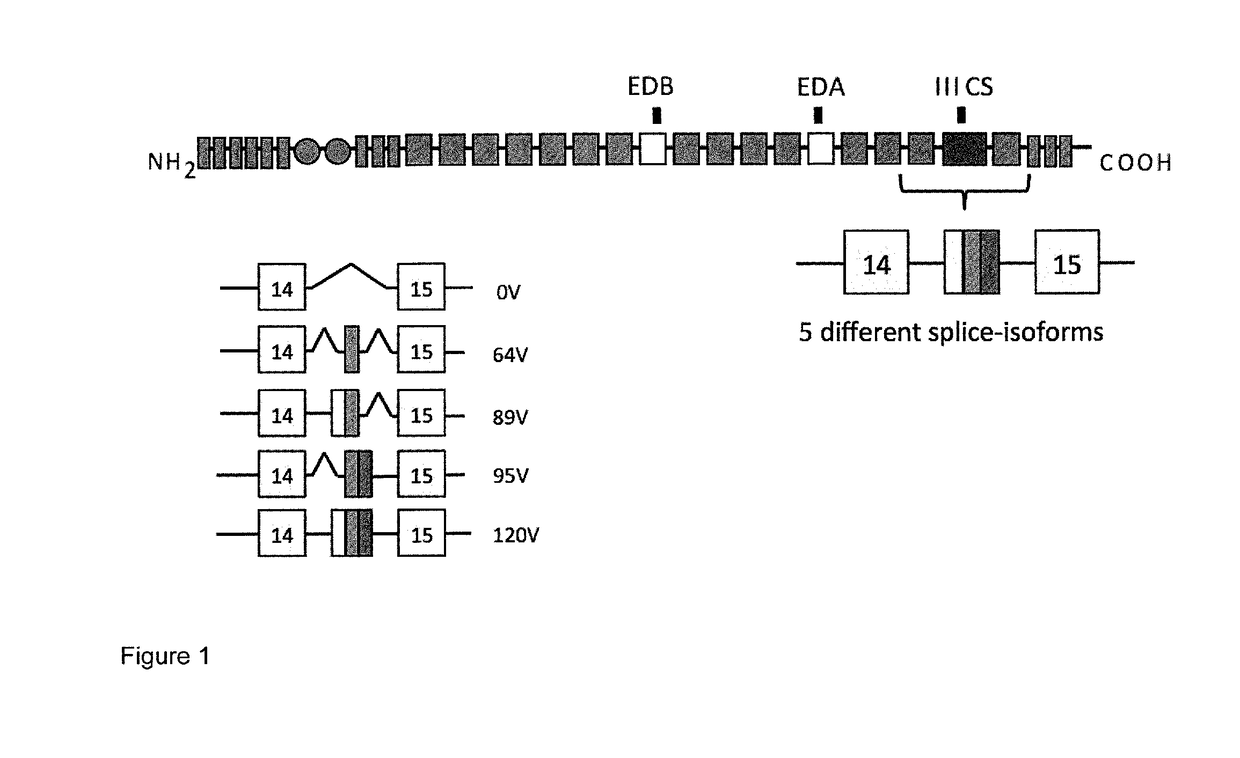
[0094]The SW01 and SW02 antibodies when used in monomeric scFv format, bind to the IIICS (89V) and IIICS (120V) isoforms (FIG. 1). Furthermore, using ELISA antibodies SW01 and SW02 were shown to recognize both the human and the rat recombinant IIICS (120V) isoform.
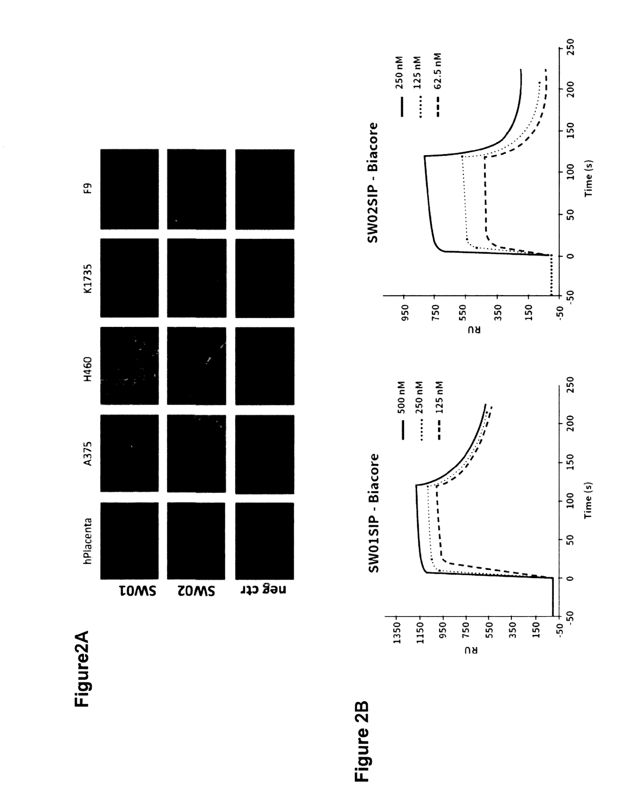
[0095]Antibodies SW01 and SW02 display good performance in immunofluorescence analyses. FIG. 2A shows staining of human placenta, human tumours (A375, H460) and mu...
example 2
on and Characterisation of a New Antibody Against MMP3
[0103]The CH01 antibody was isolated in scFv configuration from phage display libraries which include the libraries described in PCT / EP2009 / 006487, in Weber et al. (PLoS One, 2014, 9 (6) doi: 10 / 1361) and in Silacci et al. (Protein Engineering Design & Selection, 2006, 19, 471-478) according to the screening technique described by Silacci et al. (Protein Engineering Design & Selection, 2006, 19, 471-478) using a recombinant version of the catalytic domain of human MMP3 (amino acids 100-273). The antigen was produced in a bacterial expression system and biotinylated according to the standard protocol.
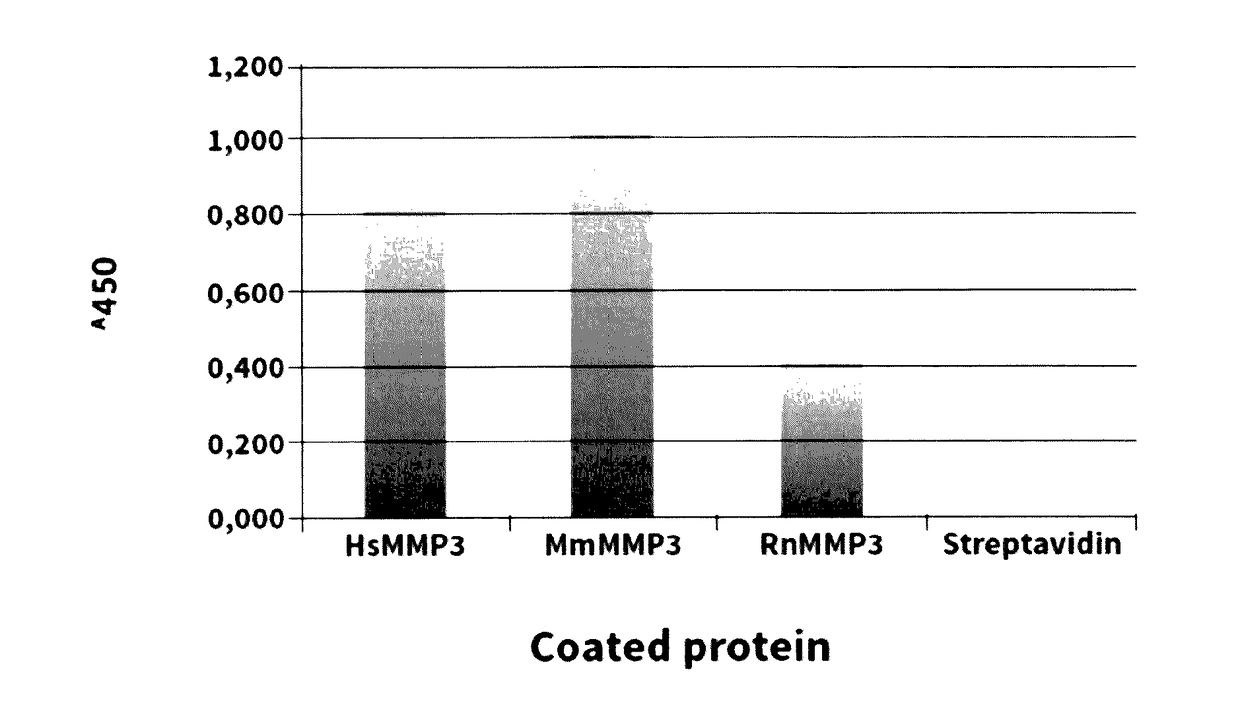
[0104]Antibody CH01 displays good staining of neovascular structures as shown by Immunofluorescence analyses. FIG. 3 shows staining of human placenta samples, in which 10 μm thick tissue samples were stained with the anti-human MMP3 antibody CH01 in scFv format. The anti-hen egg lysozyme antibody KSF in scFv format was used as an isot...
example 3
on and Characterisation of Four New Antibodies Against Periostin
[0110]The LG1, LG2, LG3 and 1E1 antibodies were isolated in scFv configuration from phage display libraries which include the libraries described in PCT / EP2009 / 006487, in Weber et al. (PLoS One, 2014, 9 (6) doi: 10 / 1361) and in Silacci et al. (Protein Engineering Design & Selection, 2006, 19, 471-478) according to the screening technique described by Silacci et al. (Protein Engineering Design & Selection, 2006, 19, 471-478). To generate antibodies against periostin, a recombinant version of the FAS domains (from 1 to 4; FIG. 5) was produced in a mammalian cell expression system. The antigen was biotinylated according to the standard protocol.
[0111]Binding of antibodies LG1, LG2 and LG3 (in scFv format) and 1E1 (in SIP format) to periostin was confirmed by Biacore analysis. The results are shown in FIGS. 6A and 6B.
Biacore Analysis:
[0112]Monomeric fractions of purified antibodies were analyzed by surface plasmon resonance...
PUM
| Property | Measurement | Unit |
|---|---|---|
| Cytotoxicity | aaaaa | aaaaa |
Abstract
Description
Claims
Application Information
 - R&D
- Intellectual Property
- Life Sciences
- Materials
- Tech Scout
- Unparalleled Data Quality
- Higher Quality Content
- 60% Fewer Hallucinations
Browse by: Latest US Patents, China's latest patents, Technical Efficacy Thesaurus, Application Domain, Technology Topic, Popular Technical Reports.
© 2025 PatSnap. All rights reserved.Legal|Privacy policy|Modern Slavery Act Transparency Statement|Sitemap|About US| Contact US: help@patsnap.com


