

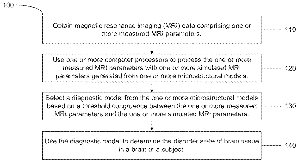
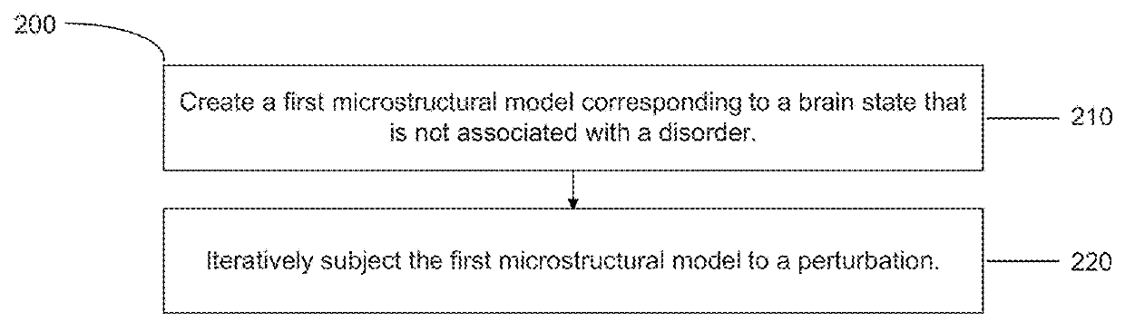
Methods and systems for identifying brain disorders
a brain disorder and brain technology, applied in the field of brain disorders, can solve the problems of a staggering direct and indirect cost, a huge societal burden, and a devastating effect on 9 million people domestically and 47 million people worldwide, and achieve the effect of reliable prediction and interpretation of health
- Summary
- Abstract
- Description
- Claims
- Application Information
AI Technical Summary
Benefits of technology
Problems solved by technology
Method used
Image

Examples
example 1
of Microstructure to MRI
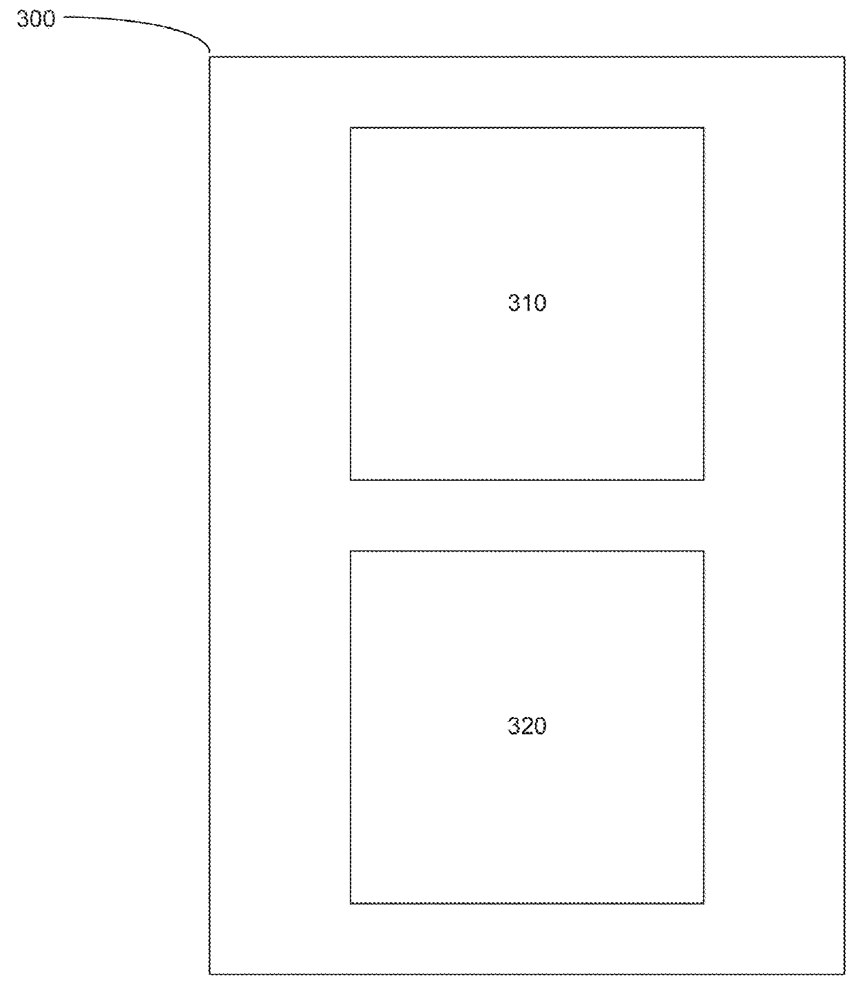
[0141]FIG. 5 shows the construction of exemplary microstructural models.
[0142]The first step is construction of a normal tissue microstructural model. The normal tissue microstructural model includes known measured or predicted values of cell density, cell shape, cell geometry, cell size, intercellular spacing, extracellular matrix heterogeneity, interstitial tortuosity, water to lipid ratio, and other tissue parameters that can influence structural and diffusion measurements. Typical ensembles consist of 1024×1024×1024 cell arrays. FIG. 5A shows a portion of a cell array for a normal tissue microstructural model. As shown in FIG. 5A, gray cubes 510 represent brain cells and green spots 520 represent molecular obstacles of the extracellular space.
[0143]The normal tissue microstructural model is then used for finite-element and Monte Carlo simulations of the tissue chemical composition, tissue micro-lattice topography, and molecular kinetics, which can subsequ...
example 2
ation of Human MRI
[0146]FIG. 6 shows the processing of human MRI images to produce neurodegeneration maps.
[0147]With a rigorous pipeline for structural prediction of MRI, each voxel of the human brain scan can be comprehensively characterized. The resulting output is a gray map with voxel-wise intensity scaling to represent the predicted deviation from normal.
[0148]Based on literature knowledge and initial experimental measurements, it is determined if the calculated tissue structure from a given combination of T1 time, T2 time, and diffusion coefficient values is within tolerance for healthy brain tissue or resides in the spectrum of abnormality. Each voxel 610 determined abnormal is coded red or a variable color, respectively denoting binary (percent neurodegeneration, PND) or quantitative (quantitative neurodegeneration, QND) abnormality in output maps. In effect, PND identifies the abnormal voxels (red) and QND defines how abnormal those voxels are. The range is from dark blue 6...
example 3
n of PND and QND Brain Maps
[0149]FIG. 7 shows exemplary MRI, PND, and QND brain maps from a diseased individual. The MRI scan shown in FIG. 7 is a diffusion-weighted image that, combined with T1-weighted and T2-weighted images (not shown in FIG. 7), is used to generate a grayscale output image. The grayscale output image serves as the underlying image for the PND and QND maps following removal of the skull and cerebrospinal fluid domains. Red voxels in the PND map represent microstructures that deviate from predicted ranges of structural tolerance for normal tissue. Colored voxels in the QND map similarly represent abnormal voxels, with color added to code for the extent of abnormality (blue=low abnormality, red=high abnormality).
[0150]To assess the degenerative state of an individual scan, the abnormality relative coverage (PND), extent of degeneration (QND), estimated total degeneration (END), variation of gray value (gRou), and variation of color value (color roughness, cRou) wer...
PUM
 Abstract
Description
Claims
Application Information