Drug delivery implants with bi-directional delivery capacity

a drug delivery and implant technology, applied in the field of implantable intraocular drug delivery devices, can solve the problems of many pathologies of the eye progress, the inability the inability to completely eliminate the ability of individuals to perceive visual images, so as to facilitate the passage of ocular fluid, facilitate the production of additional nitric oxide, and increase blood flow

Pending Publication Date: 2018-10-25
GLAUKOS CORP
View PDF4 Cites 15 Cited by
  • Summary
  • Abstract
  • Description
  • Claims
  • Application Information

AI Technical Summary

Benefits of technology

[0014]In several embodiments, implants are provided that comprise an impermeable barrier positioned within the interior lumen that divides the lumen into two compartments, each compartment housing a separate therapeutic agent. In such embodiments, there is additionally provided a drug eluting regulating membrane at both the distal and proximal regions of the implant, e.g., an elution controlling membrane for the first compartment and an elution controlling membrane for the second compartment. Depending on the embodiment, the elution controlling membranes may be the same material, or may be different materials, based on the characteristics of the drug positioned within each interior compartment. Additionally, each of the membranes may have different elution characteristics, depending on the therapeutic agent housed within each compartment. In some embodiments, the implants allow for bidirectional drug elution, e.g., drug elution from the proximal portion of the implant, as well as drug elution from the distal portion of the implant. In several embodiments, this bidirectional drug elution allows two different regions of the eye to be treated concurrently. In some embodiments two different aspects of an ocular disorder are treated, while in some embodiments a side effect of one of the therapeutic agents is lessened by the administration of the second therapeutic agent.
[0016]In some embodiments, the first drug comprises a prostaglandin analog. In several embodiments the prostaglandin analog is selected from the group consisting of bimatoprost, latanoprost, travoprost and unoprostone. In several embodiments, the prostaglandin analog is positioned within the more proximally positioned interior chamber of an implant having two chambers and thus, the prostaglandin analog is positioned such that it elutes from the implant into the anterior chamber of the eye. In several embodiments, the second drug positioned within the second interior chamber comprises a vasodilator. Vasodilator compounds that are released from the implant, in several embodiments, act on the trabecular meshwork, and its integrated vasculature, to increase blood flow in and around the trabecular meshwork. In some embodiments, this increase blood flow facilitates passage of ocular fluid through the trabecular meshwork and into a physiological fluid outflow pathway. Suitable vasodilators include, but are not limited to, endothelium derived hyperpolarizing factor (EDHF), cyclic GMP-dependent protein kinase, nitric oxide, epinephrine, histamine, prostacyclin, prostaglandin D2, prostaglandin E2, vasoactive intestinal peptide (VIP), adenosine, L-arginine, bradykinin, substance P, niacin, platelet activating factor, or precursors or derivatives of any of the agent listed above. In particular embodiments, nitric oxide, or precursor or derivatives thereof, is released from the implant. In several embodiments, a multicomponent composition suitable for generating nitric oxide in vivo is positioned within the second chamber of an implant. For example, one or more of the enzymes that functions to convert L-arginine to nitric oxide as well as L-arginine can be positioned within the second chamber of an implant. Thus, nitric oxide can be generated in vivo, close to its site of action, which in some embodiments addresses the relatively short half-life of nitric oxide. In several embodiments endothelial nitric oxide synthase, neuronal nitric oxide synthase, and / or inducible nitric oxide synthase positioned within the implant, along with L-arginine. In alternative embodiments the implant can house L-arginine as the second therapeutic agent and the natives enzymes responsible for converting L-arginine to nitric oxide within the eye are leveraged to generate nitric oxide as the L-arginine is eluted from the implant. In several embodiments, S-nitroso-N-acetylpenicillamine (SNAP), hydroxylamine, and / or sodium nitroprusside can be positioned within the implant to generate nitric oxide. Additionally, in several embodiments, supplemental compounds can be included in the implant that facilitate the production of additional nitric oxide, for example, by regeneration of L-arginine (thereby allowing further production of nitric oxide by conversion of the regenerated L-arginine).

Problems solved by technology

Numerous pathologies can compromise or entirely eliminate an individual's ability to perceive visual images, including trauma to the eye, infection, degeneration, vascular irregularities, and inflammatory problems.
Glaucoma causes pathological changes in the optic nerve, visible on the optic disk, and it causes corresponding visual field loss, which can result in blindness if untreated.
Many pathologies of the eye progress due to the difficulty in administering therapeutic agents to the eye in sufficient quantities and / or duration necessary to ameliorate symptoms of the pathology.
Due to this metabolism, systemic administration may require undesirably high concentrations of the drug to reach therapeutic levels at an ocular target site.
This can not only be impractical or expensive, but may also result in a higher incidence of side effects.
Topical administration is potentially limited by limited diffusion across the cornea, or dilution of a topically applied drug by tear-action.
Even those drugs that cross the cornea may be unacceptably depleted from the eye by the flow of ocular fluids and transfer into the general circulation.

Method used


Image

  • Drug delivery implants with bi-directional delivery capacity
  • Drug delivery implants with bi-directional delivery capacity
  • Drug delivery implants with bi-directional delivery capacity

Examples


Embodiment Construction

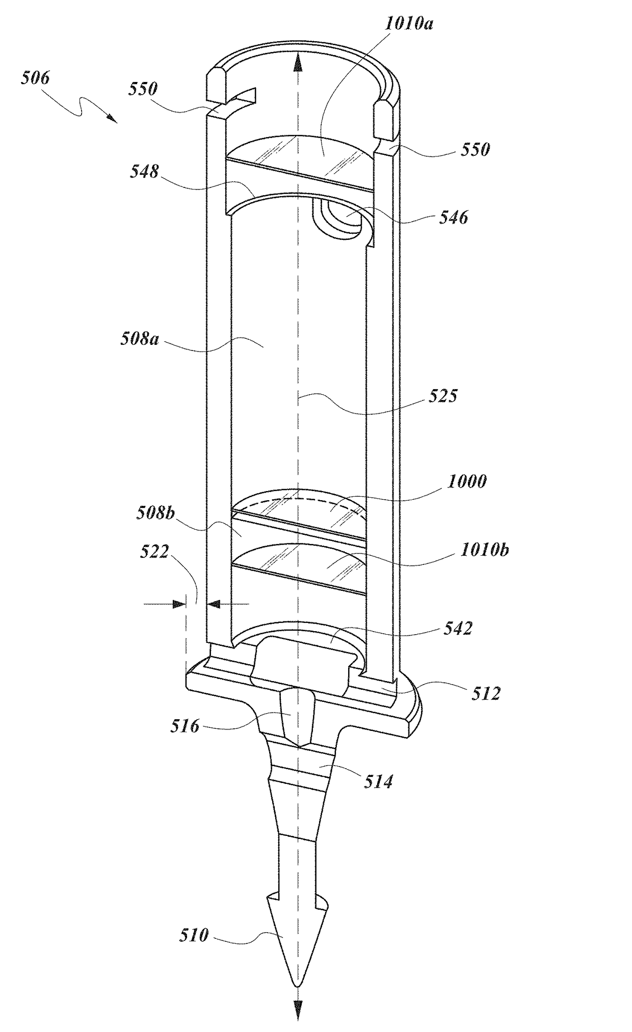
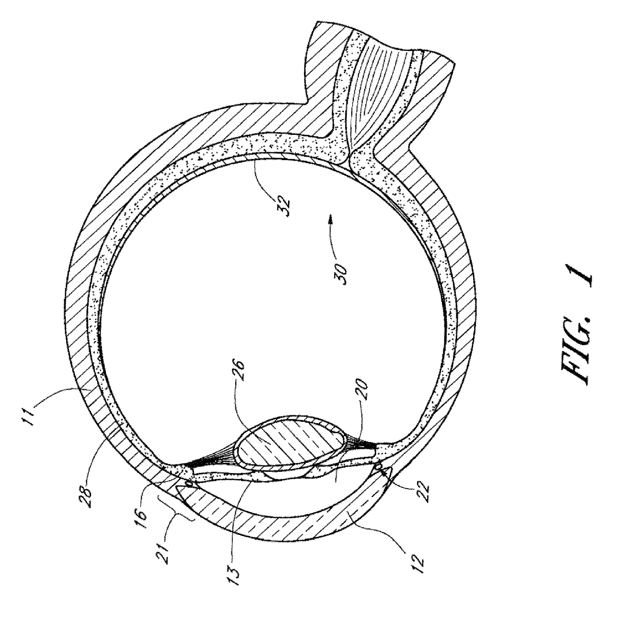
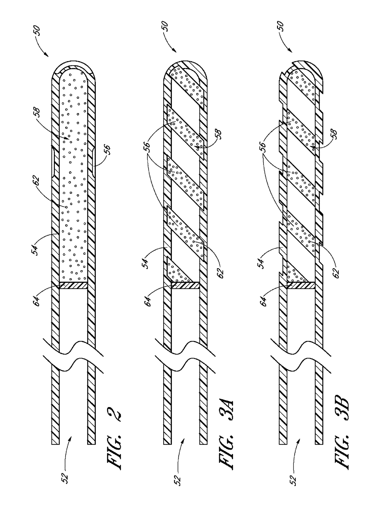
[0084]Achieving local ocular administration of a drug may require direct injection or application, but could also include the use of a drug eluting implant, a portion of which, could be positioned in close proximity to the target site of action within the eye or within the chamber of the eye where the target site is located (e.g., anterior chamber, posterior chamber, or both simultaneously). Use of a drug eluting implant could also allow the targeted delivery of a drug to a specific ocular tissue, such as, for example, the macula, the retina, the ciliary body, the optic nerve, or the vascular supply to certain regions of the eye. Use of a drug eluting implant could also provide the opportunity to administer a controlled amount of drug for a desired amount of time, depending on the pathology. For instance, some pathologies may require drugs to be released at a constant rate for just a few days, others may require drug release at a constant rate for up to several months, still others ...


PUM


Abstract

Disclosed herein are drug delivery devices and methods for the treatment of ocular disorders requiring targeted and controlled administration of a drug to an interior portion of the eye for reduction or prevention of symptoms of the disorder. In several embodiments, the devices are capable of controlled release of two or more drugs, the release of each drug into a different ocular space (e.g., an ocular compartment versus an ocular fluid outflow pathway).

Description

RELATED APPLICATIONS[0001]This application claims priority to U.S. Provisional Application No. 62 / 213,573, filed Sep. 2, 2015, which is incorporated by reference herein in it its entirety.BACKGROUNDField[0002]This disclosure relates to implantable intraocular drug delivery devices structured to provide targeted and / or controlled release of a drug to a desired intraocular target tissue and methods of using such devices for the treatment of ocular diseases and disorders. In certain embodiments, this disclosure relates to a treatment of increased intraocular pressure wherein aqueous humor is permitted to flow out of an anterior chamber of the eye through a surgically implanted pathway. In certain embodiments, this disclosure also relates particularly to a treatment of ocular diseases with drug delivery devices affixed to the eye, such as to fibrous tissue within the eye.Description of the Related Art[0003]The mammalian eye is a specialized sensory organ capable of light reception and i...

Claims


Application Information

Patent Timeline
Patent Type & Authority Applications(United States)
IPC IPC(8): A61F9/00A61K9/00
CPCA61F9/0017A61K9/0051A61F9/00781A61K9/0004A61F2250/0068A61K9/0092A61M31/002A61F2250/0067A61K9/009A61K9/0097A61K9/0024A61K9/00
Inventor HEITZMANN, HAROLD A.BURNS, THOMAS W.HAFFNER, DAVID S.
Owner GLAUKOS CORP
Why Eureka
Try Eureka