Taggable fluorescent probe for calcium ion detection
a fluorescent probe and calcium ion technology, applied in the field of fluorescent probes for calcium ion detection, can solve the problems of difficult to precisely visualize intracellular casup>2+/sup> dynamics, and difficulty in realizing the localization of such probes to any intracellular si
- Summary
- Abstract
- Description
- Claims
- Application Information
AI Technical Summary
Benefits of technology
Problems solved by technology
Method used
Image
Examples
example 1
Synthesis of bis(acetoxymethyl) 2,2′-((4-(3′-acetoxy-2′,7′-dichloro-6′-(3-((2-(2-((6-chloro hexyl)oxy)ethoxy)ethyl)carbamoyl)azetidin-1-yl)-3-oxo-3H-spiro[isobenzofuran-1,9′-xanthene]-5-carboxylic acid amido)-2-(2-(acetoxymethoxy)-2-oxoethoxy)phenyl)azanediyl) diacetate (Compound 14)
[0065]Compound 14, one of the compounds of the present invention, was synthesized according to the procedure of the following reaction scheme.
(1) Synthesis of bis(acetoxymethyl)2,2′-((2-(2-(acetoxymethoxy)-2-oxoethoxy)-4-aminophenyl)azanediyl)diacetate (Compound 3)
[0066]2-(2-(Bis(2-methoxy-2-oxoethyl)amino)-5-nitrophenoxy) acetic acid (Compound 1) (767.8 mg, 2.155 mmol) was placed in a flask and dissolved in THF (15 mL). After addition of 1N NaOH (6.7 mL) while stirring the solution, the mixture was stirred at room temperature under light-shielding conditions for 40 minutes. Then, 1N NaOH (2.1 mL) was added, and the mixture was further stirred for 30 minutes. Ethyl acetate was added to the solution, and ...
example 2
Evaluation of Chemical Characteristics of Compound 14
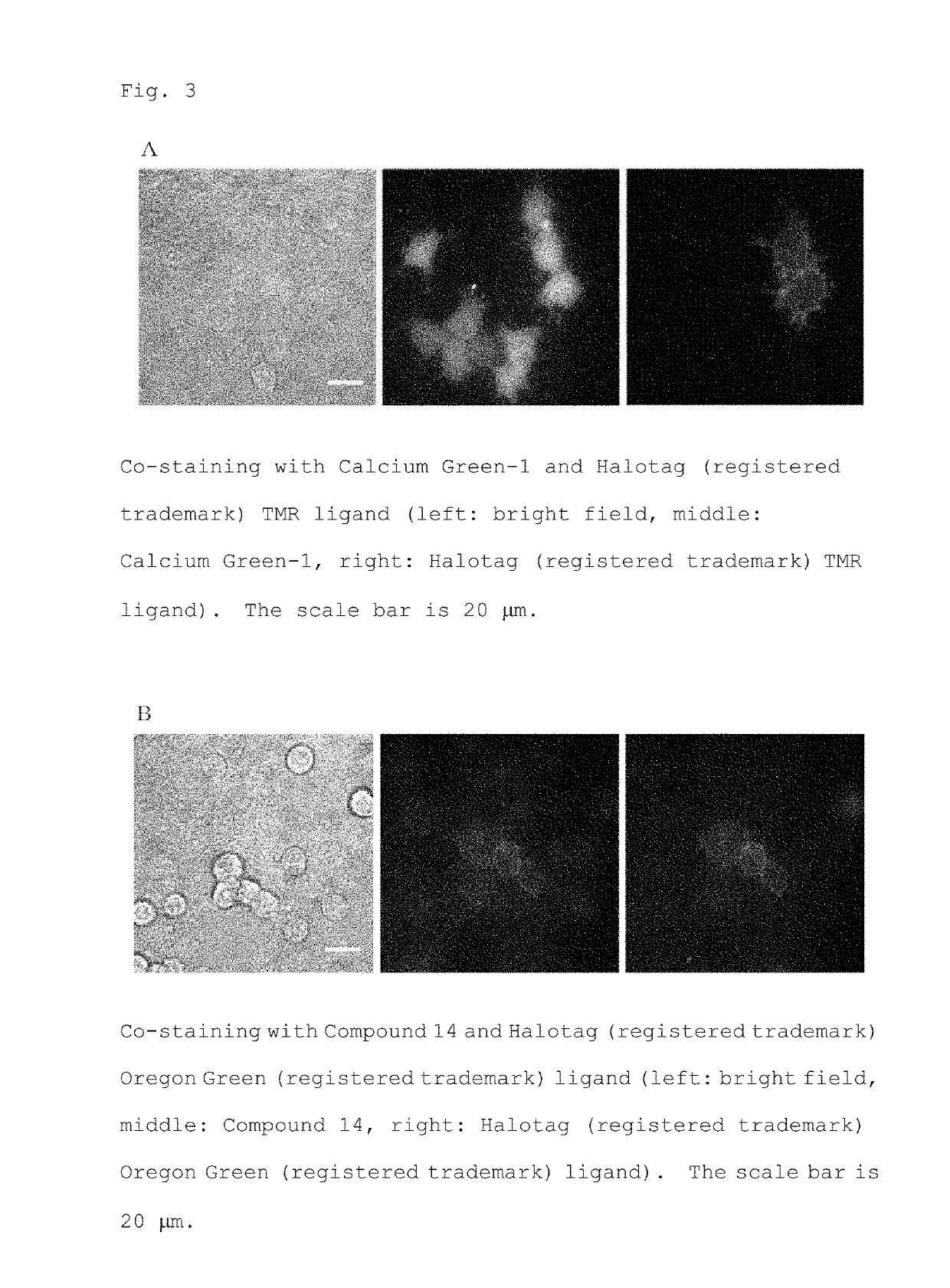
[0105]In order to evaluate the chemical characteristics of the synthesized Compound 14, a hydrolysate was obtained by esterase treatment according to the above method, and the fluorescence intensity in a buffer in which the Ca2+ concentration was adjusted was measured with a spectrofluorophotometer.
[0106]As a result, the fluorescence intensity of the probe bound to Ca2+ was about 11 times larger (Fmax / Fmin=10.8) than that of the unbound probe, and the dissociation constant (Kd) was 11.1 μM (FIG. 2). In FIG. 2, the black circles (●) indicate each data point, which showed a fit curve that is a curved line.
example 3
Evaluation of Localization and Ca2+ Response Characteristics in HEK 293T Cells
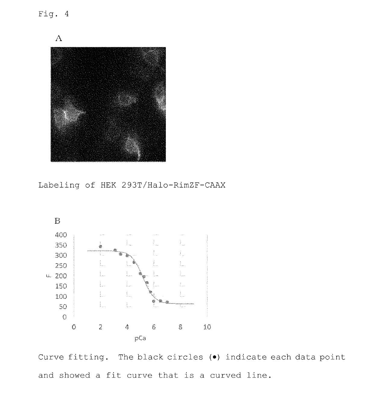
[0107]Staining with a fluorescent probe was performed using HEK 293T cells expressing a HaloTag protein on the cell membrane by introducing pcDNA Halo-RimZF CAAX gene (hereinafter referred to as HEK 293T / Halo-RimZF-CAAX). With Calcium Green, AM, the cytoplasm of all cells in the visual field was stained regardless of expression of the HaloTag protein (FIG. 3A). On the other hand, with Compound 14, unlike conventional probes, fluorescence localized on the cell membrane on which the HaloTag protein is expressed was observed (FIG. 3B). The expression site of the HaloTag protein was identified by staining with a HaloTag ligand.
[0108]Next, Ca2+ response characteristics of the labeled probe were evaluated by in situ calibration. As a result, it was found that Fmax / Fmin was 4.7 and Kd was 6.4 μM (FIG. 4). In FIG. 4B, the black circles (●) indicate each data point, which showed a fit curve that is a curved line.
[0...
PUM
 Abstract
Description
Claims
Application Information
 - R&D
- Intellectual Property
- Life Sciences
- Materials
- Tech Scout
- Unparalleled Data Quality
- Higher Quality Content
- 60% Fewer Hallucinations
Browse by: Latest US Patents, China's latest patents, Technical Efficacy Thesaurus, Application Domain, Technology Topic, Popular Technical Reports.
© 2025 PatSnap. All rights reserved.Legal|Privacy policy|Modern Slavery Act Transparency Statement|Sitemap|About US| Contact US: help@patsnap.com


