Immune adjuvant for cancer
a cancer and immune-boosting technology, applied in the field of immune-boosting drugs for cancer, can solve the problems of giving up clinical applications and insufficient response rate, and achieve the effects of mild improvement of the response rate and the effect of pd-1/pd-l1 antibody therapy
- Summary
- Abstract
- Description
- Claims
- Application Information
AI Technical Summary
Benefits of technology
Problems solved by technology
Method used
Image
Examples
example 1
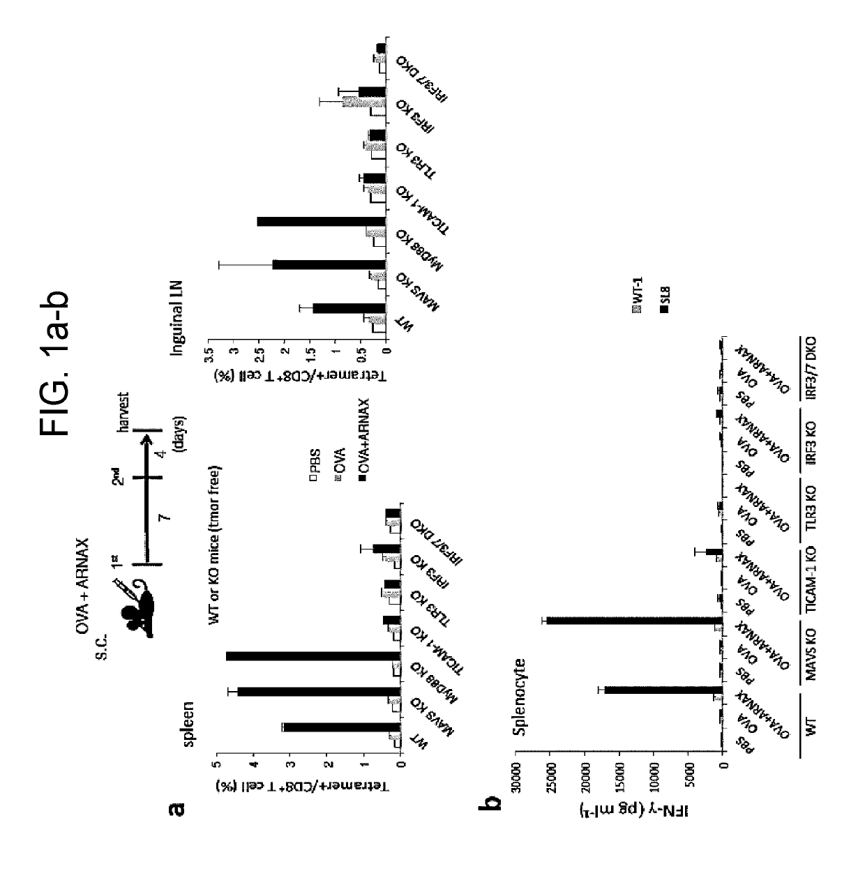
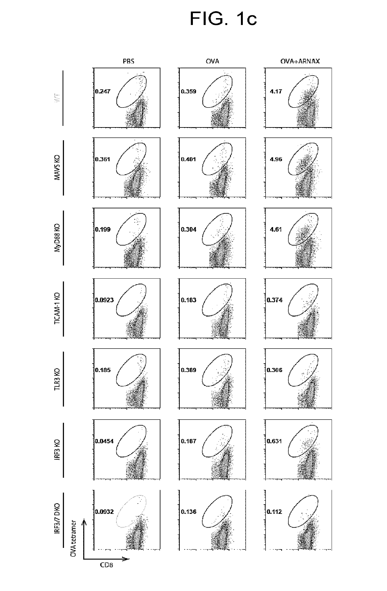
of CTLs by Administration of ARNAX and Antigen
1. Materials and Method
[0142]8 to 11-week wild type (C57BL / 6) and knockout male mice (MAVS KO, MyD88 KO, TLR3 KO, TICAM-1 KO, IRF3 KO, and IRF3 / 7 DKO) on the B57BL / 6 background were used in an experiment. PBS, OVA (100 μg), or OVA (100 μg)+ARNAX (60 μg) was subcutaneously administrated to mice on day 0 and day 7. On day 11, spleens and lymph nodes of lower limbs were collected, and the proportion of OVA-specific CD8+ T cells in CD8+ T cells was measured with an OVA-tetramer (made by MO BIO Laboratories, Inc.). Tetramer assay was performed according to the attached document. Specifically, spleens and lymph node cells (1×10⁷ / ml) were dyed with an H2-Kb OVA tetramer (PE) 50× diluted for 20 minutes, and were washed, followed by dyeing with anti-CD3e mAb (APC) and anti-CD8a mAb (FITC) to analyze the proportion of OVA-specific CD8+ T cells in CD8+ T cells with a cell sorter FACSAria (BD Biosciences). Spleen cells (1×10⁷ / ml) were stimulated wit...
example 2
n of Cytokine Production Effect and Safety of ARNAX According to Administration Route
1. Materials and Method
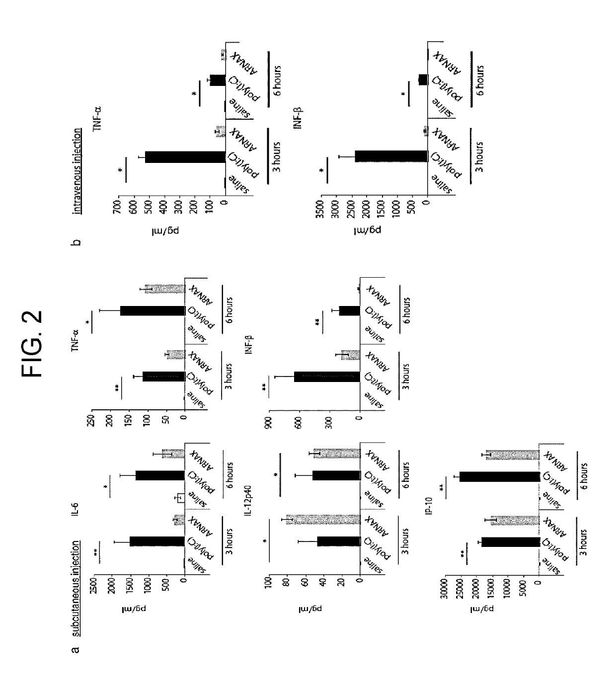
[0145]Saline, poly(I:C) (150 μg), or ARNAX (150 μg) was subcutaneously administrated to 8-week wild type (C57BL / 6) female mice, or saline, poly(I:C) (50 μg), or ARNAX (50 μg) was intravenously administrated to them. Blood cytokine amount was then measured after 3 hours and 6 hours (FIG. 2). IL-6, TNF-α, and IL-12 p40 were measured by CBA and IFN-β, and IP-10 was measured by ELISA.
2. Results
[0146]Inflammatory cytokines IL-6 and TNF-α in blood in the subdermal administration of ARNAX were less than in the administration of poly(I:C). The amount of IFN-γ was also extremely small. In contrast, production of a Th1 cytokine IL-12p40 was induced more than in poly(I:C), and production of a chemokine IP-10, which recruit NK cells, NKT cells, and T cells, was induced as much as poly(I:C). In the intravenous administration, poly(I:C) strongly induced TNF-α, IL-6, and IFN-β while ARNAX ba...
example 3
Effect by Administration ARNAX and Antigen (Thymoma)
1. Materials and Method
[0148]An OVA-expressing tumor (thymoma) line EG7 (2×10⁶ / 200 μl PBS) was subcutaneously transplanted to the lower backs of C67BL / 6 mice (7-week old, female). On day 7, PBS (n=5) or OVA (100 μg)+ARNAX (60 μg) (n=5) was subcutaneously administrated, and the proliferation of tumors was measured over time (FIG. 3c). On day 14, tumors were collected, the proportion of CD8+ T cells within the tumors, and proportion of OVA-specific CD8+ T cells and CD11c-positive CD8+ T cells within the CD8+ T cells were measured by flow cytometry (FIG. 3d). Tumor tissue sections thereof were prepared, and were immunostained with an anti-CD8a antibody. The invasion of CD8+ T cells into the tumors was observed with a confocal microscope in the tissue sections of 20 fields of a PBS group and 16 fields of an OVA+ARNAX group, and the results were converted into numeric values (FIG. 3f). Furthermore, RNAs were purified from the tumor tiss...
PUM
| Property | Measurement | Unit |
|---|---|---|
| concentration | aaaaa | aaaaa |
| concentration | aaaaa | aaaaa |
| concentration | aaaaa | aaaaa |
Abstract
Description
Claims
Application Information
 - R&D
- Intellectual Property
- Life Sciences
- Materials
- Tech Scout
- Unparalleled Data Quality
- Higher Quality Content
- 60% Fewer Hallucinations
Browse by: Latest US Patents, China's latest patents, Technical Efficacy Thesaurus, Application Domain, Technology Topic, Popular Technical Reports.
© 2025 PatSnap. All rights reserved.Legal|Privacy policy|Modern Slavery Act Transparency Statement|Sitemap|About US| Contact US: help@patsnap.com


