

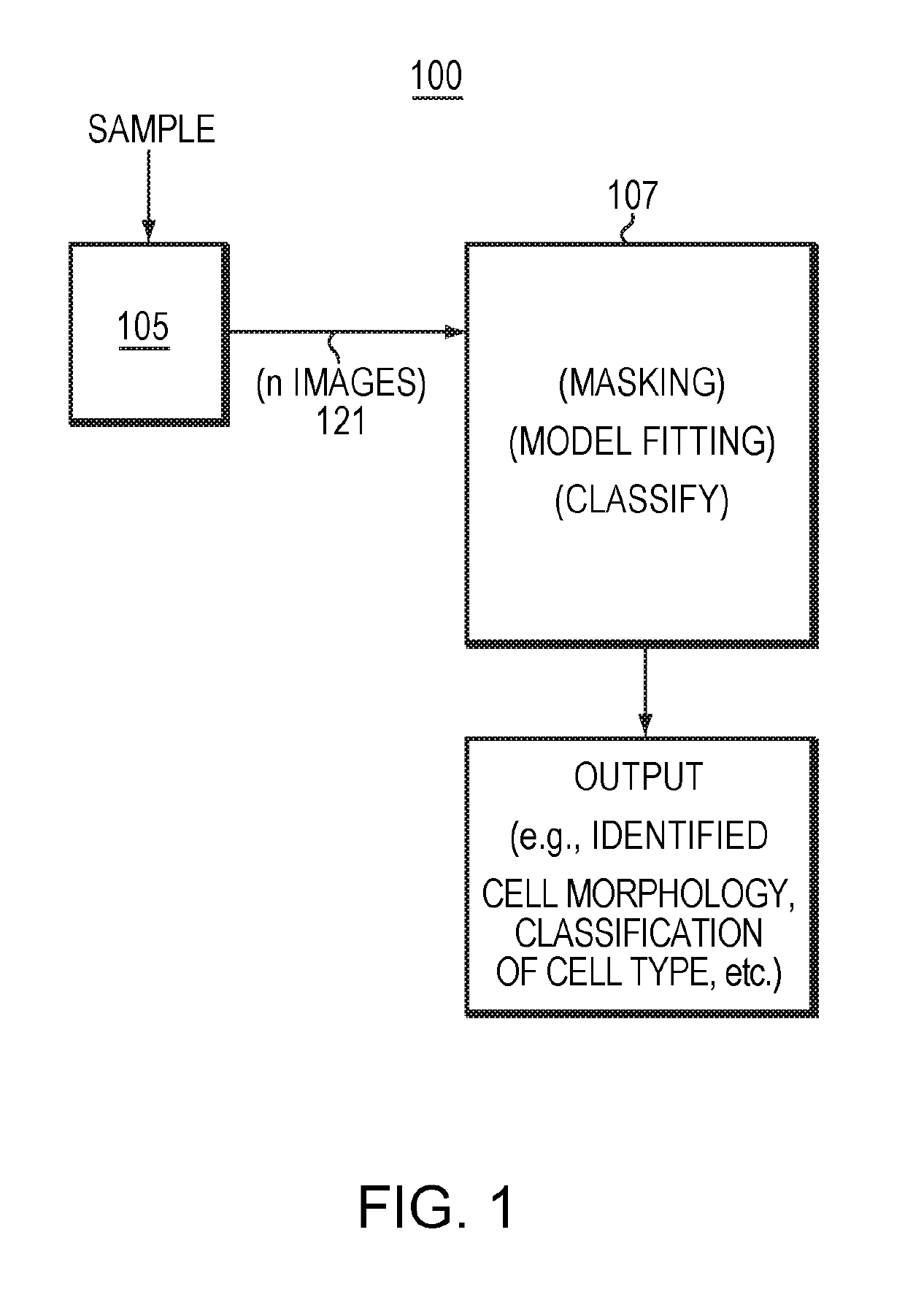
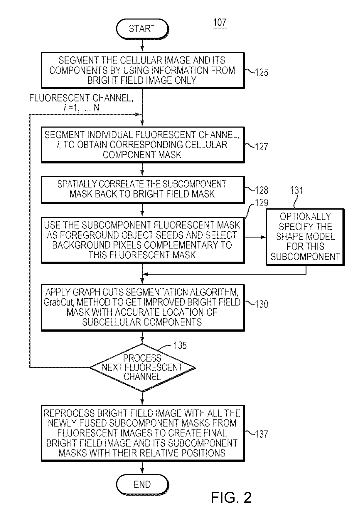
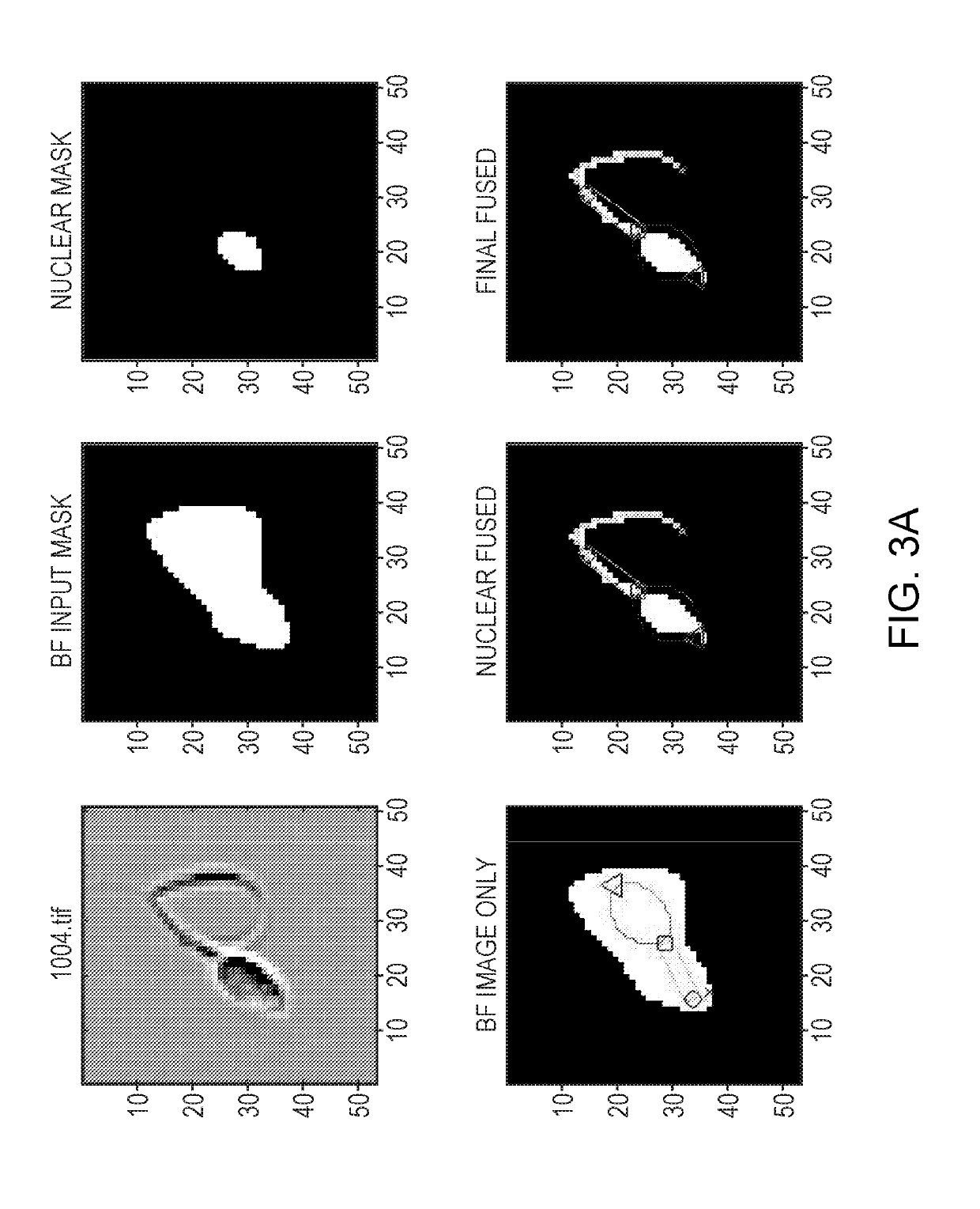
A Method To Combine Brightfield And Fluorescent Channels For Cell Image Segmentation And Morphological Analysis Using Images Obtained From Imaging Flow Cytometer (IFC)
a morphological analysis and cell image technology, applied in image enhancement, instruments, medical/anatomical pattern recognition, etc., can solve the problems of difficult to accurately classify complex morphologies such as sickle cells, diatoms, spermatozoa, etc., and achieve accurate extraction of salient morphological features at high speed and resolution.
- Summary
- Abstract
- Description
- Claims
- Application Information
AI Technical Summary
Benefits of technology
Problems solved by technology
Method used
Image

Examples
example
[0053]Assessment of sperm morphology is one of the most important steps in the evaluation of sperm quality. A higher percentage of morphologically abnormal sperm is strongly correlated to lower fertility. Morphological image analysis can be used to segment sperm morphology, extract associated quantitative features, and classify normal and abnormal sperms in large quantities. In the majority of human infertility clinics and livestock semen laboratories, brightfield microscopy is used to evaluate sperm morphology. However, since sperm morphology contains a variety of shapes, sizes, positions, and orientations, the accuracy of the analysis can be less than optimal as it is based on a limited number of cells (typically 100-400 cells per sample). To overcome this challenge, we used ImageStream imaging flow cytometry to acquire brightfield, side-scatter and Propidium Iodide (PI) images of boar sperm samples at 60× magnification. We developed novel image algorithms to perform image segment...
PUM
 Abstract
Description
Claims
Application Information