Automated Cell Identification Using Shearing Interferometry

a technology of shearing interferometry and cell identification, applied in the field of systems and methods for automated cell identification/classification, can solve the problems of high cost and cumbersome analysis

Active Publication Date: 2019-07-25
UNIV OF CONNECTICUT
View PDF0 Cites 9 Cited by
  • Summary
  • Abstract
  • Description
  • Claims
  • Application Information

AI Technical Summary

Benefits of technology

[0006]The present disclosure provides improved systems and methods for automated cell identification/classification. More particularly, the present disclosure pr

Problems solved by technology

However, such analysis can be expensive and cum

Method used


Image

  • Automated Cell Identification Using Shearing Interferometry
  • Automated Cell Identification Using Shearing Interferometry
  • Automated Cell Identification Using Shearing Interferometry

Examples


example 1

nd Field-Portable 3D Printed Shearing Digital Holographic Microscope for Automated Cell Identification

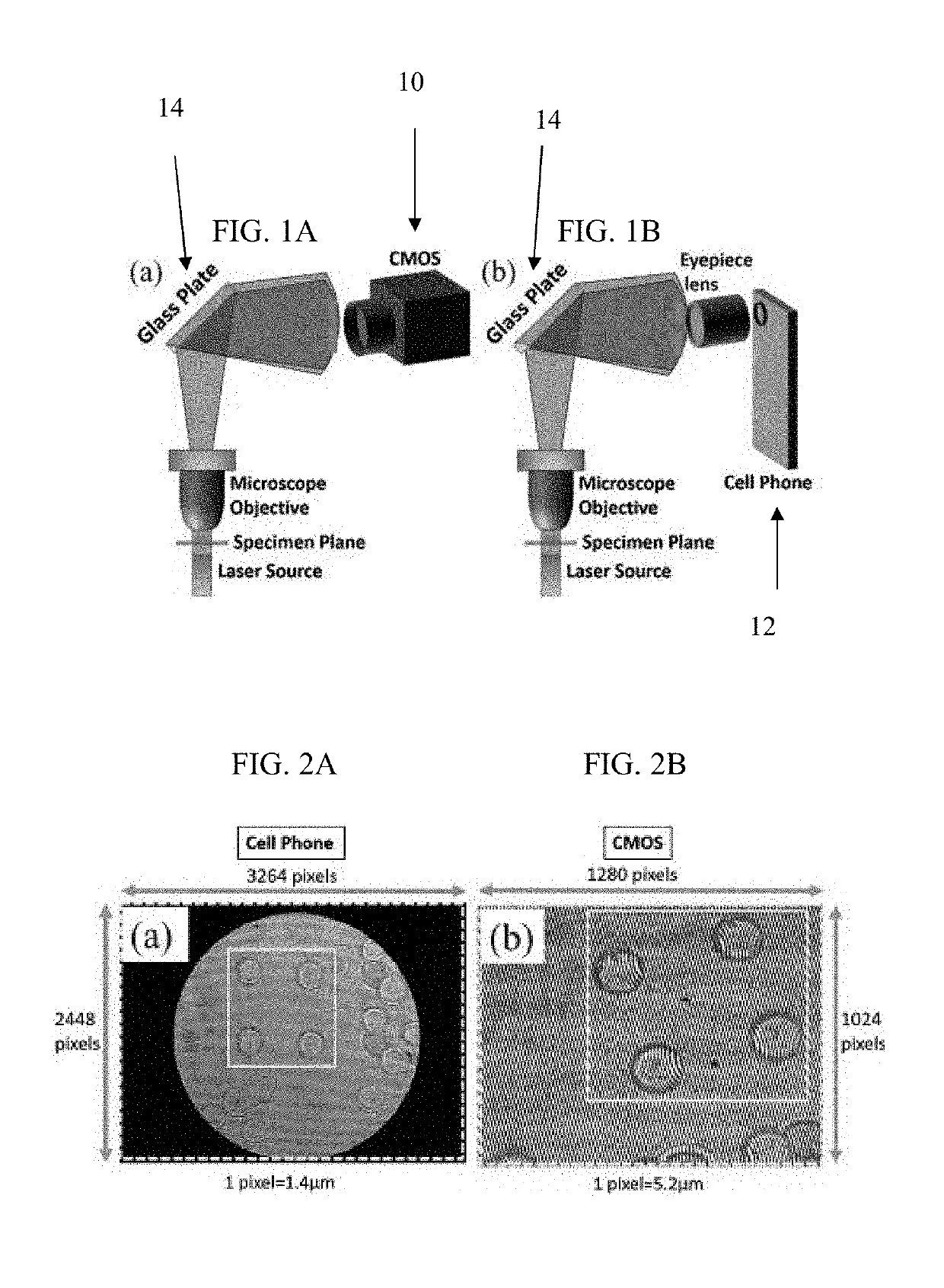
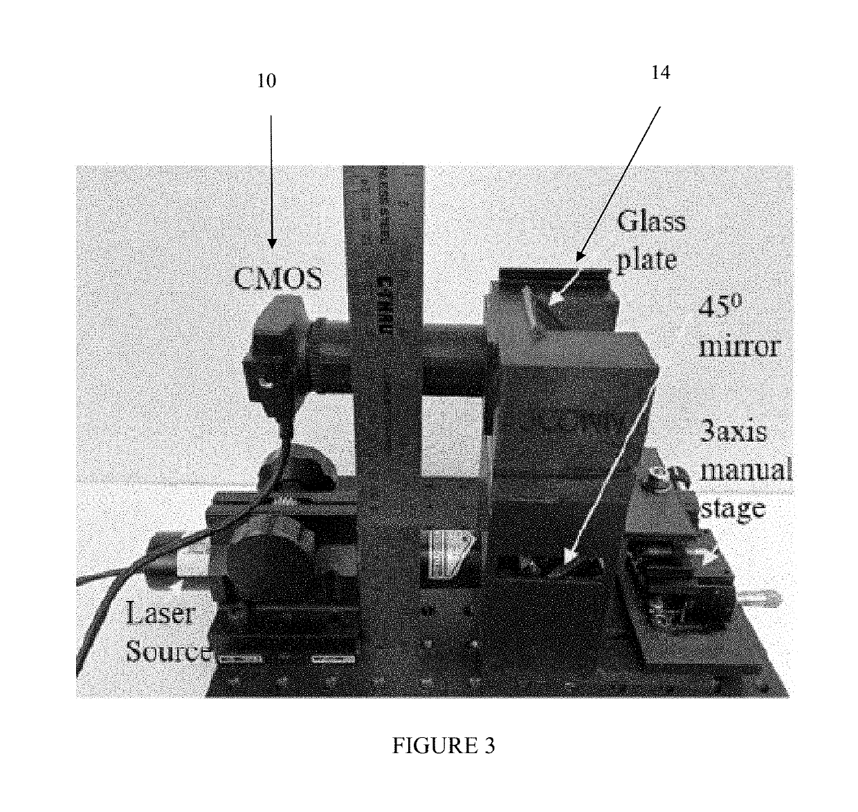
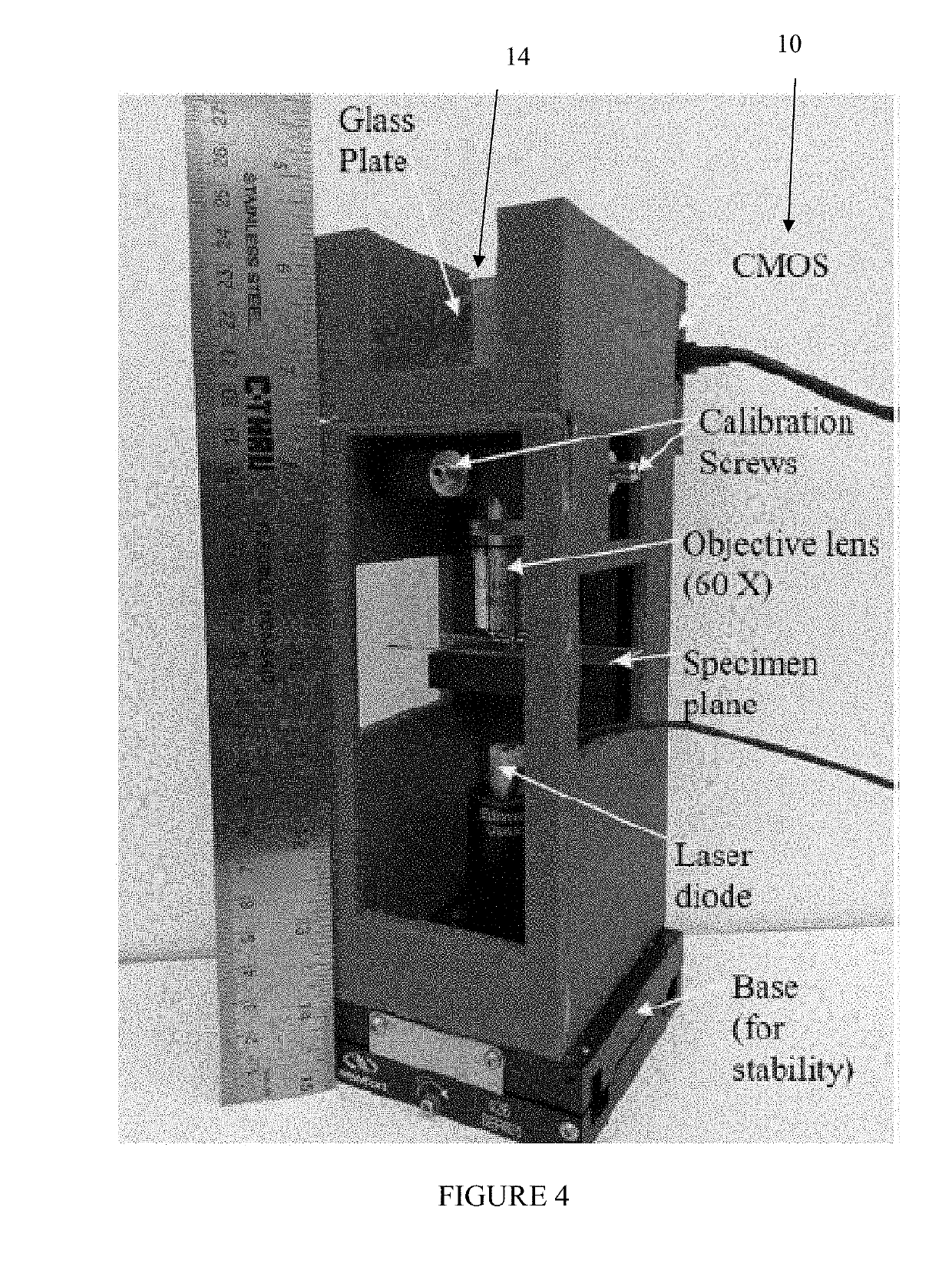
[0118]In exemplary embodiments, the present disclosure provides a low-cost, compact, and field-portable 3D printed holographic microscope for automated cell identification / classification based on a common path shearing interferometer setup. Once a hologram is captured from the portable setup, a 3D reconstructed height profile of the cell is created. One can extract several morphological cell features from the reconstructed 3D height profiles, including mean physical cell thickness, coefficient of variation, optical volume (OV) of the cell, projected area of the cell (PA), ratio of PA to OV, cell thickness kurtosis, cell thickness skewness, and the dry mass of the cell for identification using the random forest (RF) classifier. The 3D printed prototype can serve as a low-cost alternative for the developing world, where access to laboratory facilities for disease diagnosis are limited...

example 2

ll Disease Diagnosis Based on Spatio-Temporal Cell Dynamics Analysis Using 3D Printed Shearing Digital Holographic Microscopy

[0162]This Example provides a spatio-temporal analysis of cell membrane fluctuations to distinguish healthy patients from patients with sickle cell disease. A video hologram containing either healthy red blood cells (h-RBCs) or sickle cell disease red blood cells (SCD-RBCs) was recorded using a low-cost, compact, 3D printed shearing interferometer. Reconstructions were created for each hologram frame (time steps), forming a spatio-temporal data cube. Features were extracted by computing the standard deviations and the mean of the height fluctuations over time and for every location on the cell membrane, resulting in two-dimensional standard deviation and mean maps, followed by taking the standard deviations of these maps. The optical flow algorithm was used to estimate the apparent motion fields between subsequent frames (reconstructions). The standard deviati...

example 3

[0227]Digital holographic microscopy (DHMIC) is a label-free imaging modality that enables the viewing of microscopic objects without the use of exogenous or contrast agents. DHMIC provides high axial accuracy; however, the lateral resolution is dependent on the magnification of the objective lens used. DHMIC overcomes two problems associated with conventional microscopy: the finite depth of field, which is inversely proportional to the magnification of the objective, and low contrast between the cell and the surrounding media. Cells alter the phase of the probe wave front passing through the specimen, depending on the refractive index and thickness of the object. Several methods have been developed to transform the phase information of the object into amplitude or intensity information, but these methods only provide qualitative information and lack quantitative information. Staining methods, such as the use of exogenous contrast agents, can enhance the image contrast, but it might...


PUM


Abstract

The present disclosure provides improved systems and methods for automated cell identification/classification. More particularly, the present disclosure provides advantageous systems and methods for automated cell identification/classification using shearing interferometry with a digital holographic microscope. The present disclosure provides for a compact, low-cost, and field-portable 3D printed system for automatic cell identification/classification using a common path shearing interferometry with digital holographic microscopy. This system has demonstrated good results for sickle cell disease identification with human blood cells. The present disclosure provides that a robust, low cost cell identification/classification system based on shearing interferometry can be used for accurate cell identification. For example, by combining both the static features of the cell along with information on the cell motility, classification can be performed to determine the type of cell present in addition to the state of the cell (e.g., diseased vs. healthy).

Description

CROSS-REFERENCE TO RELATED APPLICATIONS[0001]This application claims priority to U.S. Provisional Application Ser. No. 62 / 631,140, filed on Feb. 15, 2018, and entitled “Automated Cell Identification Using Shearing Interferometry” and to U.S. Provisional Application Ser. No. 62 / 631,268, filed on Feb. 15, 2018, and entitled “Portable Common Path Shearing Interferometry-Based Holographic Microscopy System.” The content of each of the foregoing provisional applications is incorporated herein in their entireties.STATEMENT OF GOVERNMENT SUPPORT[0002]This invention was made with Government support under Grant 1545687 awarded by the National Science Foundation. The government has certain rights in the invention.FIELD OF THE DISCLOSURE[0003]The present disclosure relates generally to systems and methods for automated cell identification / classification and, more particularly, to systems and methods for automated cell identification / classification using shearing interferometry with a digital h...

Claims


Application Information

Patent Timeline
IPC IPC(8): G01N15/10G01B9/02G06K9/00G06T7/00
CPCG01N15/10G01B9/02G06K9/00147G06T7/0012G01N2015/1486G01N2015/1006G01N2015/1093G01N2015/1087G01N2015/1075G06T2207/30024G06T2207/10056G06T2207/10152G06T2207/10016G06T2207/20081G06T7/62G03H1/0443G01N15/1468G02B21/14G03H2001/005G03H2001/0452G03H1/041G03H2226/11G06V20/693G06V20/698
Inventor JAVIDI, BAHRAMMARKMAN, ADAMRAWAT, SIDDHARTHANAND, ARUN
Owner UNIV OF CONNECTICUT
Why Eureka
Try Eureka