
Computer aided diagnosis system for classifying kidneys

a technology kidney, applied in the field of computer aided diagnosis system and automatic method to classify kidney, can solve the problems of patient development of chronic kidney disease, complications can still arise, and the kidney function is gradually lost, so as to achieve accurate identification and classification of renal transplants, reliable non-invasive diagnostic tools

Active Publication Date: 2019-08-01
UNIV OF LOUISVILLE RES FOUND INC
View PDF0 Cites 22 Cited by
  • Summary
  • Abstract
  • Description
  • Claims
  • Application Information

AI Technical Summary

Benefits of technology

The present invention is a non-invasive diagnostic tool that uses magnetic resonance imaging (MRI) to detect abnormalities in kidney transplants. The initial results on 66 subjects using a specific approach showed a high accuracy in classifying abnormal and normal kidney transplants. This suggests that the invention can reliably detect and locate areas of damage or disease in kidneys without causing harm to the patient.

Problems solved by technology

These can result in a gradual loss of kidney function leading to waste build up in the body and cause the patient to develop chronic kidney disease (CKD).
Though this has greatly improved the outcome of patients diagnosed with stage 5 CKD, complications can still arise.
Traditional blood tests and urine sampling to evaluate renal transplant dysfunction cannot assess the function of individual kidneys.
Biopsies are invasive, expensive, have a high morbidity rate, and may over- or under-estimate the extent of inflammation (an indicator of rejection) in the entire graft.
However, functional abnormalities inside different parts of the kidney (e.g., cortex and medulla) cannot be discriminated precisely due to scintigraphy's limited spatial resolution.
Also, radionuclide imaging includes radiation exposure, thus limiting the range of its applications.
However, the contrast agents used are still nephrotoxic and the information gathered by CT to detect acute rejection (AR) is unspecific, thus limiting the CT role in AR diagnosis.
The aforementioned shortcomings make those imaging techniques impractical for clinical use and has led to the use of alternative modalities (e.g., the ultrasound (US) and MRIs) to assess renal transplant functionality.
However, shadowing artifacts, low signal-to-noise ratios (SNRs), and speckles notably decrease the quality of US image and diagnostic confidence.
Moreover, conventional US parameters are not exact indicators of renal graft dysfunction, and it can only provide a prognostic graft marker or even a similar indication like other diagnostic possibilities, such as ATN.
In addition, their methods are unable to compensate for the motion of the kidney since they did not account for the whole kidney.
Furthermore, none of the aforementioned studies integrates both image—and clinical—based biomarkers to evaluate the kidney function.
Moreover, the studies mentioned above did not investigate the fusion of multiple MR imaging modalities (e.g. DCE- and / or DW- with BOLD-MRI) to evaluate kidney function.
Finally, none of these studies investigated neither the differentiation between AR and different renal disease (DRD) nor between the different AR types (e.g., T-cell mediated-rejection (TMR) or anti-body mediated-rejection (AMR)).

Method used


Image

  • Computer aided diagnosis system for classifying kidneys
  • Computer aided diagnosis system for classifying kidneys
  • Computer aided diagnosis system for classifying kidneys

Examples


Embodiment Construction

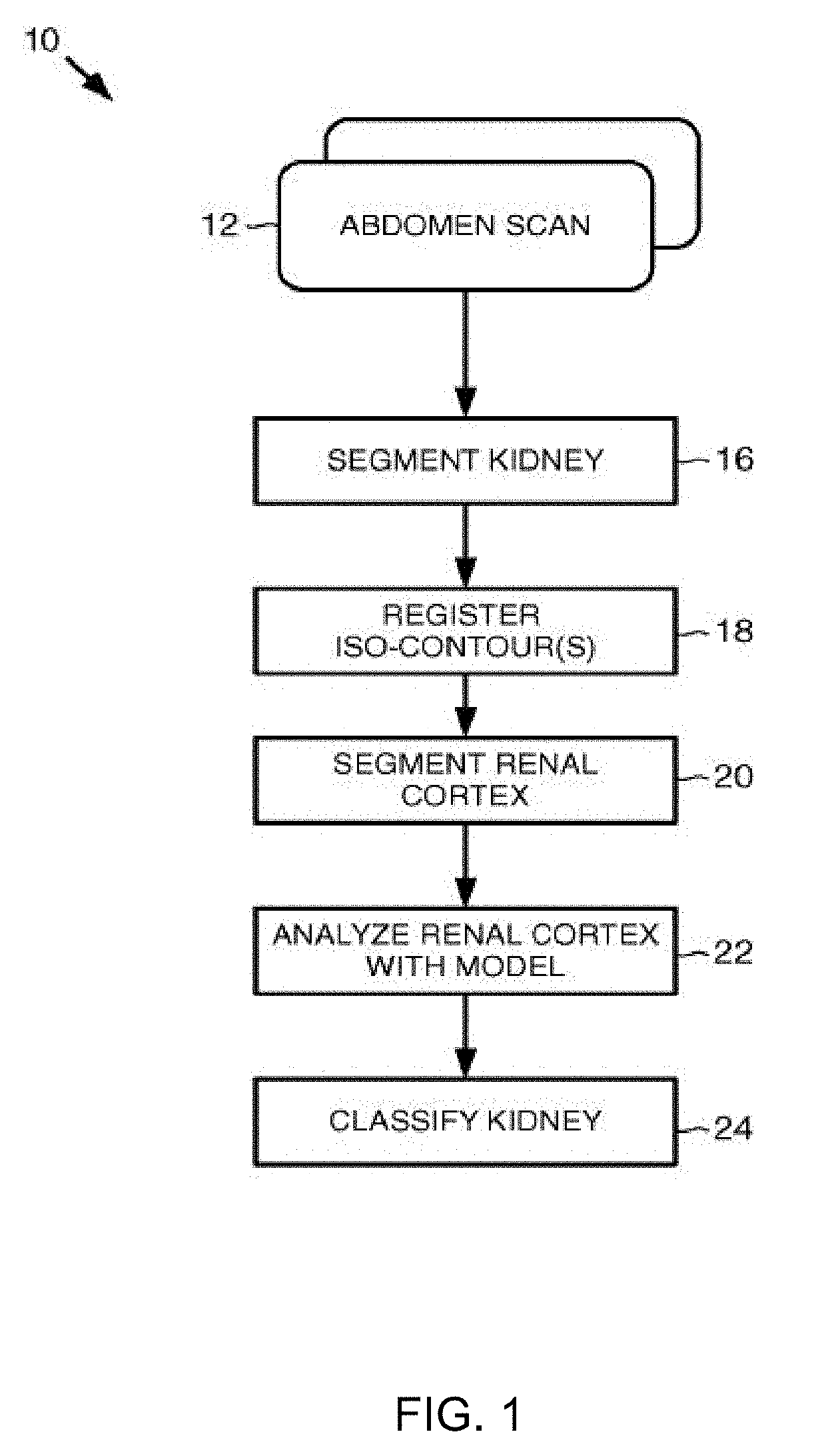
[0052]Embodiments consistent with the invention provide for automated classification of a kidney based on image analysis of at least a portion of the kidney that is in image data of a medical scan, such as an abdomen scan. Consistent with embodiments of the invention, a transplanted kidney may be classified as acutely rejected or non-rejected based at least on image analysis performed on a medical scan associated with the transplanted kidney. For example, embodiments of the invention may receive an abdomen scan that comprises a plurality of time sliced dynamic contrast-enhanced magnetic response imaging (DCE-MRI) data that includes kidney image data. Embodiments of the invention may analyze the kidney image data, and based on a learned model that classifies kidneys, the transplanted kidney may be classified based on one or more features determined from the analyzed kidney image data.

[0053]Further details are provided in [1] F. Khalifa, M. El-Ghar, B. Abdollahi, H. Frieboes, T. EI-Di...


PUM


Abstract

A computer aided diagnostic system and automated method to classify a kidney utilizes medical image data and clinical biomarkers in evaluation of kidney function pre- and post-transplantation. The system receives image data from a medical scan that includes image data of a kidney, then segments kidney image data from other image data of the medical scan. The kidney is then classified by analyzing at least one feature determined from the kidney image data and the at least one clinical biomarker.

Description

CROSS REFERENCE TO RELATED APPLICATION[0001]This application is a continuation-in-part of U.S. application Ser. No. 15 / 903,769, entitled COMPUTER AIDED DIAGNOSTIC SYSTEM FOR CLASSIFYING KIDNEYS, filed Feb. 23, 2018, which is a continuation of U.S. application Ser. No. 14 / 676,111, entitled COMPUTER AIDED DIAGNOSTIC SYSTEM FOR CLASSIFYING KIDNEYS, filed Apr. 1, 2015, which claims the benefit of U.S. application Ser. No. 61 / 974,134, entitled COMPUTER AIDED DIAGNOSTIC SYSTEM FOR CLASSIFYING KIDNEYS, filed Apr. 2, 2014. This application further claims the benefit of U.S. application Ser. No. 62 / 633,917, entitled NON-INVASIVE DIAGNOSIS PLATFORM FOR EVALUATING KIDNEY FUNCTION BY INTEGRATING CLINICAL-BIOMARKERS WITH MULTI-MODALITY MR IMAGE-MARKERS, filed Feb. 22, 2018. The contents of each of these related applications are hereby incorporated by reference in their entireties.FIELD OF THE INVENTION[0002]A computer aided diagnostic system and automated method to classify a kidney utilizes med...

Claims


Application Information

Patent Timeline
Patent Type & Authority Applications(United States)
IPC IPC(8): G16H30/40G06T7/143G06T7/12G06T7/33G06T7/00A61B5/20G16H30/20G16H50/20
CPCG16H30/40G06T7/143G06T7/12G06T7/33G06T7/0012A61B5/201G16H30/20G16H50/20G06T2207/30084G06T2207/10088G06T2207/10096G06T2207/20081A61B2576/02A61B5/7264A61B2505/05A61B5/4848A61B5/14546A61B5/0261A61B5/0295G16H50/30
Inventor EL-BAZ, AYMAN S.DWYER, AMYSOLIMAN, AHMEDSHEHATA, MOHAMEDABDELTAWAB, HISHAMKHALIFA, FAHMI
Owner UNIV OF LOUISVILLE RES FOUND INC
Features
  • Generate Ideas
  • Intellectual Property
  • Life Sciences
  • Materials
  • Tech Scout
  • Unparalleled Data Quality
  • Higher Quality Content
  • 60% Fewer Hallucinations
Learn More