
Structured illumination microscopy system using digital micromirror device and time-complex structured illumination, and operation method therefor

a microscopy system and structure technology, applied in the direction of fluorescence/phosphorescence, instruments, optical elements, etc., can solve the problems of inability to make high-speed measurement, inability to change the diffraction lattice pattern, and generation of vibration and speed limitations

Active Publication Date: 2020-03-12
TOMOCUBE INC
View PDF2 Cites 6 Cited by
  • Summary
  • Abstract
  • Description
  • Claims
  • Application Information

AI Technical Summary

Benefits of technology

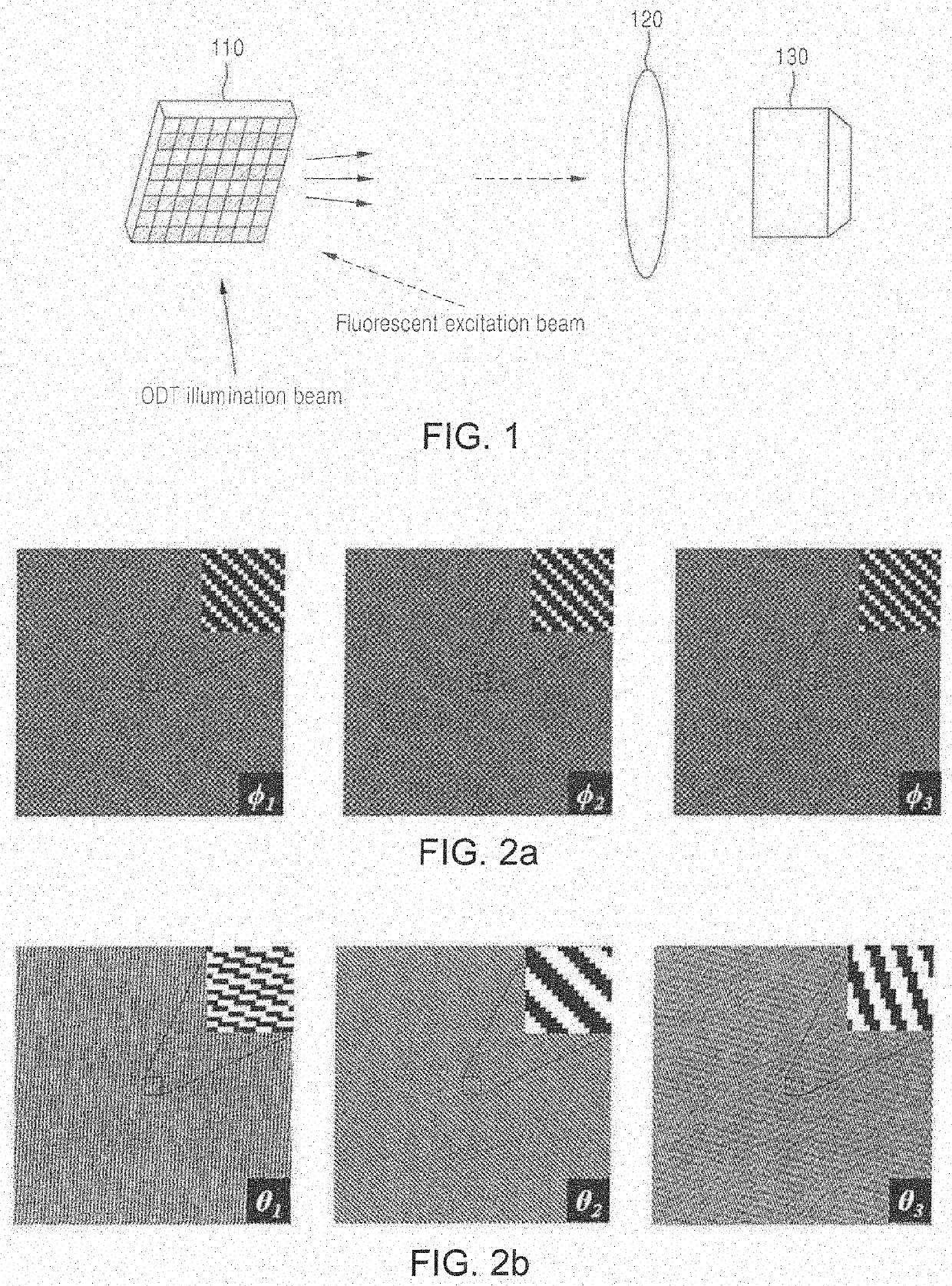
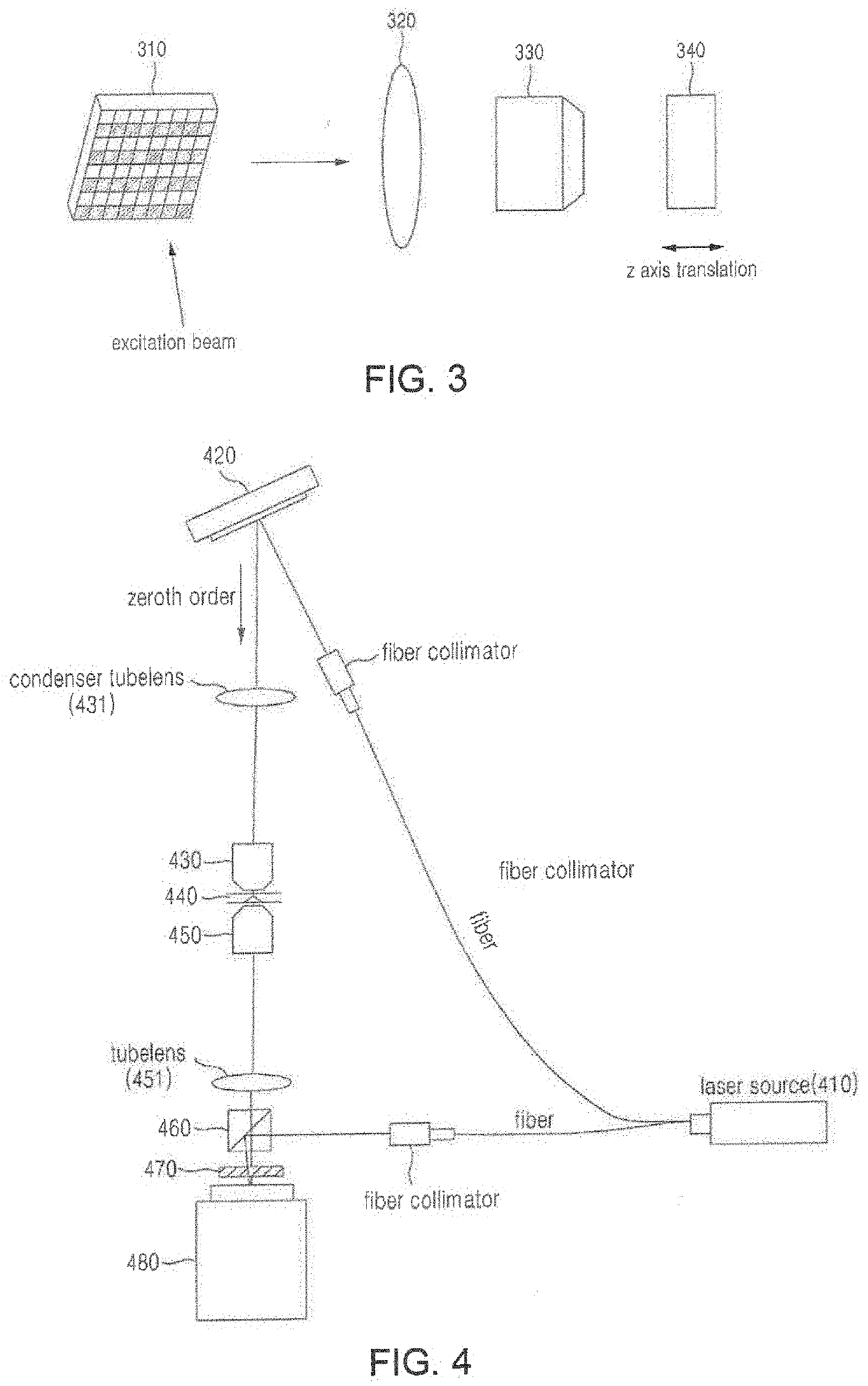
This patent is about a system and method for high-resolution 3D fluorescence imaging using a digital micromirror device and time-complex structured illumination. The system is capable of implementing time-complex structured illumination by using the digital micromirror device and extracting a high-resolution 3D fluorescence image.

Problems solved by technology

However, a technique using a physical diffraction lattice (non-patent document 2) has a disadvantage in that vibration and speed limitation are generated and the diffraction lattice pattern cannot be changed because the diffraction lattice pattern has to be mechanically moved.
In addition, since a technique using a conventional digital micromirror device (DMD) (non-patent document 4) is a method of inputting a binary pattern into a digital micromirror device (DMD), an incoherent light source such as an LED is required to generate a sinusoidal pattern so that it is impossible to make high-speed measurement.
When using a coherent light source such as a laser to increase the speed, an undesired noise pattern occurs due to the diffraction that occurs in the binary pattern, so that the image analysis is impossible.

Method used


Image

  • Structured illumination microscopy system using digital micromirror device and time-complex structured illumination, and operation method therefor
  • Structured illumination microscopy system using digital micromirror device and time-complex structured illumination, and operation method therefor
  • Structured illumination microscopy system using digital micromirror device and time-complex structured illumination, and operation method therefor

Examples


Embodiment Construction

[0192]While a few exemplary embodiments have been shown and described with reference to the accompanying drawings, it will be apparent to those skilled in the art that various modifications and variations can be made from the foregoing descriptions. For example, adequate effects may be achieved even if the foregoing processes and methods are carried out in different order than described above, and / or the aforementioned elements, such as systems, structures, devices, or circuits, are combined or coupled in different forms and modes than as described above or be substituted or switched with other components or equivalents.

[0193]Therefore, other implements, other embodiments, and equivalents to claims are within the scope of the following claims.


PUM


Abstract

Presented are a structured illumination microscopy system using a digital micromirror device and a time-complex structured illumination, and an operation method therefor. A structured illumination microscopy system using a digital micromirror device and a time-complex structured illumination according to an embodiment may comprise: a light source; a digital micromirror device (DMD) for receiving light irradiated from the light source, implementing a time-complex structured illumination, and causing a controlled structured illumination to enter a sample; and a fluorescence image measurement unit for extracting a high-resolution 3D fluorescence image of the sample.

Description

TECHNICAL FIELD[0001]The following embodiments relate to a structured illumination microscopy system using a digital micromirror device and a time-complex structured illumination, and an operation method therefor.BACKGROUND ART[0002]In order to obtain a fluorescence image of a cell, a fluorescent protein is expressed in a specific organ (molecule) of the cell or dye is attached thereto. If an excitation light source is incident into the expressed fluorescence material, the fluorescent protein emits a fluorescence signal of another wavelength after absorbing the excitation light source. An image of an inner structure of a cell may be distinguished and measured through the fluorescence signal.[0003]Recently, a structured illumination microscopy is applied to a method of increasing the resolution of a fluorescence image. The structured illumination microscopy is a method of obtaining an image of an ultrahigh-resolution, which exceeds the value of a diffraction limit by making an excita...

Claims


Application Information

Patent Timeline
IPC IPC(8): G02B21/00G02B21/26G02B21/36G06T11/00G06T3/40H04N13/365
CPCG02B21/0084G02B21/0056G02B21/367G06T3/4053H04N13/365G02B21/26G02B21/0076G02B21/0032G06T11/003G02B21/0048G02B6/32G02B6/35G01B9/02G01N21/17G01N21/45G01N21/64G02B21/06G02B26/0833
Inventor PARK, YONGKEUN
Owner TOMOCUBE INC
Learn More