Dual image system suitable for oncological diagnoses and real time guided biopsies

a biopsies and image technology, applied in the field of image technology, can solve the problems of system capability, reduced detection sensitivity of the system, heavy equipment and difficult operation, etc., and achieve the effect of improving event statistics, maximizing results, and increasing detection efficiency

Pending Publication Date: 2020-04-02
UNIV DE VALENCIA +1
View PDF9 Cites 0 Cited by
  • Summary
  • Abstract
  • Description
  • Claims
  • Application Information

AI Technical Summary

Benefits of technology

[0035]Thus, it is possible, to adjust the number of events measured and the uncertainty in the detection, as needed from the information provided by the ultrasound image to maximize the result based on the damage position.
[0036]In another preferred embodiment of the invention, at least one Compton detector comprises three or more individual gamma detectors, substantially aligned in the same detection direction and with detection surfaces substantially parallel between them. Thus, it is achieved to provide a higher detection efficiency, which improves the event statistics, and, hence, reduces the time needed for reconstructing the functional image of the patient's tissue.

Problems solved by technology

That is, gamma ray detectors that employ a collimator limit the system capabilities in terms of optimum energy range for the image reconstruction and, above all, they substantially reduce detection sensitivity of the system (considering only the detected events of gamma rays that must necessarily have passed through the collimator) and also make the equipment very heavy and difficult to operate.

Method used


Image

  • Dual image system suitable for oncological diagnoses and real time guided biopsies
  • Dual image system suitable for oncological diagnoses and real time guided biopsies
  • Dual image system suitable for oncological diagnoses and real time guided biopsies

Examples


Embodiment Construction

[0052]An exemplary preferred embodiment of the present invention is described for purposes of illustration, but not limitation thereof, will now be described.

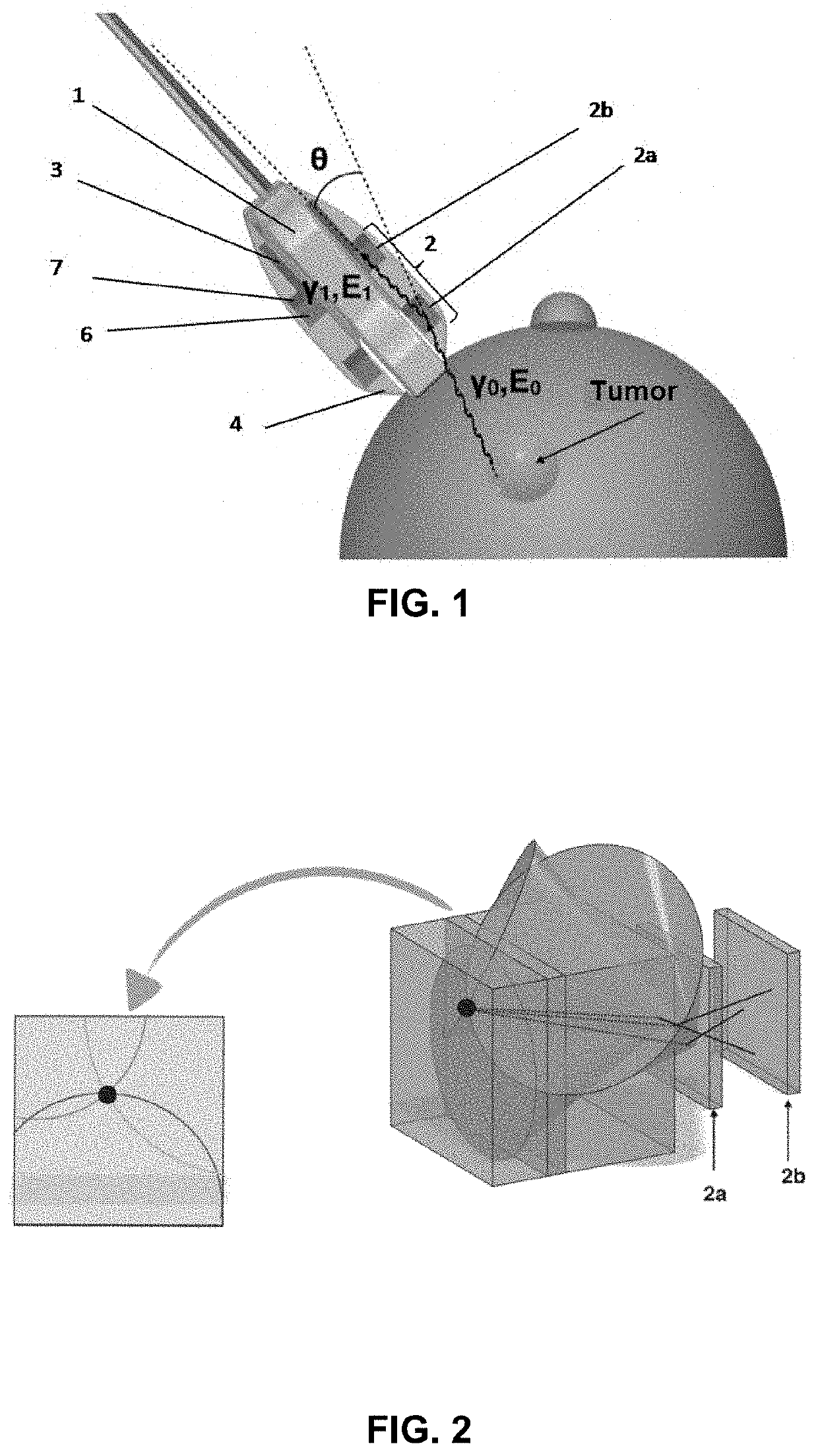
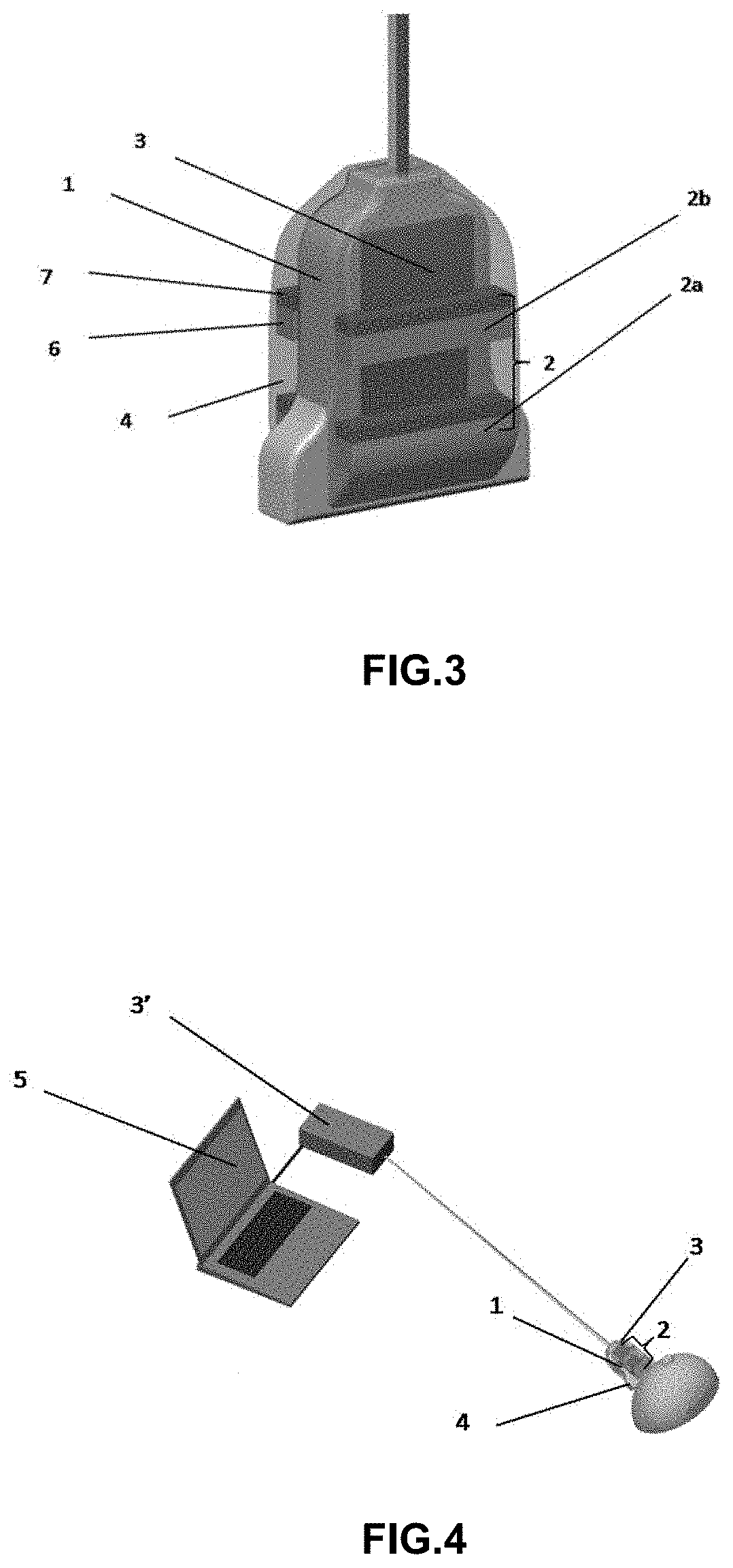
[0053]As described in the previous sections, a principal object of the invention is related to a dual imaging system, suitable for the performance of guided biopsies through images based on ultrasound imaging for the providing of signals associated to one or more anatomical images and gamma radiation device for oncological diagnostics.

[0054]In a preferred embodiment of the invention, as shown in the FIGS. 1-5, the dual imaging system comprises an ultrasound imaging device (1), at least one gamma radiation Compton detector (2), an electronic module (3), a signal processing module (3′), a casing (4) and a computational module (5).

[0055]Preferably, each gamma radiation Compton detector (2), as shown in the FIG. 2, comprises at least two gamma detection regions, substantially aligned in the same detection direction. These two gamma...


PUM


Abstract

The present invention relates to a dual image system, suitable for oncological diagnoses and real time guided biopsies, comprising: an ultrasound imaging device (1) for obtaining associated signals with one or more anatomical images; a gamma ray detector (2) for obtaining associated signals with one or more functional images; an electronic module (3) for processing the obtained signals by the detectors (1, 2); a casing (4) housing the detectors (1, 2); characterized in that the gamma ray detector (2) is a gamma ray Compton scattering detector, comprising at least two individual gamma detectors (2a, 2b) substantially aligned in the same detection direction and with the detection surfaces substantially parallel to each other; the ultrasound imaging device (1) and the gamma ray detector (2) have the same detection directions substantially aligned with each other.

Description

FIELD OF THE INVENTION[0001]The present invention is reported within the technical field of the oncological surgery, and more specifically, to the image technologies, diagnosis and treatment of tumor diseases. The invention relates without limitation to the development of a portable and light dual h gamma rays and ultrasound imaging system, suitable for medical use in oncological diagnoses, which allows to perform a guided biopsy, providing a functional and anatomic real time visualization of the tumor zone, while tissue extraction is performed. Said system comprises an ultrasonic camera, and a gamma radiation camera based on Compton dispersion of the gamma rays.BACKGROUND OF THE INVENTION[0002]In the past, diagnoses, detection and treatments of tumors have typically been based on nuclear medicine technologies and methods, such Positron Emission Tomography (PET) or Single Photon Emission Computed Tomography (SPECT) or planar gammagraphy. These technologies, based on the gamma ray em...

Claims


Application Information

Patent Timeline
Patent Type & Authority Applications(United States)
IPC IPC(8): A61B6/00A61B8/00A61B8/08A61B6/12A61B10/02G01T1/161
CPCA61B8/4416A61B6/12A61B2017/3413A61B8/463G01T1/161A61B6/463A61B10/0233A61B6/4291A61B8/0841A61B6/50A61B8/565A61B8/085A61B6/563A61B6/4258A61B6/4417A61B6/4405A61B6/5205A61B8/00G01T1/1603
Inventor CABALLERO ONTANAYA, LUISDOMINGO PARDO, CÉSARALBIOL COLOMER, FRANCISCO JAVIER
Owner UNIV DE VALENCIA
Why Eureka
Try Eureka