
Method for the Study of Embryo Mutations in IN VITRO Reproduction Processes

Pending Publication Date: 2021-11-04
JOURNEY GENOMICS SL
View PDF0 Cites 0 Cited by
  • Summary
  • Abstract
  • Description
  • Claims
  • Application Information

AI Technical Summary

Benefits of technology

This patent describes a method for analyzing the DNA of embryos using a technique called PGD-A and PGD-M. This method is more efficient and economical than previous methods based on karyotyping or karyomapping. It can be adapted to the number of samples being analyzed and allows for the combination of these two techniques using massive sequencing. This method is faster and more cost-effective than previous technologies.

Problems solved by technology

Despite being very different, all methods share common features:Firstly, they require prior amplification.
This method produces long DNA fragments with a low error rate.Secondly, they have a high Allele-Dropout (ADO) rate.
Thus, for example, if it can be established that a specific number of repetitions of a specific STR is always present in relatives with the pathogenic mutation, said polymorphism may be associated with the pathological allele and those embryos that possess it may be discarded.Lastly, the direct analysis of the mutation is not always possible.
However, this is only possible if it is a point mutation.
With this kind of technique, it is not possible to detect other types of alterations, such as deletions.
In turn, Karyomapping is not able to detect the direct mutation in any case.
Although technology has evolved in the improvement of these PGD techniques, there are currently certain limitations on the analysis technique.
The main limitations of the technique are:It is an expensive technique, such that many couples who would truly benefit from it, for example, older mothers since a high number of their oocytes would be aneuploidies, would not be able to choose them.A PCR-based amplification method is used, which has a much higher percentage of ADO than the MDA-based methods, which hinders the integration with techniques for the study of monogenic diseases, as indicated above.It requires specific equipment, such as a microarray scanner.It does not allow for the analysis of mutations, such that it cannot be used to select embryos free from genetic pathology when the parents are carriers.It does not allow distinction between normal embryos and those that carry a balanced translocation.It does not allow identification of those embryos that carry numerical anomalies in mosaic.Mini sequencing.
The main limitations are:It requires the design of specific primers in a very specific region, which can hinder the analysis.It requires previous amplification through MDA, making it difficult to combine with techniques for the screening of aneuploidies.Prior knowledge of the mutation to be analysed and a long set-up process is needed.It requires a capillary sequencer.It does not enable structural anomalies of any kind to be detected.It is only useful in the case of point mutations.Fragment analysis.
Its limitations are the following:Although it detects aneuploidies, it is not able to detect mitotic errors, which means that it is not capable of determining the presence of mosaicism.It requires samples in trio (father, mother and child previously affected) to determine the segregation of the alleles.
This is because it only carries out an indirect study.In no case is it possible to detect the mutation itself, such that the risk of recombination is never excluded.It is expensive.The protocol is long, and thus cannot be used for cycles with fresh transfer.
Furthermore, the parental origin of the error is identified in each case, which is not possible by some karyotyping methods.
However, it has not yet been possible to successfully establish a quick, effective and economical method that enables PGD-A and PGD-M to be combined with a single biopsy.

Method used


Image

  • Method for the Study of Embryo Mutations in IN VITRO Reproduction Processes
  • Method for the Study of Embryo Mutations in IN VITRO Reproduction Processes
  • Method for the Study of Embryo Mutations in IN VITRO Reproduction Processes

Examples


example 1

Diagnosis of Aneuploidies

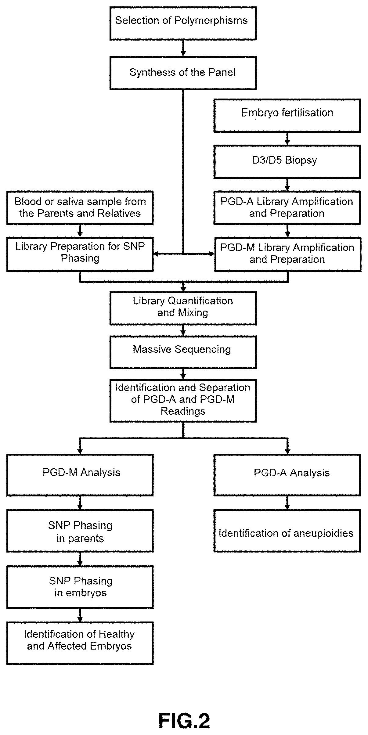
[0076]In FIG. 2 the complete outline for use in the diagnosis of aneuploidies is shown. It is considered a European population couple wherein a member of the couple is a carrier of the autosomal dominant pathogenic variant VHL:c.233A>G p.(Asn78Ser) (chromosomal position chr13:10183764) causing a condition known as Von Hippel-Lindau syndrome, which has an autosomal dominant mode of inheritance.

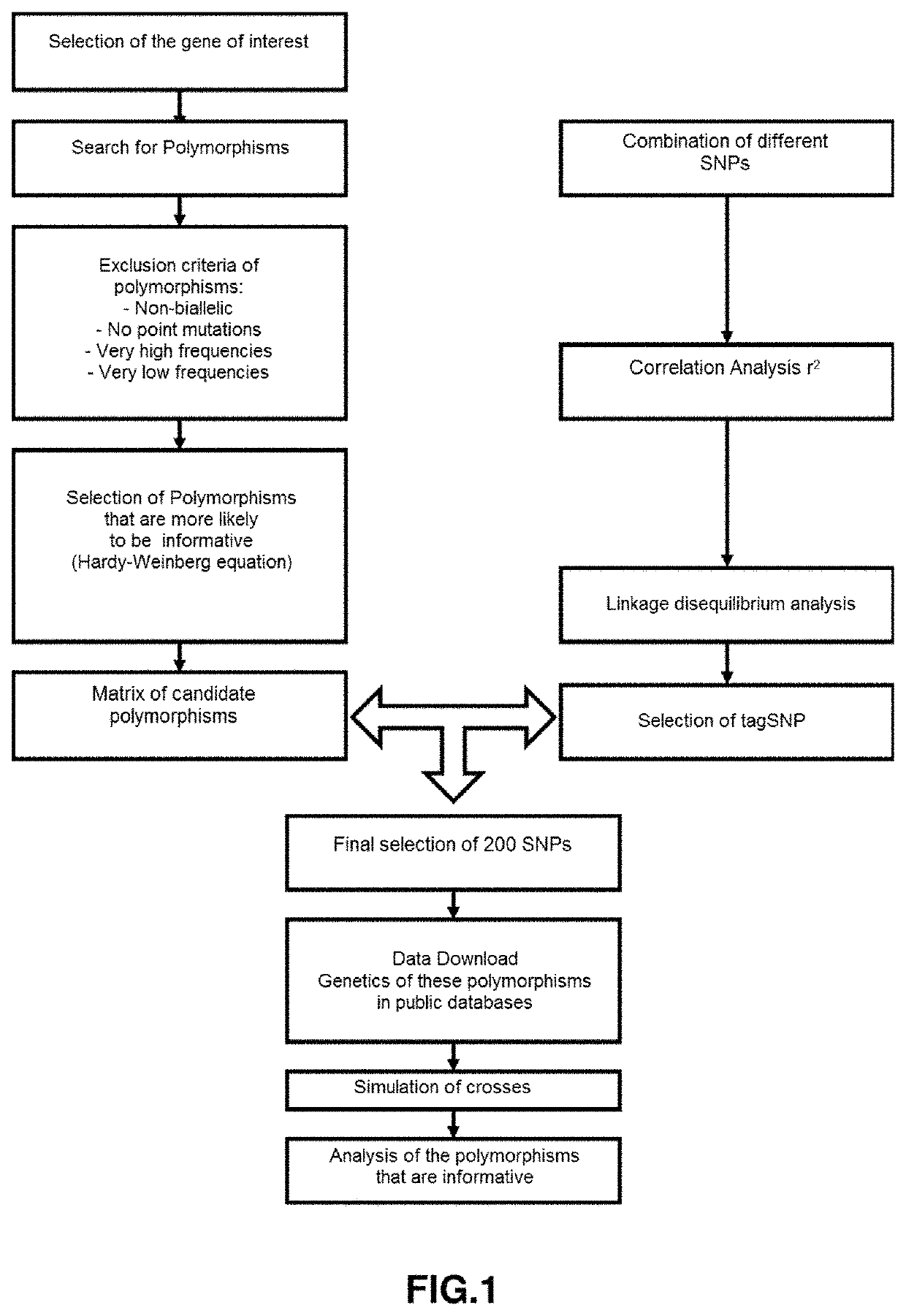
[0077]The input of the software will be the 69473 SNPs contained in the block-region chr13:9181319-11681319. The output of this algorithm will be a matrix M of 1625 candidate SNPs, which will act as input for the SNP selection algorithm, whose output will be a panel of 283 tagSNPs. In the validation phase, it was found that on average 49% of tagSNPs in the panel were informative.

[0078]Wet laboratory protocol. Once selected the polymorphisms that are to be sequenced, positions are entered into the corresponding enrichment platform. Preferably, Ion Ampliseq. This platform...

example 2

Identification of Triploid Embryos

[0090]Triploid embryos are a major problem in any IVF cycle. They account for 15% of miscarriages due to chromosomal abnormalities. Triploid embryos should always be discarded from any in vitro fertilisation cycle, but it is difficult to identify them because there are no differences in embryo quality with respect to normal embryos. Sometimes, it is possible to distinguish them because in D+1 three pronuclei are observed, but it is not always possible. The triploid embryos may be of a dyspermic origin (in cases of IVF) or be originated by an oocyte failure when the second polar corpuscle is not extruded.

[0091]Triploid embryos cannot be identified by ordinary PGD-A techniques, despite being a numerical anomaly. Sometimes, through visual inspection, it is possible to detect embryos 46, XXY when observing an abnormal distribution of the readings of the sex chromosomes, but it is not always possible and requires trained personnel.

[0092]The method herein...

example 3

Identification of Embryos with Balanced Translocations

[0093]Sometimes, some couples decide to undergo in vitro fertilisation cycles because one of them is a carrier of a balanced translocation. In these cases, these parents have a high reproductive risk, since 50% of their embryos will have an unbalanced translocation as a result of inheriting one of the altered chromosomes. Furthermore, there will be a 25% chance of producing completely normal embryos, and a 25% chance of producing embryos with the balanced alteration. FIG. 6 shows an outline of the possible embryos produced. Current techniques enable those embryos with unbalanced alterations to be distinguished, most of the time by simply using PGD-A. Nevertheless, it is not possible to differentiate those embryos with the balanced alteration from those that are completely normal, since there are no changes in copy number. Through the present development, it is possible to map the entire chromosome by different polymorphisms and, ...


PUM

PropertyMeasurementUnit
response timeaaaaaaaaaa
lengthaaaaaaaaaa
minor allele frequencyaaaaaaaaaa

Abstract

The invention relates to a method for the study of embryo mutations in in vitro reproduction processes with the particular feature that it combines the detection techniques of Aneuploidy (PGD-A) and the study of monogenic diseases in embryos (PGD-M), and wherein the method comprises a SNP selection process wherein the values of some n candidate SNPs (t1 . . . tk) of each subject x, in a chromosomal region of interest and specifically extracted for a study population, are taken as an input; a SNP selection process wherein all the SNP combinations are evaluated to obtain a minimum set t of tagSNPs from the matrix M obtained in the first SNP selection process; and an in-silico validation process of the tagSNP panel obtained in the second process.

Description

[0001]The object of the present invention is a method for the study of embryo mutations in in vitro reproduction processes with the particular feature that it combines the detection techniques of Aneuploidy (PGD-A) and the study of monogenic diseases in embryos (PGD-M) according to claim 1.TECHNICAL FIELD[0002]The present invention relates to a method for the study of mutations in embryos of couples undergoing in vitro reproductive cycles by means of SNP (single nucleotide polymorphism) analysis by massive sequencing that combines the detection of Aneuploidies (PGD-A Preimplantation Genetic Diagnosis of Aneuploidies) and the study of monogenic diseases in embryos (PGD-M, Preimplantation Genetic Diagnosis for Monogenic Diseases) with a single biopsy.PRIOR ART[0003]A single nucleotide polymorphism or SNP is a variation in the DNA sequence that affects a single base (adenine (A), thymine (T), cytosine (C) or guanine (G)) of a genome sequence.[0004]Preimplantation genetic diagnosis (PGD...

Claims


Application Information

Patent Timeline
Patent Type & Authority Applications(United States)
IPC IPC(8): G16B20/20C12Q1/6869G16B20/10C12Q1/6827C12N15/10
CPCG16B20/20C12Q1/6869C12Q2600/156C12Q1/6827C12N15/1072G16B20/10C12Q1/6883
Inventor ALCARAZ MAS, LUIS A.ANTON SALAS, ANDRES
Owner JOURNEY GENOMICS SL
Learn More