
Oct-based retinal artery/vein classification

a retinal artery and classification technology, applied in image analysis, medical science, diagnostics, etc., can solve the problems of inability to classify the smaller caliber vessels in the fundus photograph, inability to take care, and inability to solve many real-world situations. , to achieve the effect of improving the segmentation of the retinal layer, fast scanning speed, and enhancing the resolution of current oct devices

Pending Publication Date: 2020-12-17
CARL ZEISS MEDITEC INC
View PDF0 Cites 5 Cited by
  • Summary
  • Abstract
  • Description
  • Claims
  • Application Information

AI Technical Summary

Benefits of technology

This patent is about a method called OCT-based imaging that can show the blood vessels in the retina. This technique is useful for studying diseases like age-related macular degeneration and glaucoma. The patent also explains how new algorithms have improved the resolution of the images. Overall, the patent describes a way to see the tiny blood vessels in the retina, which could help with the diagnosis and treatment of retinal and optic nerve disorders.

Problems solved by technology

In clinical practice, however, fundus photographs are often inadequate to classify the smaller caliber vessels, particularly in eyes with media opacities or other pathology obscuring differentiating features.
It is noted that care must be taken since the fluorescent dye may lead to a life-threatening allergic reaction in a portion of the population.
Furthermore, if the transit phase is lost, or if blood vessel classification in both eyes is needed, a repeated FA examination is required on a subsequent day, which is impractical in many real-world situations.
Because FA is an invasive procedure, it is rarely used as a primary method for classifying retinal vessels.
Although OCTA can identify regions of blood flow, it generally cannot identify the direction of blood flow.
This technique was found to be time-consuming and of limited utility in routine clinical care.
This approach, however, is not commercially available and is generally limited to research facilities.
Thus, none of these methods allow for immediate visual evaluation of retinal vessel classification without additional scans, and many of them cannot currently be used in clinical practice due to delivery barriers.

Method used


Image

  • Oct-based retinal artery/vein classification
  • Oct-based retinal artery/vein classification
  • Oct-based retinal artery/vein classification

Examples


Embodiment Construction

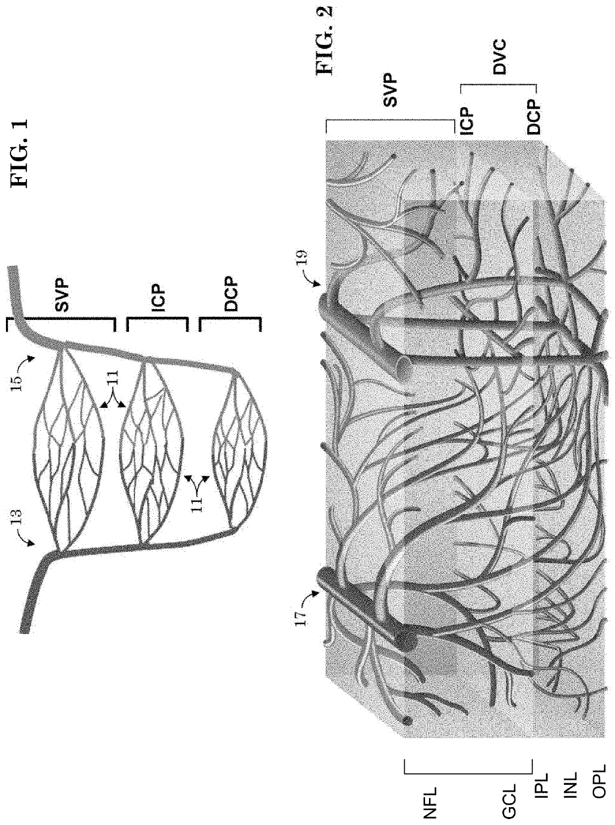
[0055]As is discussed more fully below, optical coherence tomography (OCT) and optical coherence tomography angiography (OCTA) enable noninvasive, depth-resolved (e.g., A-scan), volumetric (e.g., C-scan) and two-dimensional (e.g., en face or cross-sectional / B-scan) visualization of retinal vasculature. OCT provides structural images of vasculature whereas OCTA provides functional images of vasculature. For example, OCTA may image vascular flow by using the motion of flowing blood as an intrinsic contrast. However, there are various types of ophthalmic imaging systems, such as discussed below in section “Fundus Imaging System” and in section “Optical Coherence Tomography (OCT) Imaging System.” Unless otherwise stated, aspects of the present invention(s) may apply to any, or all, such ophthalmic imaging systems. For example, the methods / systems presented herein for differentiating arteries and veins in vasculature images may be applied to OCT structural images and / or OCTA functional i...


PUM


Abstract

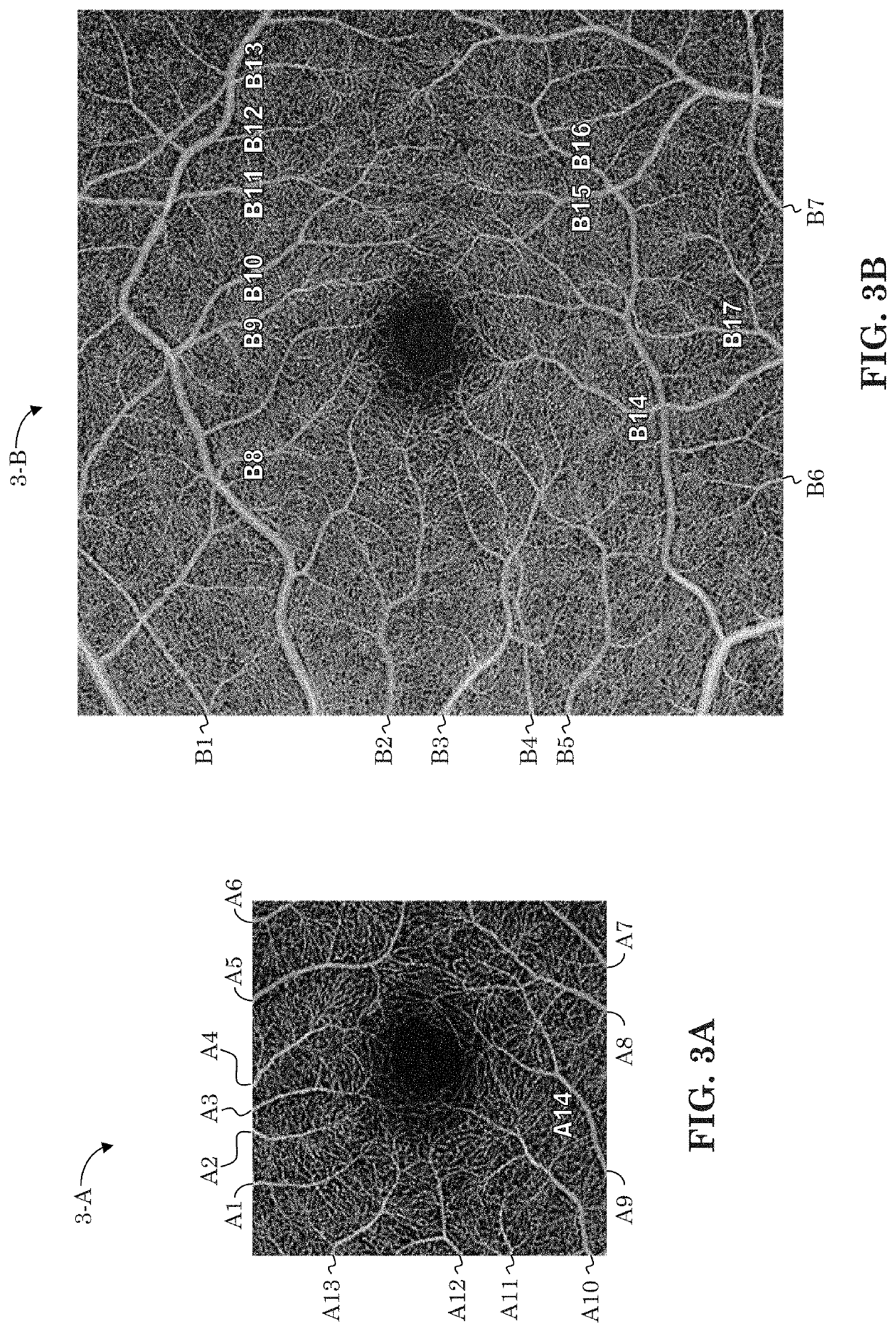
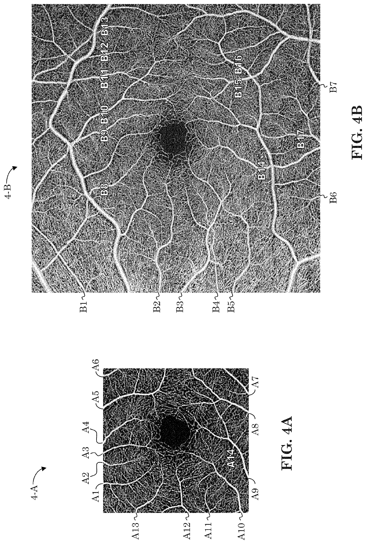
An optical coherence tomography (OCT) unit provides a mechanism / method for automatic artery and vein identification. The method may be embodied by specialized algorithm or by a machine learning model, such as a support vector machine or neural network. The present method may identify a vascular configuration that forms a vortex structure in the deep capillary plexus (DCP) of an OCT-based (e.g., OCTA) image of a retina. The identified vortex may then be designated a vein, and other vascular structures from other plexuses, such as from the superficial vascular plexus (SVP), that connect to the identified vortex structure are likewise designated veins. Additional criteria, some based on the vein designations, may be used to identify arteries.

Description

PRIORITY[0001]The present application claims priority to Provisional Application Serial No. 62 / 860,660 filed Jun. 12, 2019, the contents of which are hereby incorporated by reference in their entirety.FIELD OF INVENTION[0002]The present invention is generally directed to the field of ophthalmic imaging systems. More specifically, it is directed to techniques for identifying arterial and venous systems in images of retinal vasculature. Still more specifically, it is directed to techniques for classification of retinal vessels as arteries or veins in optical coherence tomography images irrespective of whether the optic nerve is not within the image.BACKGROUND[0003]There are various types of ophthalmic examination systems, including ophthalmoscopes, optical coherence tomography, and other ophthalmic imaging systems (see for example U.S. Pat. Nos. 4,170,398, 4,732,466, PCT Publication No. 2012059236, US Patent Application No. 2014 / 0232987, and US Patent Publication No. 2015 / 0131050). Th...

Claims


Application Information

Patent Timeline
IPC IPC(8): G06T7/00A61B3/10
CPCG06T2207/30101G06T2207/10028G06T2211/404G06T2207/10101G06T7/0012G06T2207/20081A61B3/102G06T2207/20084G06T2207/30041A61B3/1233A61B3/1241
Inventor FREUND, KENNETH BAILEYXU, XIAOYUDURBIN, MARY K.
Owner CARL ZEISS MEDITEC INC
Learn More