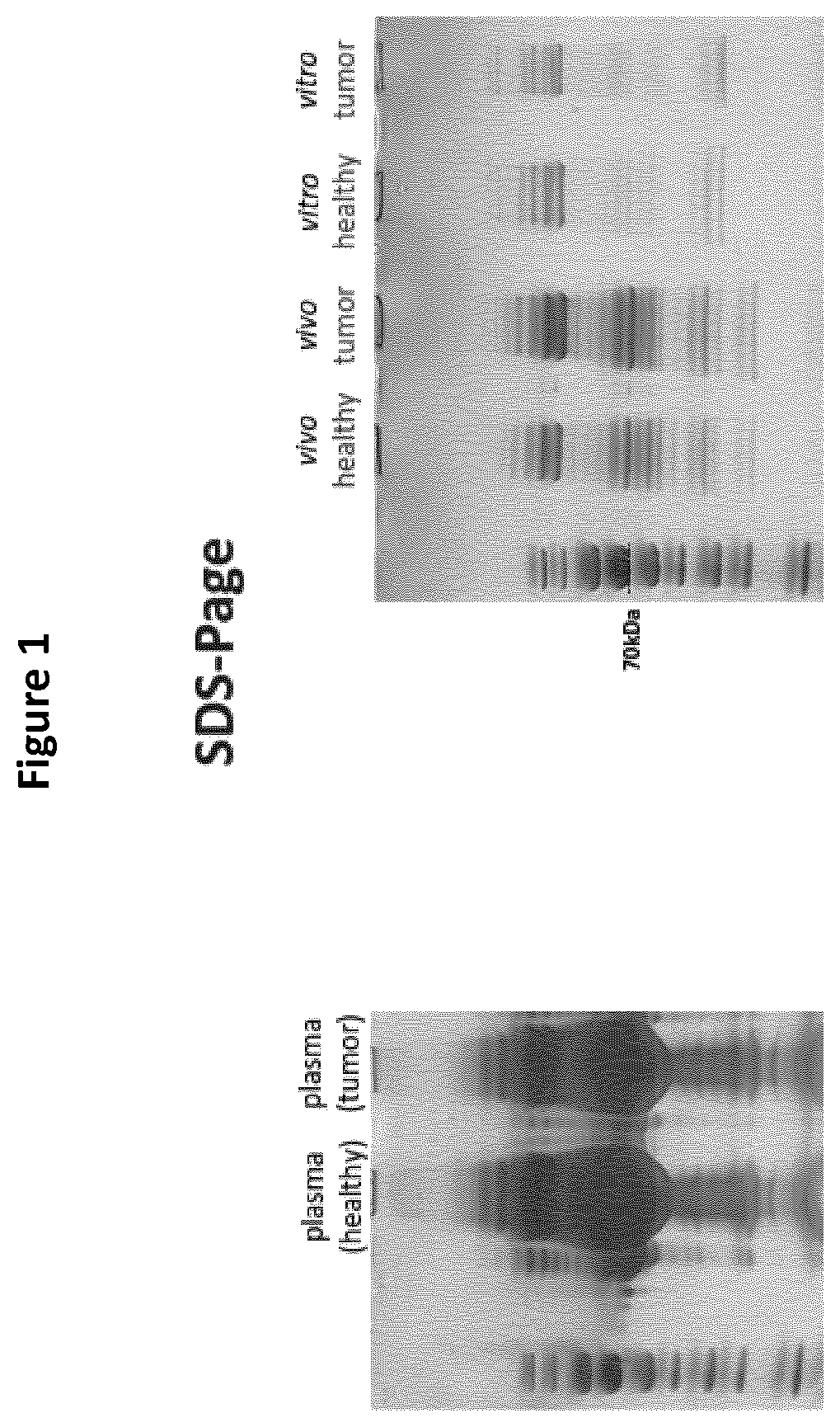
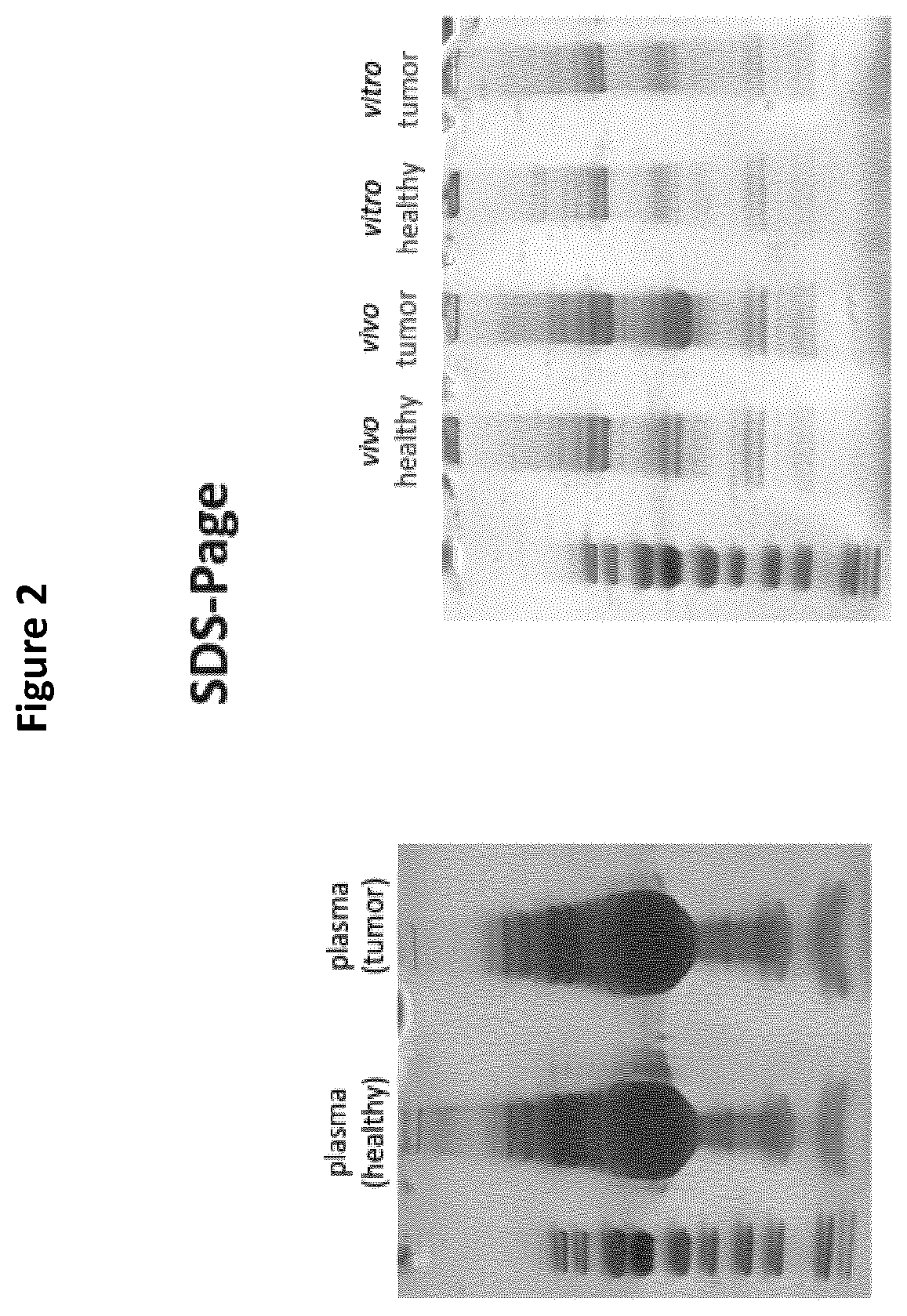
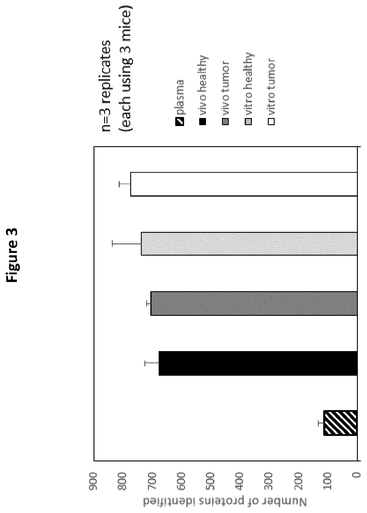
Detection of cancer biomarkers using nanoparticles

Active Publication Date: 2021-05-06
UNIV OF MANCHESTER
View PDF0 Cites 2 Cited by
  • Summary
  • Abstract
  • Description
  • Claims
  • Application Information

AI Technical Summary

Benefits of technology

[0013]The methods result in an interaction between the nanoparticles and a greater number of different types of proteins than can be detected by direct analysis of biofluids taken from a subject in a diseased state. It is to be understood that the method involves identification of a biomarker that provides a measurable indicator of some biological state or condition. This includes, but is not limited to, the discovery of unique disease-specific biomolecules (those biomolecules that are only present in a diseased state) but also includes detection of changes in biomolecule(s) that are present in both healthy and diseased states, for example upregulation or down regulation of biomolec

Problems solved by technology

Such biomarkers can exist at extremely low concentrations, for example in early stage cancer, and accurate determination of such low concentration biomarkers has remained a significant challenge.
Research into nanoparticle-based technologies for biomarker detection has been carried out but so far has failed to provide suitable methods to accurately identify/discover and detect biomarkers.
One particular problem is that currently available laboratory tests detect only a minute fraction of potential biomarkers, due to their extremely low concentration in biofluids.
In addition to the ‘swamping’ effect, cau

Method used


Image

  • Detection of cancer biomarkers using nanoparticles
  • Detection of cancer biomarkers using nanoparticles
  • Detection of cancer biomarkers using nanoparticles

Examples


example 1

Mice Models

[0068]Six to eight week old male nude SCID beige mice were purchased from Charles River (UK). Five to six week old female C57BL / 6 mice were purchased from Charles River (UK). Animal procedures were performed in compliance with the UK Home Office Code of Practice for the Housing and Care of Animals used in Scientific Procedures. Mice were housed in groups of five with free access to water and kept at temperature of 19-22° C. and relative humidity of 45-65%. Before performing the procedures, animals where acclimatized to the environment for at least 7 days.

[0069]Lung Adenocarcinoma Model:

[0070]Six to eight weeks old male nude SCID beige mice were intravenously injected (via tail vein) with a549-luc cells (5E6 cell / 200 ul of PBS).

[0071]Melanoma Model:

[0072]Five to six week old female C57BL / 6 mice were subcutaneously injected (to the left leg) with 0.5×10⁶ of B 16F10-luc melanoma cells in a volume of 50 μl of PBS.

[0073]Preparation of Liposome Nanoparticles

[0074]Materials

[0075...

example 2

Human Experiments

[0102]Subjects

[0103]Eligible patients included women with recurrent ovarian cancer commencing liposomal doxorubicin (Caelyx) treatment for the first time.

[0104]Nanoparticles

[0105]Dynamic light scattering (DLS), ζ-potential measurements and negative stain transmission electron microscopy (TEM) data showing physicochemical characteristics of the PEGylated doxorubicin-encapsulated liposomes (Caelyx®) employed in this study before and after dosing are summarised in FIG. 12.

[0106]Dosing and Blood Sample Collection

[0107]Patients were intravenously infused with Caelyx (diluted in 5% dextrose) at a dose of 40 mg / m2 for approximately 1.5 h. Collection of paired plasma samples (i.e. before and after cycle 1 infusion) were collected into commercially available anticoagulant-treated tubes (K2 EDTA BD Vacutainer). Plasma was then prepared by inverting the collection tubes 10 times to ensure mixing of blood with EDTA and subsequent centrifugation for 12 minutes at 1300 RCF at 4° ...


PUM


Abstract

Methods for identifying and detecting potential disease specific biomarkers from biofluids. The methods involve in vivo administration of nanoparticles to a subject in a diseased state or incubating nanoparticles in a biofluid sample taken from a subject in a diseased state and analysis of the biomolecule corona formed on said nanoparticles. The methods distinguish between a healthy and diseased state in a subject, such as, for example, the presence of a tumor in a human subject.

Description

FIELD OF THE INVENTION[0001]The present invention relates to methods for identifying and detecting potential disease specific biomarkers from biofluids. In particular, the methods involve administration of nanoparticles to a subject in a diseased state or incubation of nanoparticles in a biofluid sample taken from a subject in a diseased state and subsequent analysis of the biomolecule corona formed on said nanoparticles. In addition, the present invention also relates to methods that provide a means to distinguish between healthy and diseased states in a subject, such as for example the early detection of a tumor in a human subject or the presence or monitoring of the growth and / or response to treatment of a tumor in a human subject.BACKGROUND OF THE INVENTION[0002]A biomarker, or biological marker, generally refers to a qualitative and / or quantitative measurable indicator of some biological state or condition. Biomarkers are typically molecules, biological species or biological ev...

Claims


Application Information

Patent Timeline
IPC IPC(8): G01N33/574G01N33/543
CPCG01N33/57488G01N33/5432G01N33/574
Inventor KOSTARELOS, KOSTASHADJIDEMETRIOU, MARILENA
Owner UNIV OF MANCHESTER
Why Eureka
Try Eureka