
Apparatus and methods for x-ray imaging

a technology of x-ray imaging and apparatus, applied in the field of digital xray imaging, can solve the problems of reducing the clarity of the image formed by primary x-ray measurements, time-consuming, and generally not portable, and achieve the effect of improving x-ray imaging

Pending Publication Date: 2021-08-12
XENSELAB LLC
View PDF0 Cites 11 Cited by
  • Summary
  • Abstract
  • Description
  • Claims
  • Application Information

AI Technical Summary

Benefits of technology

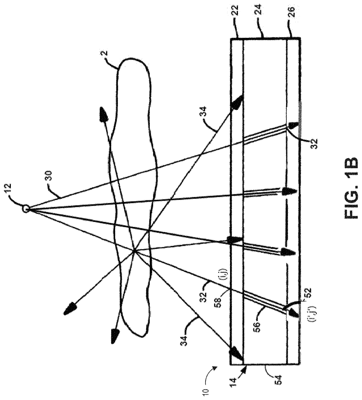
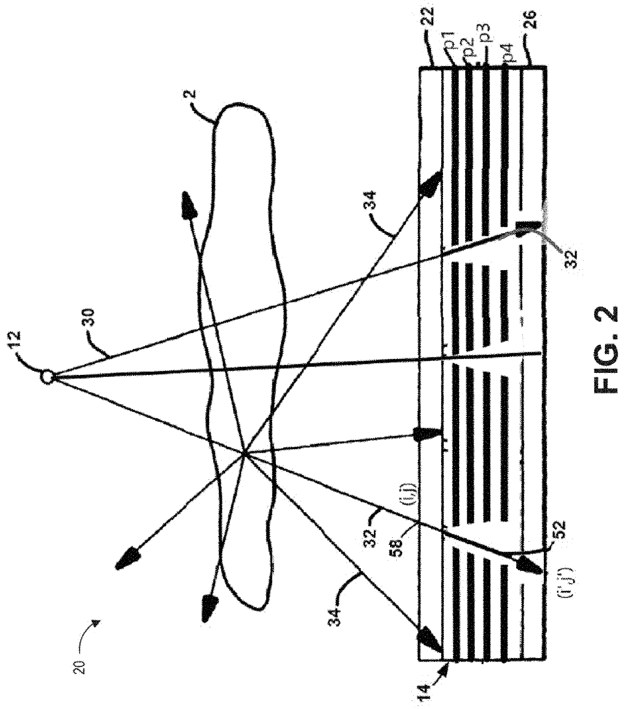
The patent describes a filter that can be used in a medical imaging system to improve the speed of energy and spectral sensitive measurements. The filter can consist of a coded aperture, which can be located between the subject and the detector or the x-ray source and the subject. The collimator can be used to guide the x-rays to the filter. The detector can be a flat panel detector or a detector with a unique feature such as a spectral measurement detector or a detector with a varied frame rate. The filter can help the system to measure energy and spectral information quickly and accurately.

Problems solved by technology

It is difficult to correlate the scattered signal collected by the detector quantitatively with different densities of the content present internal to the subject, and the measurement of scatter is not easily correlated to a precise 3D spatial location of the internal content of the subject which generates the scatter signal, and can reduce clarity of the image formed by primary x-ray measurements.
Rotational computed tomography (CT) can provide quantitative imaging data, but can be time consuming, generally not portable, requiring high radiation, and / or generally having a low molar sensitivity, in the order of 10−1-10−3 molar.
Non-rotational CT and 2D imaging are cheaper, faster, and / or requiring lower radiation compared to rotational CT, but is typically not quantitative.
As a result, the process is time-consuming.
Due to high radiation level of conventional 3D CT scanners, the frequency of such imaging method, especially in diagnostic and therapeutic processes, is limited.
Current x-ray imaging technology does not have the capability of obtaining a clear two-dimensional projection x-ray image of a human body and internal organs due to the overlapping image of the plaster cast.
Because the chemical composition of the plaster cast or fiberglass is close to that of the bone material and because of the thickness and irregular structure of the casts, practically no image information can be obtained regarding the status of the injured bone or tissue inside the cast.
Consequently, the cast needs to be removed before each x-ray images can be taken to monitor the recovery and the patient's treatment and / or post operation care cannot be more timely personalized or administered.
However, current imaging modalities, such as x-ray imaging, have not been able to achieve the sensitivity needed.

Method used


Image

  • Apparatus and methods for x-ray imaging
  • Apparatus and methods for x-ray imaging
  • Apparatus and methods for x-ray imaging

Examples


Embodiment Construction

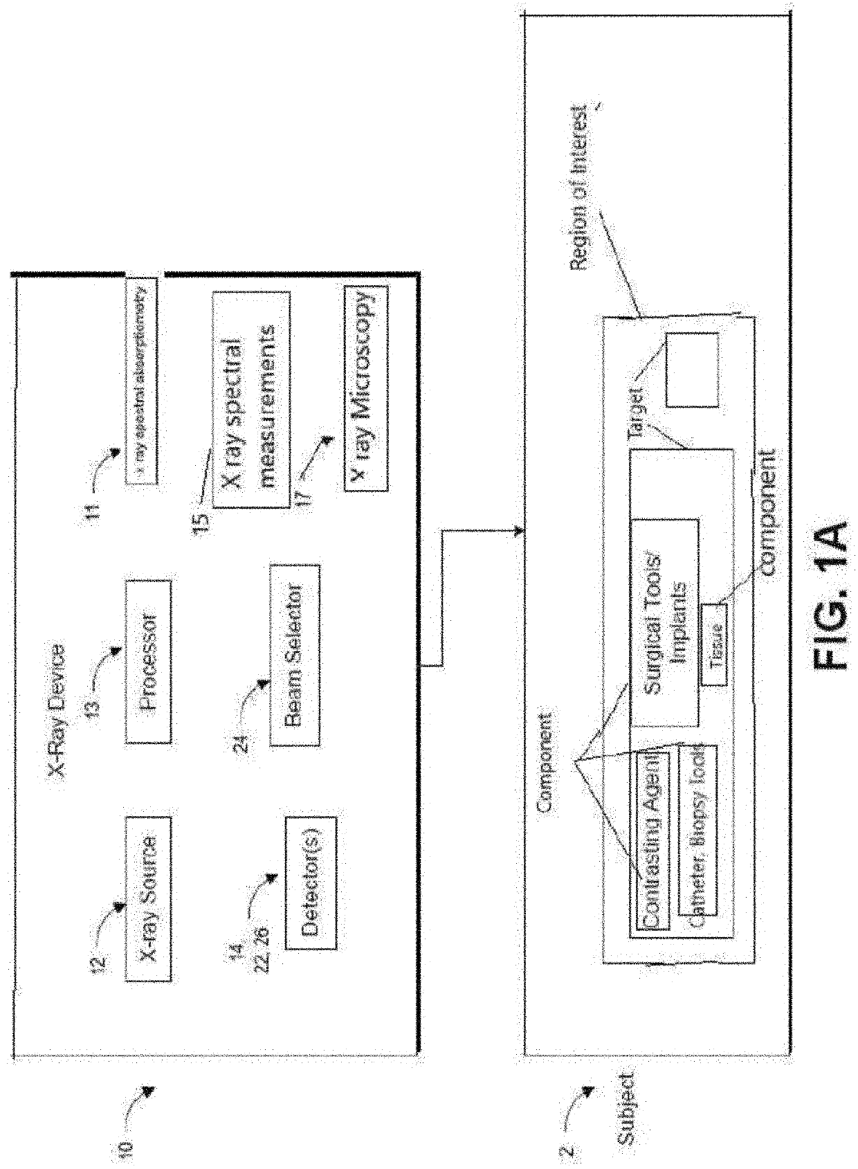
[0084]Aspects of the disclosure are provided with respect to the figures and various embodiments. One of skill in the art will appreciate, however, that other embodiments and configurations of the apparatus and methods disclosed herein will still fall within the scope of this disclosure even if not described in the same detail as some other embodiments. Aspects of various embodiments discussed do not limit scope of the disclosure herein, which is instead defined by the claims following this description.

Overview

[0085]The apparatuses, and methods and materials disclosed herein can be used for x-ray measurements of components, especially in some cases when various components of the subject being x-rayed are not easily differentiated using x-ray measurements of conventional 2D radiography due to scatter noise and / or when conventional CT scanner may not be used routinely due to high radiation, and / or the CT scanner being too time consuming, not real time, and / or infeasible.

[0086]As illus...


PUM


Abstract

An x-ray apparatus and method can improve x-ray imaging in a variety of ways. For example, the improve x-ray apparatus can reduce scatter from x-ray images acquired by two-dimensional detectors. An improved 2D x-ray apparatus can provide 3D imaging for medical and / or industrial applications. An improved 2D x-ray apparatus and method can produce separate material imaging, and composition analysis for characterization and correlation of image, densitometry, and composition information of individual component or individual material within a single subject. Non-rotational 3D microscopy, combining 2D or 3D full field x-ray imaging and high resolution 2D or 3D x-ray microscopy or spectral absorptiometry and spectroscopy can achieve a higher resolution and wider field of view in x-ray imaging and quantitative analysis in 3D and real time. The x-ray apparatus can improve tracking and / or surgical guidance in time and / or space.

Description

INCORPORATION BY REFERENCE TO ANY PRIORITY APPLICATIONS[0001]This application claims the benefit under 35 U.S.C. § 120 and 35 U.S.C. § 365(c) as a continuation of International Application No. PCT / US2019 / 044226, designating the United States, with an international filing date of Jul. 30, 2019, titled “APPARATUS AND METHODS FOR X-RAY IMAGING,” which claims priority benefit of U.S. Provisional Patent Application No. 62 / 713,554, filed on Aug. 2, 2018, U.S. Provisional Patent Application No. 62 / 803,613, filed on Feb. 11, 2019; U.S. Provisional Patent Application No. 62 / 853,050, filed on May 26, 2019; U.S. Provisional Patent Application No. 62 / 712,058, filed on Jul. 30, 2018; U.S. Provisional Patent Application No. 62 / 745,369, filed on Oct. 14, 2018; U.S. Provisional Patent Application No. 62 / 729,433, filed on Sep. 11, 2018; U.S. and U.S. Provisional Patent Application No. 62 / 755,425, filed on Nov. 3, 2018, the entire disclosure of each of which is hereby incorporated by reference and ma...

Claims


Application Information

Patent Timeline
IPC IPC(8): A61B6/00A61B6/06
CPCA61B6/5282A61B6/486A61B6/4085A61B6/06A61B6/482A61B6/481A61B6/4266A61B6/4452A61B6/4241A61B6/4007A61B6/4071A61B6/4078A61B6/483A61B6/025A61B6/032A61K49/0409A61B6/5205
Inventor ZHAO, YING
Owner XENSELAB LLC
Learn More