Biomarker for Early Diagnosis and Preoperative Assessment of Pheochromocytoma/Paraganglioma, and Application thereof

a biomarker and pheochromocytoma technology, applied in the field of tumor diagnosis and treatment, can solve the problems of limiting the preoperative evaluation and early diagnosis of genetic testing, research is a technical blank, and the exosomes of pcc and pgl patients carry genetic information that reflects the parental tumor cells, etc., and achieve the effect of effective methods

Active Publication Date: 2021-09-09
FIRST AFFILIATED HOSPITAL OF DALIAN MEDICAL UNIV
View PDF4 Cites 0 Cited by
  • Summary
  • Abstract
  • Description
  • Claims
  • Application Information

AI Technical Summary

Benefits of technology

[0017]1. The present invention is the first to reveal and provide the evidence for the main existence of double-stranded DNA fragments in the serum exosomes of PCCs and PGLs.
[0018]2. The double-stranded DNA from PCC and PGL exosomes in the present invention could be used to identify mutations that exist in tumor cells, and be used as a noninvasive molecular markers for clinical diagnosis and preoperative evaluation of PCCs and PGLs.
[0019]3. According to the analysis of measuring data in the invention implementation, RET, HIF2A, VHL, and SDHB mutations detected in the exosome DNA, by the method described in the present invention, are completely consistent with the tumor somatic mutations of PCCs and PGLs. Exosome DNA is highly consistent with the corresponding tumor gene DNA. Therefore, the detection technology provided by the present invention can offer an effective method for preoperative screening of pheochromocyte somatic mutation.
[0020]FIG. 1 The SEM pictures of exosomes from the PC12 cell line, serum samples from nude mice with mutated xenografts, and PCC and PGL patients were determined by electron microscopy; wherein, the exo-DNA I, exo-DNA II, and exo-DNA III represent exosome DNA separated from PC12 cell line, serum of nude mice with mutated xenografts, and serum of PCC and PGL patients, respectively.
[0021]FIG. 2 The results of exosomes from the PC12 cell line, serum samples from nude mice with mutated xenografts, and PCC and PGL patients, for the expression of exosome-specific marker TSG101, CD9 and CD63, by western blotting assay.
[0022]FIG. 3 Detection results of exosome double-stranded DNA from a) PC12 cell line, b) serum of nude mice with mutated xenografts, and c) serum of PCC and PGL patients, respectively.

Problems solved by technology

However, relative to the detection of germline mutations, somatic mutation detection still relies on tumor tissue samples, which greatly restricts the genetic testing for preoperative evaluation and early diagnosis.
In the existing technologies, the related research is a technical blank, which needs to be solved urgently.
However, whether exosomes from PCC and PGL patients carry genetic information that reflects the parental tumor cells remains unknown.

Method used


Image

  • Biomarker for Early Diagnosis and Preoperative Assessment of Pheochromocytoma/Paraganglioma, and Application thereof
  • Biomarker for Early Diagnosis and Preoperative Assessment of Pheochromocytoma/Paraganglioma, and Application thereof
  • Biomarker for Early Diagnosis and Preoperative Assessment of Pheochromocytoma/Paraganglioma, and Application thereof

Examples


example 1

[0028]1 Patients and Tissue Samples Collection

[0029]15 clinical cases of sporadic PCC and PGL and tumor samples were collected for gene testing. Among them, three of the tumors did not have germline or somatic mutations in the 12 known susceptibility genes, the other 12 tumors had RET, VHL, HIF2A, and SDHB somatic mutations. There was no adjuvant radiotherapy or chemotherapy before tumor resection. Peripheral blood (10 ml) was collected from each patient. The blood samples were subjected to centrifugation at 3,000 g for 10 min at 4° C., to isolate serum for the extraction and analysis of exosome and DNA.

[0030]2 Cell Culture

[0031]During PC12 cell culture, to avoid contamination from the exosomes in serum, the fetal bovine serum (FBS) and horse serum (HS) was subjected to ultracentrifugation for 11 h at 100,000 g. The supernatant was taken. PC12 cells were cultured in basic medium RPMI 1640 (Gibco) with 10% HS (exosome-depleted) and 5% FBS (exosome-depleted), with medium changes every...

example 2

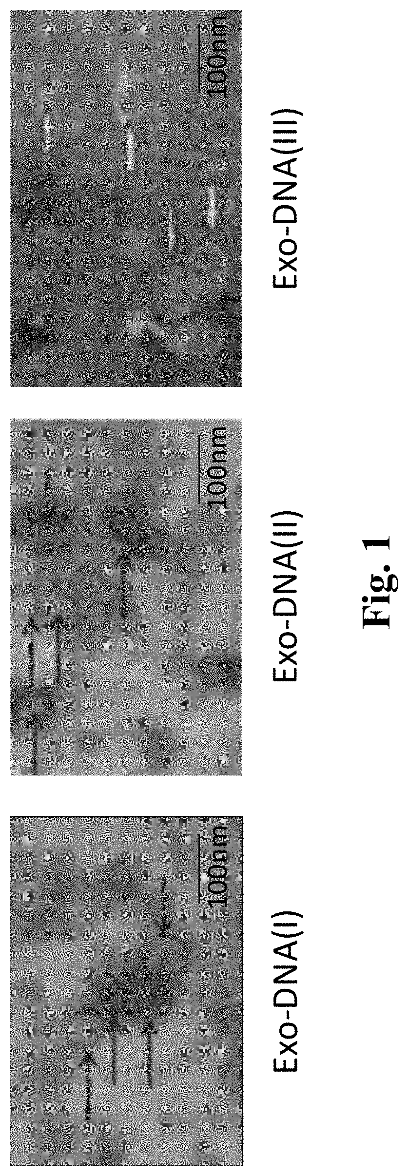
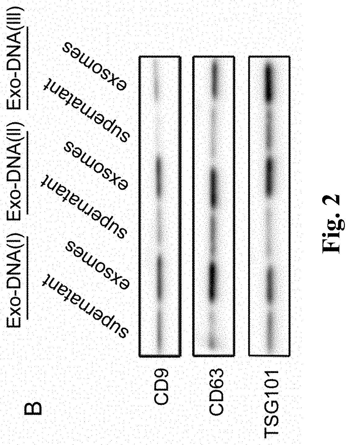
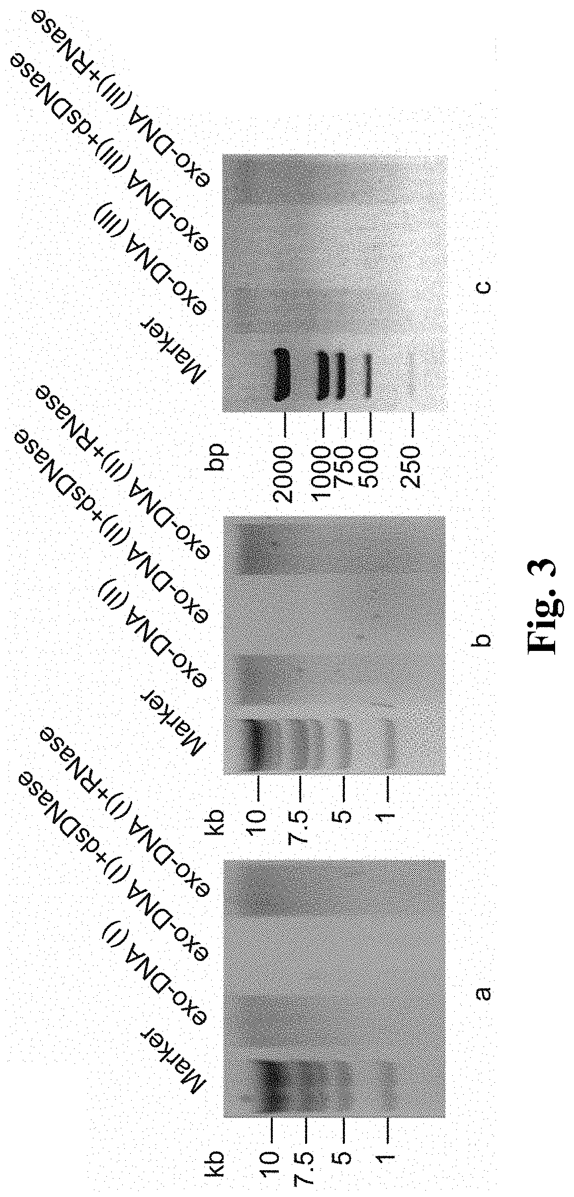
[0037]1 Isolation and Characterization of Exosomes from Cells and Serum

[0038]Exosomes were isolated from the medium collected from PC12 cells, the serum of nude mice implanted with mutated xenografts, and PCC and PGL patient serum, respectively.

[0039]1) PC12 cells in a logarithmic growth period were chose, and its medium was replaced with medium containing 10% HS and 5% FBS that had been depleted of exosomes. Then, the conditioned medium was subjected to centrifugation at 3,000 rpm for 10 min at 4° C. to remove cellular debris and dead cells. The supernatants were subjected to differential centrifugation at 100,000 g for 2.5 h. The exosome pellets were washed with 1×PBS one time and subjected to centrifugation at 100,000 g for 70 min. The final exosome pellets were re-suspended in 1×PBS.

[0040]2) To isolate exosomes from the peripheral blood, samples were subjected to centrifugation at 3,000 rpm for 15 min at 4° C. to separate the serum. The collected serum was centrifuged at 3,000 r...


PUM

PropertyMeasurementUnit
volume ratioaaaaaaaaaa
temperatureaaaaaaaaaa

Abstract

A double-stranded DNA biomarker derived from exosomes is useful in early diagnosis and preoperative evaluation of pheochromocytoma and paraganglioma, and applications thereof. The biomarker is genome double-stranded DNA fragment specifically derived from exosomes in blood serum of pheochromocytoma and paraganglioma patients. The double-stranded DNA can represent variations of RET, VHL, and HIF2A with high frequency of somatic cell mutation, and metastatic phenotype-related SDHB, which are susceptibility genes of PCCs and PGLs. The circulating exosome DNA in patient's peripheral blood contains 97% of the same chromosomal point mutation information as the tumor cells from which the DNA originated. There is evidence off the existence of double-stranded DNA in the serum exosomes of PCCs and PGLs. The double-stranded DNA can be used to identify mutations in tumor cells and provide a noninvasive molecular marker for the clinical diagnosis and preoperative evaluation of PCCs and PGLs.

Description

FIELD OF TECHNOLOGY[0001]The present invention belongs to the field of tumor diagnosis and treatment, and specifically relates to a double-stranded DNA biomarker derived from exosomes for the early diagnosis and preoperative assessment of pheochromocytoma and paraganglioma.BACKGROUND ART[0002]Pheochromocytomas (PCCs) and paragangliomas (PGLs) are rare endocrine tumors that arise from adrenal medullary pheochromocyte. Approximately 50-80% of PCCs and PGLs are related to germline or somatic mutations of at least 12 genes of NF1, RET, VHL, SDHA, SDHB, SDHC, SDHD, SDHAF2, TMEM127, MAX, HIF2A, and FH. Furthermore, accompanied by the continuous accumulation of mutation detection data, recent genetic studies have identified 21 additional genes that are involved in PCC and PGL tumorigenesis. Compared with other tumors, PCCs and PGLs are characterized by remarkable genetic determinism. Specific genotype-phenotype correlations have been established between susceptibility gene mutations and th...

Claims


Application Information

Patent Timeline
Patent Type & Authority Applications(United States)
IPC IPC(8): C12Q1/6886
CPCC12Q1/6886C12Q2600/156
Inventor LIU, JINGWANG, LIANGMA, JINGYUN
Owner FIRST AFFILIATED HOSPITAL OF DALIAN MEDICAL UNIV
Why Eureka
Try Eureka