
Cardiosphere-derived cells and exosomes secreted by such cells in the treatment of heart failure with preserved ejection fraction

a technology of exosomes and cells, applied in the direction of skeletal/connective tissue cells, medical preparations, pharmaceutical delivery mechanisms, etc., can solve the problems of increasing challenge of hfpef, no treatment has been proven to reduce morbidity and mortality in hfpef, and the underlying pathophysiological mechanisms have not been completely elucidated

Pending Publication Date: 2021-12-30
CEDARS SINAI MEDICAL CENT
View PDF0 Cites 3 Cited by
  • Summary
  • Abstract
  • Description
  • Claims
  • Application Information

AI Technical Summary

Benefits of technology

The patent describes a method for treating heart failure with preserved ejection fraction (HFpEF) using a composition containing cardiosphere-derived cells (CDCs) or exosomes isolated from those cells. The treatment can improve cardiac performance, reduce fibrosis, inflammation, and blood flow obstacles in the heart. The CDCs or exosomes can be administered as a primary or adjunctive therapy for HFpEF. The technical effect of the patent is to provide a novel treatment for HFpEF that targets the underlying causes of the disease.

Problems solved by technology

Unlike heart failure with reduced ejection fraction (HFrEF), where numerous pharmacological and device options have been proven to be effective, no treatments have been proven to reduce morbidity and mortality in HFpEF.
The challenge of HFpEF is increasing as the population ages and comorbidities become more prevalent.
Compounding the problem is the fact that underlying pathophysiological mechanisms have not been completely elucidated.
However, HFpEF constitutes a distinct clinical syndrome refractory to routine medical approaches.
No effective treatment has been identified and HFpEF has become a major public health concern.
Outcomes of HFpEF are poor, and, so far, no treatment has been shown to decrease morbidity or mortality.
For example, treatment to date has focused on the renin-angiotensin-aldosterone system and the adrenergic nervous system, but clinical trials have failed to show any significant benefit to their blockade.
The net result is impaired diastolic relaxation and filling of the left ventricle (LV), increased myocardial stiffness, impaired vascular compliance and increased diastolic pressure.
Cardiac hypertrophy indeed has little in common with limited myocardial infarction, and in both conditions, mechanisms driving LV remodeling are likely to be dissimilar.

Method used


Image

  • Cardiosphere-derived cells and exosomes secreted by such cells in the treatment of heart failure with preserved ejection fraction
  • Cardiosphere-derived cells and exosomes secreted by such cells in the treatment of heart failure with preserved ejection fraction
  • Cardiosphere-derived cells and exosomes secreted by such cells in the treatment of heart failure with preserved ejection fraction

Examples


example 1

CDC Culture

[0074]CDCs were prepared as described in U.S. Patent Application Publication No. 2012 / 0315252, the disclosures of which are herein incorporated by reference in their entirety.

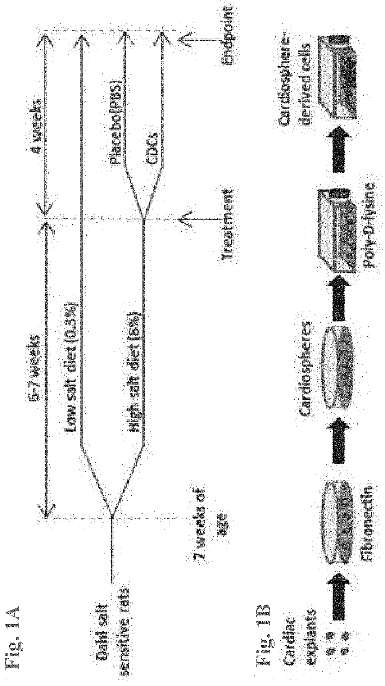
[0075]In brief, heart biopsies were minced into small fragments and briefly digested with collagenase. Explants were then cultured on 20 mg / mL fibronectin-coated dishes. Stromal-like flat cells and phase-bright round cells grew out spontaneously from tissue fragments and reached confluency by 2-3 weeks. These cells were harvested using 0.25% trypsin and were cultured in suspension on 20 mg / mL poly-d-lysine to form self-aggregating cardiospheres. CDCs were obtained by plating and expanding the cardiospheres on a fibronectin-coated flask as an adherent monolayer culture. All cultures were maintained at 5% O2, 5% CO2 at 37° C., using IMDM basic medium supplemented with 10% FBS, 1% penicillin / streptomycin, and 0.1 mL 2-mercaptoethanol. CDCs were grown to 100% confluency on a fibronectin-coated flask to p...

example 2

Isolation of Exosomes from CDCs

[0076]When the CDCs reached the desired confluency, the flask was washed three times with PBS. CDCs were treated with serum-free medium (IMDM) and were incubated at 37° C. at 5% O2, 5% CO2 for 15 days. After 15 days, the conditioned medium was collected in 225 mL BD Falcon polypropylene conical tubes (BD 352075—Blue Top) and centrifuged at 2,000 rpm for 20 minutes at 4° C. to remove cells and debris (care was taken not to disturb the pellet). The conditioned medium was run through a 0.45 μm membrane filter. The conditioned medium was concentrated using centrifugal filter. A 3 KDa Centricon Plus-70 Centrifugal Filter was pre-rinsed with 10-25 mL of molecular grade water and was centrifuged at 3220 g for five minutes at 18° C. Once the filter was rinsed, all remaining water was carefully removed without touching the filter. 15 mL of the conditioned medium was added to the filter and was centrifuged at 3220 g for 45 minutes at 18° C. After the initial spi...

example 3

Exosome Precipitation with 25% Polyethylene Glycol (PEG)

[0077]The appropriate volume of 25% PEG was added to the filtered concentrated conditioned medium. The samples were incubated at 4° C. for 12-16 hours on an orbital shaker. Once incubation was complete, the samples were centrifuged at 1500 g for 30 minutes at 4° C. The supernatant was carefully removed without disrupting the pellet. The pellet was resuspended in the desired volume of serum-free medium and sampled for particle count.


PUM

PropertyMeasurementUnit
diameteraaaaaaaaaa
diameteraaaaaaaaaa
diametersaaaaaaaaaa

Abstract

Heart failure with preserved ejection fraction (HFpEF) is a disease condition characterized by heart failure (HF) signs and symptoms, but with normal or near normal left ventricular ejection fraction (LVEF) and is not responsive to standard therapy for treatment of HF. Described herein are compositions and methods related to use of cardiosphere derived cells (CDCs) and their exosomes to improve left ventricular structure, function and overall outcome. Administration of CDCs led to improved LV relaxation, lower LV end-diastolic pressure, decreased lung congestion and enhanced survival. Lower risk of arrhythmias in HFpEF was also observed following CDC administration. Improvement of diastolic dysfunction following administration of CDC-derived exosomes was observed, along with decreased mortality. In view of these salutary effects, CDCs and CDC-derived exosomes are beneficial in the treatment of HFpEF.

Description

CROSS REFERENCE TO RELATED APPLICATIONS[0001]The present application is a division of U.S. patent application Ser. No. 16 / 066,014, filed Jun. 25, 2018, which is a U.S. National Phase entry under 35 U.S.C. § 371 of International Patent Application No. PCT / US2017 / 013054, filed Jan. 11, 2017, which claims the benefit of priority to U.S. Provisional Patent Application No. 62 / 277,359, filed Jan. 11, 2016, the entirety of each of which is hereby incorporated by reference herein. Any and all applications for which a foreign or domestic priority claim is identified in the Application Data Sheet as filed with the present application are hereby incorporated by reference under 37 CFR 1.57.STATEMENT REGARDING FEDERALLY-SPONSORED RESEARCH[0002]This invention was made with government support under HL083109 awarded by the National Institutes of Health. The government has certain rights in the invention.FIELD OF THE INVENTION[0003]Described herein are methods and compositions related to use of cell...

Claims


Application Information

Patent Timeline
Patent Type & Authority Applications(United States)
IPC IPC(8): A61K35/34C12N5/077A61P9/04A61K9/00
CPCA61K35/34A61K9/0019A61P9/04C12N5/0657
Inventor MARBAN, EDUARDOGALLET, ROMAIN
Owner CEDARS SINAI MEDICAL CENT
Learn More