

Composition for use in treatment of allergic diseases
- Summary
- Abstract
- Description
- Claims
- Application Information
AI Technical Summary
Benefits of technology
Problems solved by technology
Method used
Image

Examples
example 1
Exome Analysis of Atopic Dermatitis Model Mouse
[0120]In this example, exome analysis of NC / Nga mice as model mice of atopic dermatitis was performed to identify causative genes of atopic dermatitis.
[0121]The NC / Nga mice were purchased from Charles River, Japan. DNA was extracted from blood from the NC / Nga mice under conditions suitable for extraction of mouse DNA using QIAamp DNA blood Mini Kit (Qiagen, Venlo, Netherlands). The exome analysis of the resulting DNA was performed as follows. DNA libraries were obtained using SureSelect Library prep kit (post-pool version 4; Agilent Technologies, Santa Clara, Calif.) and SureSelect Mouse All Exon Kit (Agilent Technologies) according to the manufacturer's manual. The resulting DNA libraries were subjected to emulsion PCR (SOLiD EZ Bead Emulsifier kit; Thermo Fisher Scientific, Waltham, Mass.) to generate clonal DNA fragments on beads, which were then subjected to bead enrichment (SOLiD EZ Bead Enrichment kit; Thermo Fisher Scientific, Wa...
example 2
Clec10a Expression Analysis and Functional Analysis
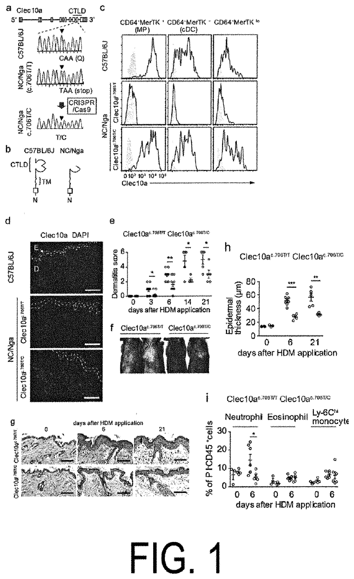
[0125]Clec10a is a member of the type II C-type lectin receptor family and detects galactose moieties at terminals of foreign and endogenous antigens. c.706C>T of Clec10a in the NC / Nga mice is present in the coding region of the C-type lectin-like domain (FIG. 1, panels a and b). The flow cytometric analysis revealed that Clec10a was expressed on the cell surface of macrophages (MP) (CD64+MerTK+), known dendritic cells (cDC) (CD64−MerTK−), and mononuclear cell-derived DCs (CD64−MerTKlo), as well as macrophages in the peritoneal cavity of C 57BL / 6J (not expressed in the peritoneal cavity of NC / Nga mice), in skin CD45+MHCII+Lineage−EpCAM−cells (see panel c in FIG. 1 and panels b and e in FIG. 5). No expression of Clec10a was observed on non-hematopoietic cells (CD45−), CD45+MHCII− cells, CD45+MHCII+(Lin, EpCAM)+ cells, and neutrophils (CD45+CD11b+Ly-6G+) of the skin of the C57BL / 6J mice (see panels c and d in FIG. 5). Similar results ...
example 3
Analysis of Clec10a Ligand
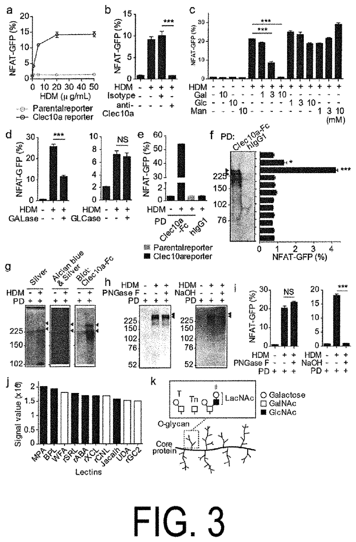
[0132]To test whether Clec10a recognized glycosylated proteins contained in HDM, NFAT-GFP reporter cells (Clec10a reporter cells) were made which expressed chimeric fusion proteins inch ling an extracellular and transmembrane portions of Clec10a fused to a cytoplasmic portion of CD3. Mouse Clec10a reporter cells expressed GFP in response to Lewis X (Clec10a oligosaccharide ligand) but did not respond to Lewis Y (see panels a and b in FIG. 8). The mouse Clec10a reporter cells also expressed GFP in response to the HDM-coated plate dose-dependently (panel a in FIG. 3). In addition, pretreatment of the reporter cells with anti-Clec10a monoclonal antibodies (mAb) or galactose inhibited GFP expression, but glucose or mannose pretreatment did not suppress GFP expression (panels b and c in FIG. 3). Furthermore, treatment of HDM with galactosidase reduced their ability to cause GFP expression in the reporter cells, but no such reduction was observed when glucosidase...
PUM
 Abstract
Description
Claims
Application Information