
System and Method for Free-Breathing Quantitative Multiparametric MRI

a multiparametric and free-breathing technology, applied in the field of systems and methods for free-breathing quantitative multiparametric mri, can solve the problems of temperature error, inability to measure temperature change in adipose tissues, and the proton resonance frequency of fat spin only displays a minute temperature dependen

Pending Publication Date: 2022-06-09
RGT UNIV OF CALIFORNIA
View PDF5 Cites 2 Cited by
  • Summary
  • Abstract
  • Description
  • Claims
  • Application Information

AI Technical Summary

Benefits of technology

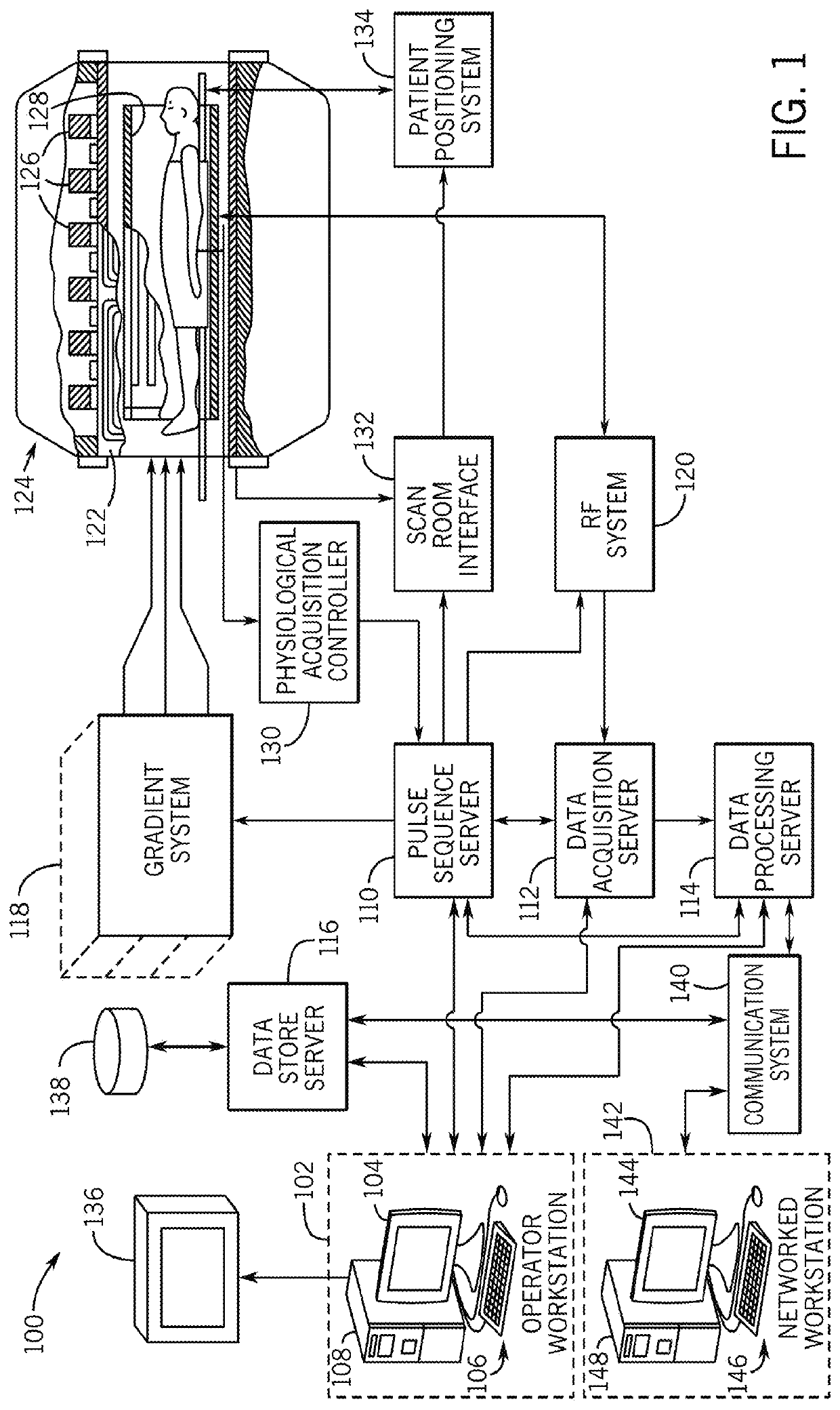
The patent describes a method and system for magnetic resonance imaging (MRI) that can measure various parameters of a subject's body. The system uses a special pulse sequence that collects data from a region of interest in multiple directions, allowing for the simultaneous measurement of T1 (the time it takes for the magnetic resonance signal to relax) and PRF (the frequency shift of the magnetic resonance signal caused by temperature changes) in the same image. This technology can be used to generate maps of fat, water, T1, proton-density fat fraction, and R2* (the relaxation time of the magnetic resonance signal) in the body, providing valuable information for medical diagnosis and treatment. The system includes a magnet, magnetic gradient system, RF system, and computer system, which work together to acquire and analyze the MRI data.

Problems solved by technology

However, PRF temperature mapping is susceptible to phase errors and image artifacts caused by motion, such as respiratory, cardiac and peristaltic motion or bulk patient movement, which may cause a mismatch between the heated and baseline maps and can lead to temperature errors.
In moving organs, such as the liver, respiratory and cardiac motion can cause mis-registration in phase images, and lead to erroneous temperature results.
A second drawback of PRF thermometry is that the proton resonance frequency of fat spins only displays a minute temperature dependence due to the lack of hydrogen bonds, and cannot be used to measure temperature change in adipose tissues.
Due to the lengthened acquisition time for multiple flip angles, VFA PRF / T1 methods are typically either confined to a single 2D slice or a thin 3D slab with limited through-plane coverage to achieve the desired temporal resolution.
This shortcoming renders it difficult to achieve a field-of-view (FOV) that encompasses larger lesions and surrounding tissues such as in the liver, or to monitor and avoid unwanted heating in both the near- and far-field of the HIFU transducer.
However, DWI faces challenges such as low image quality and cardiac and breathing motion artifacts.
Moreover, the measured apparent diffusion coefficient has not been shown to be a reliable indicator of the fibrosis stage.
While MRE is a valuable clinical tool, MRE can encounter measurement difficulties in patients with increased hepatic iron content, which is present in up to one-third of patients with diffusive liver diseases such as non-alcoholic fatty liver disease (NAFLD).
Iron in itself can contribute to liver fibrosis, and may cause a decrease in the accuracy of stiffness measurements due to the effects of T2* shortening on gradient-echo based MRE protocols.
In addition, the specialized hardware required for MRE may not be available at all institutions or practical for young children and infants.
However, the administration of contrast agents requires intravenous injection and takes additional time.
Moreover, it may not be feasible in patients with renal deficiency and diseases.
Moreover, it has been reported that combined fatty liver and iron overload can lead to worse outcomes than fatty liver alone.
However, breath-holding is challenging.
Sick, young, and elderly patients often cannot breath-hold or have imperfect breath-holding, which can result in poor image quality and quantification errors.
While a high acceleration factor can be used to facilitate Cartesian acquisition during a short breath-hold duration, this may lead to artifacts and quantification errors.

Method used


Image

  • System and Method for Free-Breathing Quantitative Multiparametric MRI
  • System and Method for Free-Breathing Quantitative Multiparametric MRI
  • System and Method for Free-Breathing Quantitative Multiparametric MRI

Examples


example 1

[0080]In various example studies discussed below, the accuracy of the disclosed VFA 3D stack-of-radial technique for T1 quantification was evaluated in a reference T1 / T2 phantom. In vivo non-heating experiments were conducted in healthy subjects to evaluate the stability of PRF and T1 in the brain, prostate, and breast. In addition, the proposed VFA 3D stack-of-radial technique was used to monitor high intensity focused ultrasound (HIFU) ablation in ex vivo porcine fat / muscle tissues and compared to temperature probe readings.

[0081]In this example, the VFA 3D stack-of-radial technique achieved 3D coverage with 1.1×1.1 to 1.3×1.3 mm2 in-plane resolution and 2-5 s temporal resolution. During 20-30 minutes of non-heating in vivo scans, the temporal coefficient of variation for T1 was 1 were consistent with temperature probe readings, with an absolute mean difference within 2° C.

[0082]In this example study, to evaluate the accuracy of T1 measurements the proposed VFA 3D stack-of-radial ...

example 2

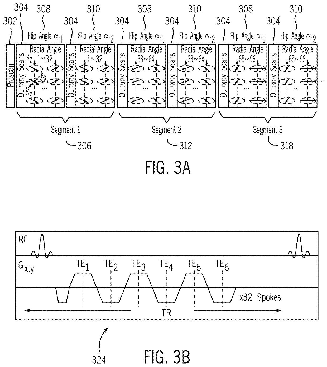
[0095]In another example study, non-heating free-breathing scans were carried out in human subjects to evaluate the reliability of both T1 and PRF measurements in a moving organ, in particular, the liver. In this study, N=10 healthy subjects (6 males, 4 females) with age of 33±11 years and body mass index of 28.4±8.8 kg / m2 underwent non-heating free-breathing abdominal scans at 3T with body and spine arrays for 5˜10 minutes. The scanning parameters for the example in vivo dynamic PRF / T1 thermometry were: Field of View (FOV) 320×320×48 mm3, matrix size 192×192×16, resolution 1.67×1.67×3 mm3, flip angles 3° and 15°, targeted T1 range 8001200 ms, number of echoes / TE1 / ΔTE 4 / 1.21 / 1.21 ms, TR 6.32 ms, number of angles / segment 32, number of segments 48, total running time 6:36 (minutes:seconds), number of radial spokes in center / total spokes 8 / 144, temporal resolution / temporal footprint of PRF maps 0.91 / 16.4 s, temporal resolution / temporal footprint of T1 maps 0.91 / 20.5 s.

[0096]To characte...

example 3

[0101]In another example study, the VFA 3D stack-of-radial multi-echo gradient echo MRI technique (“radial VFA”) was used for simultaneous free-breathing 3D liver T1, PDFF, and R2* mapping. In this example, healthy adults (n=18) and children (n=2) were imaged at 3 Tesla using radial VFA in 3-min. B1+ maps were acquired during end-expiration breath-holds for VFA T1 calculation. PDFF / R2* were calculated from multi-echo data acquired with the smaller flip angle. Self-navigated soft-gating was performed to reconstruct radial VFA images at end-expiration. Breath-holding VFA 3D Cartesian multi-echo gradient echo and breath-holding 2D Cartesian Modified Look-Locker Inversion Recovery (MOLLI) sequences were acquired as references. Bland-Altman analysis was conducted to evaluate agreement between the sequences. Intraclass correlation coefficients (ICC) were calculated to assess the repeatability of radial VFA.

[0102]To assess the T1 mapping accuracy, a system T1 / T2 phantom was placed in a 3T ...


PUM


Abstract

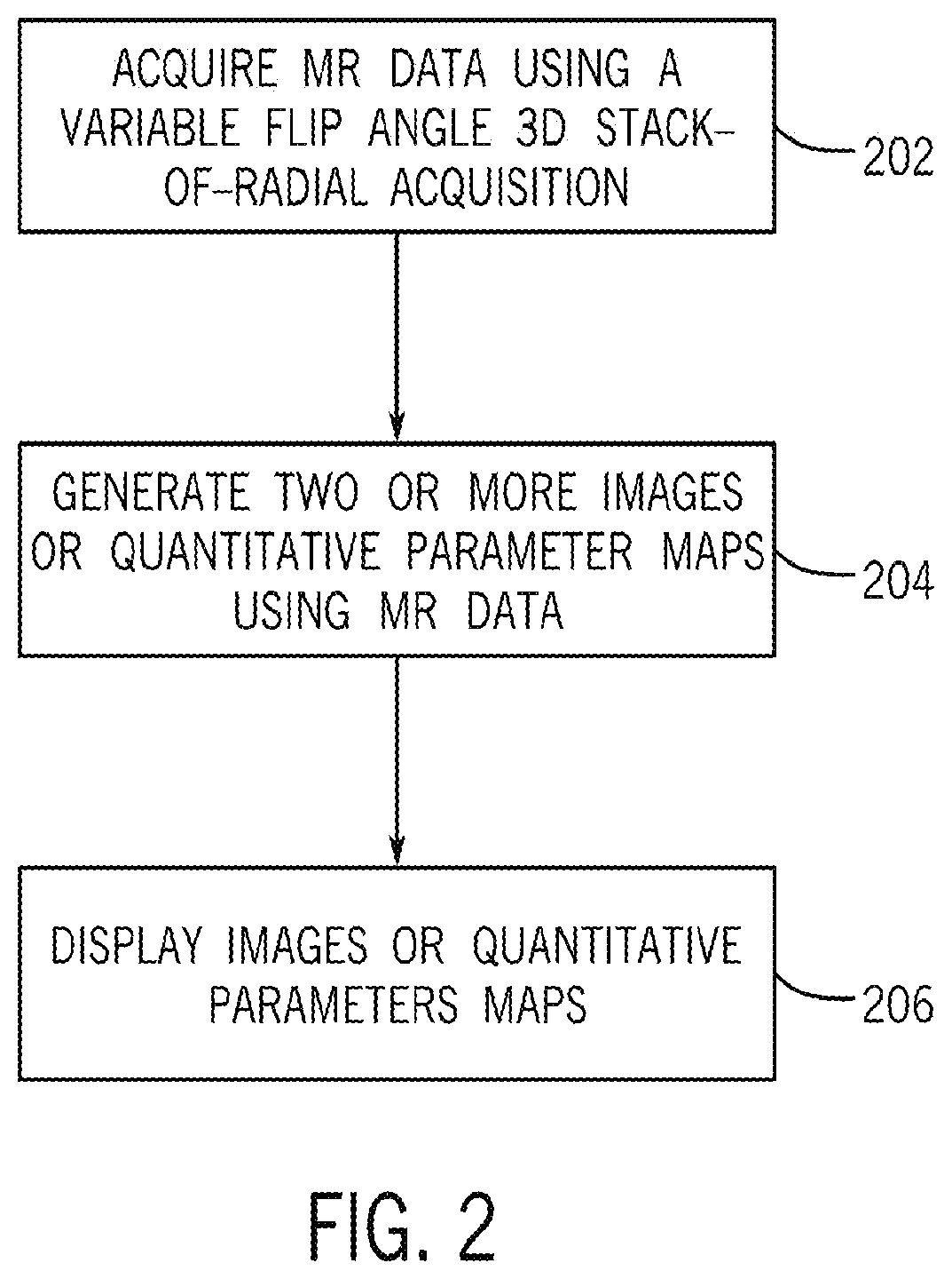
A method for proton resonance frequency shift (PRF) and T1-based temperature mapping using a magnetic resonance imaging (MRI) system includes acquiring, using the MRI system, a set of magnetic resonance (MR) data from a region of interest of a subject by performing a variable-flip-angle multi-echo gradient-echo 3D stack-of-radial pulse sequence. The pulse sequence is configured to acquire radial k-space data in a plurality of segments, each segment acquired with each of a plurality of flip angles. The method further includes generating at least one T1 map based on the set of MR data, generating at least one PRF temperature map based on the set of MR data, generating at least one T1-based temperature map based on the set of MR data and displaying the PRF temperature map and the T1-based temperature map. In another embodiment, the MR data may be used to generate a plurality of quantitative parameter maps for each of the plurality of MR parameters such as T1, proton-density fat fraction (PDFF), and R2*.

Description

CROSS-REFERENCE TO RELATED APPLICATION[0001]This application is based on, claims priority to, and incorporates herein by reference in its entirety U.S. Ser. No. 62 / 833,894 filed Apr. 15, 2019 and entitled “Method And Apparatus For Quantitative Multiparametric MRI.”BACKGROUND[0002]Magnetic resonance imaging (MRI) provides crucial information to plan, guide, monitor, and assess minimally invasive thermal therapy modalities, including high-intensity focused ultrasound (HIFU), radiofrequency (RF) ablation, microwave ablation, and laser interstitial thermal therapy (LITT). These thermal therapy modalities may be used, for example, to treat an array of cancers by inducing tissue necrosis with sustained treatment. Among these modalities, MRI-guided HIFU is especially attractive since both MRI and HIFU are non-invasive and can access tissues deep in the body for treating various conditions such as breast and prostatic cancers. The anatomical information from MRI can facilitate treatment pla...

Claims


Application Information

Patent Timeline
Patent Type & Authority Applications(United States)
IPC IPC(8): G01R33/48G01R33/50G01R33/561A61B5/00A61B5/01A61B5/055
CPCG01R33/4826G01R33/4804G01R33/4828G01R33/50A61B2576/00A61B5/0036A61B5/015A61B5/055G01R33/5616A61B5/7285A61B5/0816A61B5/1135A61B5/113A61B5/0037A61B5/318G01R33/5676G01R33/543
Inventor WU, HOLDEN H.ZHANG, LEARMSTRONG, TESS
Owner RGT UNIV OF CALIFORNIA
Learn More