hNT-neuron human neuronal cells to replace ganglion cells

a human neuronal cell and neuronal cell technology, applied in the field of human transplantation, can solve the problems of insidious progressive vision loss, eye and face pain, nausea, vomiting, etc., and achieve the effect of treating vision loss in a mammal

Inactive Publication Date: 2000-12-19
LAYTON BIOSCI
View PDF1 Cites 16 Cited by
  • Summary
  • Abstract
  • Description
  • Claims
  • Application Information

AI Technical Summary

Benefits of technology

Vision loss in a mammal can be treated by administering an effect

Problems solved by technology

Often the disease is asymptomatic, as the patient painlessly and gradually loses vision.
Symptoms include severe eye and face pain, nausea, vomiting, colored halos around lights, and loss of vision.
Compression of the optic nerve causes insidious progressive vision loss and visual field loss.
Vision may be lost if papilledema is not promptly treated.
Vision also can be lost due to higher visual pathway lesions.
Lesions at the optic chiasma tend to cause bilateral vision loss.
Thus, vision is vulnerable to a number of different pathologies in a variety of intracranial locations.
Even if the patient obtains appropriate treatment, treatment today is l

Method used


Image

  • hNT-neuron human neuronal cells to replace ganglion cells
  • hNT-neuron human neuronal cells to replace ganglion cells
  • hNT-neuron human neuronal cells to replace ganglion cells

Examples


example 1

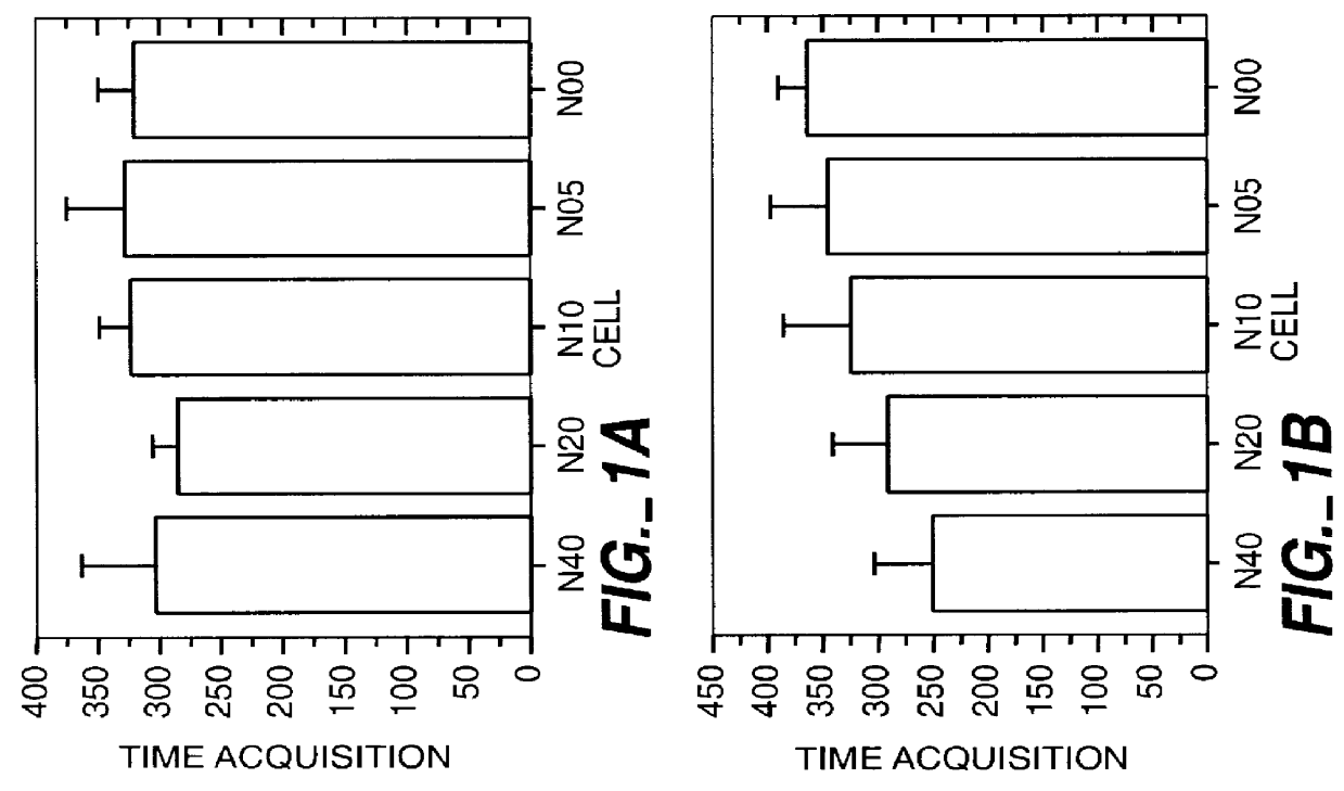
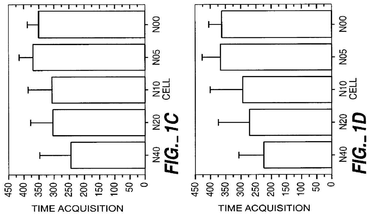
This study was performed to investigate the minimum effective dose of cryopreserved hNT cells that would product significant behavioral recovery in ischemic rats. Transplantation was carried out at one month after ischemic surgery. Ischemic rats were tested in motor asymmetry and passive avoidance learning task at one month after ischemic surgery. Only animals that displayed significant behavioral deficits were used as subjects and transplanted with specific doses (6-8 per group). In addition to a control group (seven received only vehicle), rats were given four different doses of cryopreserved hNT neurons in 3 .mu.l injections: N40 (4.times.10.sup.4 cells); N20 (2.times.10.sup.4 cells); N10 (1.times.10.sup.4 cells); and N05 (0.5.times.10.sup.4 cells). All animals were immunosuppressed throughout the 3-month post-transplant period with cyclosporine-A (CsA; i.p. 10 mg / kg). At one, two and three months after transplant, all animals were tested in EBST and passive avoidance. At the end...

example 2

In this study, rats were administered not only fresh and cryopreserved hNT-Neuron cells but also rat fetal cells as positive and negative controls. One month after stroke surgery, animals that showed significant behavioral deficits compared to control sham or normal animals were randomly assigned to stroke-surgery transplant groups. They were transplanted with (1) rat fetal striatal cells, (2) rat fetal cerebellar cells, (3) hNT-Neuron cells (fresh or cryopreserved, with or without cyclosporine (CsA) treatment), or (4) medium as described in detail elsewhere (Borlongan et al., 1995a, ibid.; Kleppner et al., 1995, ibid.; Mantione et al., 1995, ibid.; Trojanowski et al., 1993, ibid.). Additional animals (normal and sham-surgery rats) were added to serve as controls. The study groups also included controls of the two surgical procedures (stroke and transplantation) and are summarized in the following table:

Selected rats were also immunosuppressed with Cyclosporine (CsA) (Sandoz) which ...

example 3

A dose study is disclosed for treatment of vision loss due to chemical retinal damage in rats. A small amount of caustic chemical is injected into one eye. After the eye heals, the eye is injected with one of several doses of hNT-Neuron cells. Some of the rats are controls, while others receive cyclosporine-A. Vision of the rats is tested at intervals after the transplant of hNT-Neuron cells. After a suitable interval, rats are sacrificed and histologic studies, as detailed above, are performed to observe the state of the transplanted hNT-Neuron cells. Assuming that a similar effect to that of Examples 1 and 2 is observed, a study of glaucomatous Beagles is contemplated next.


PUM

PropertyMeasurementUnit
Areaaaaaaaaaaa

Abstract

Disclosed herein is the treatment of vision loss in a mammal by transplanting an effective amount of hNT-Neuron cells. The treatment can be accomplished by injecting the cells into the retinal area of the eye. Additionally, the cells can be injected into the visual cortex of the brain. Conditions to be treated are vision loss due to optic nerve damage, including glaucoma, optic nerve sheath meningioma and glioma, Graves' ophthalmopathy, benign or malignant orbital tumors, metastatic lesions, tumors arising from the adjacent paranasal sinuses or middle cranial fossa, giant pituitary adenomas, brain tumors or abscesses, cerebral trauma or hemorrhage, meningitis, arachnoidal adhesions, pseudotumor cerebri, cavernous sinus thrombosis, dural sinus thrombosis, encephalitis, space-occupying brain lesions, severe hypertensive disease or pulmonary emphysema.

Description

FIELD OF USEThe present invention is in the field of human transplantation and more particularly in the field of intraocular and intracranial transplantation of specially treated human cells which reestablish neuronal connections between the retina and the ocular cortex, which neurons having been damaged by glaucoma or other compression-causing injuries and diseases.BACKGROUND INFORMATIONGlaucoma is the occurrence of elevated intraocular pressure which causes progressive blindness in the form of gradual loss of peripheral fields of vision. It is an important cause of blindness and occurs in 1-2% of individuals over the age of 60. Often the disease is asymptomatic, as the patient painlessly and gradually loses vision. Before a diagnosis is made, the patient may have lost half of the one million optic nerve fibers in one eye. Today, intervention is focused on early detection, which depends on a routine eye examination which includes intraocular pressure measurement (tonometry), fundus...

Claims


Application Information

Patent Timeline
IPC IPC(8): A61K35/30C12N5/06A61K35/12A61L27/00A61F2/14A61P27/00A61P27/02C12N5/0797
CPCC12N5/0623A61K35/30A61K35/12A61P25/00A61P27/00A61P27/02
Inventor SNABLE, GARY L.
Owner LAYTON BIOSCI
Why Eureka
Try Eureka