

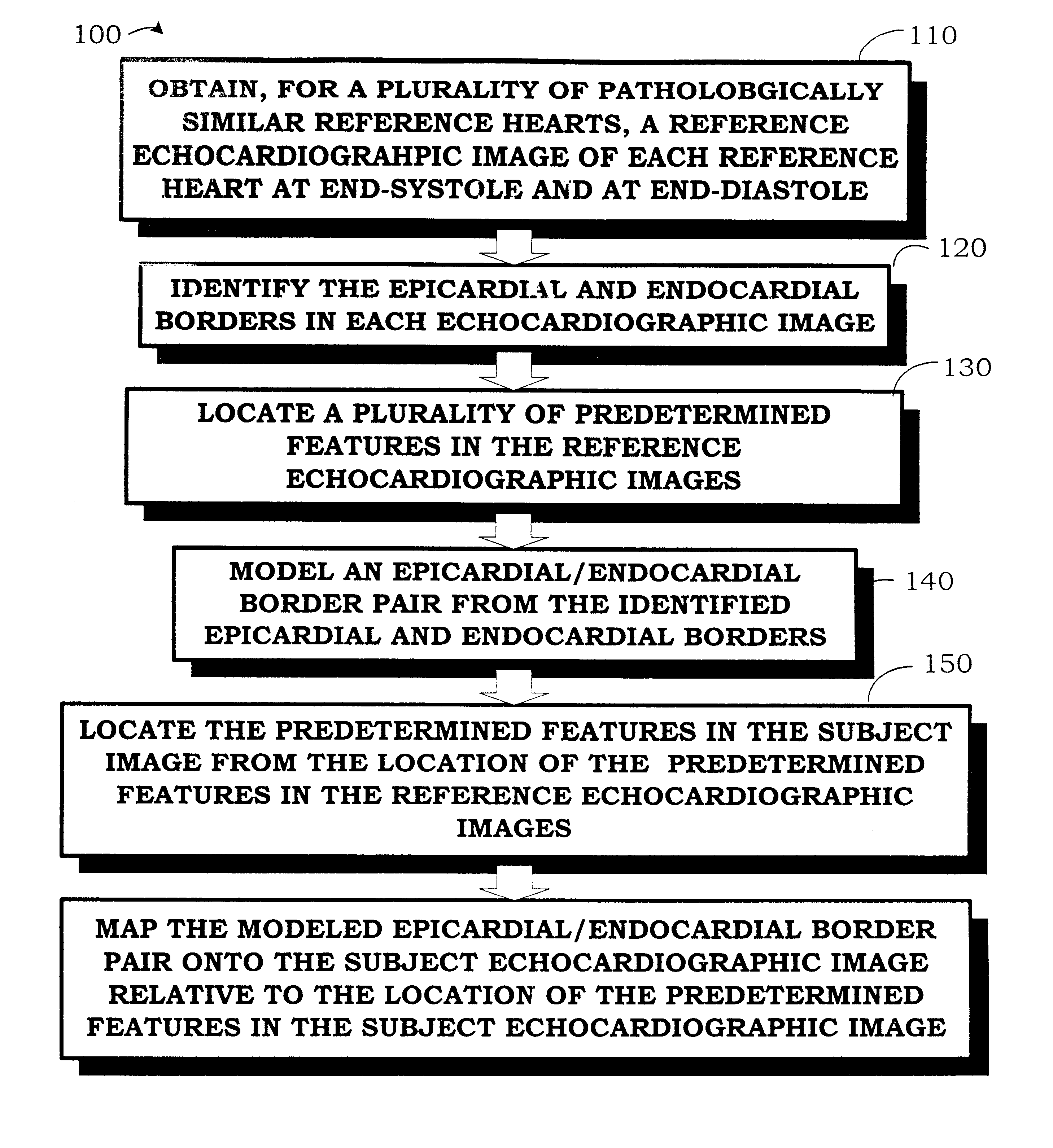
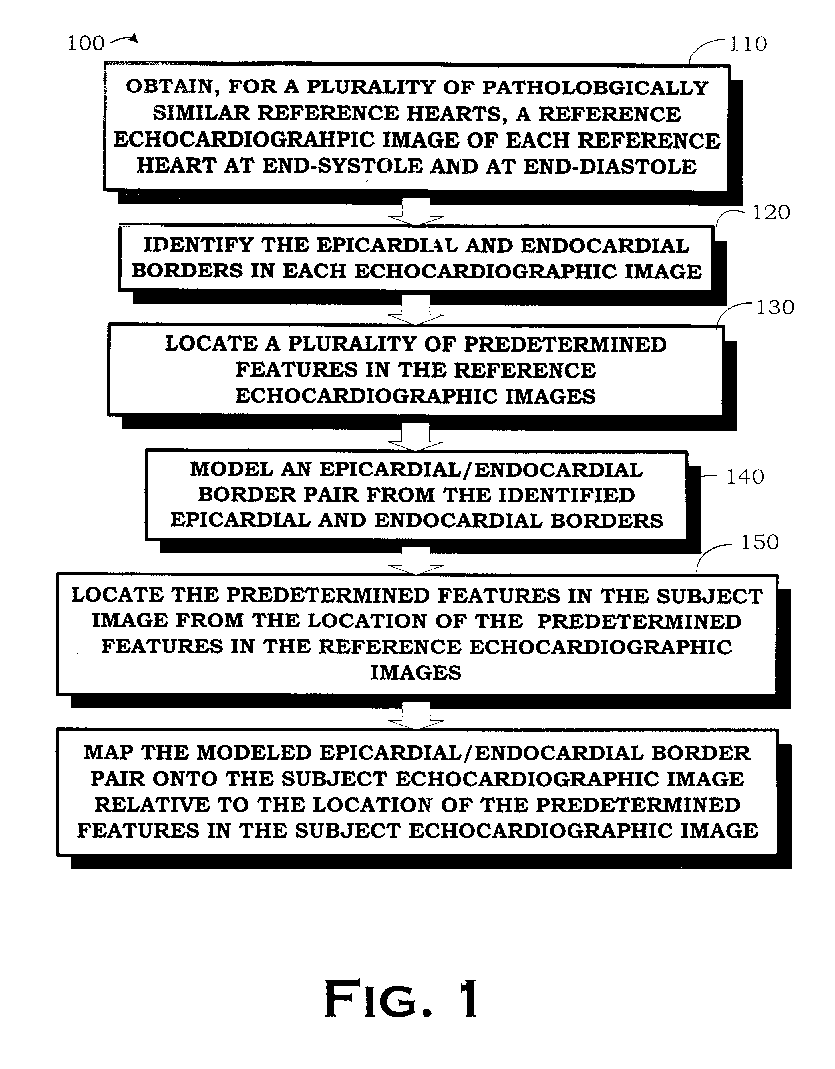
Autonomous boundary detection system for echocardiographic images
a detection system and echocardiography technology, applied in image enhancement, image analysis, medical science, etc., can solve the problems of large operator variability, time-consuming measurements, and insufficient use of ultrasonics for assessing physiology or organ function
- Summary
- Abstract
- Description
- Claims
- Application Information
AI Technical Summary
Problems solved by technology
Method used
Image

Examples
example 1
5.1 Example 1
Autonomously Detecting Boundaries in a Subject Echocardiographic Image of a Subject Heart Using an Apical Four-chamber View
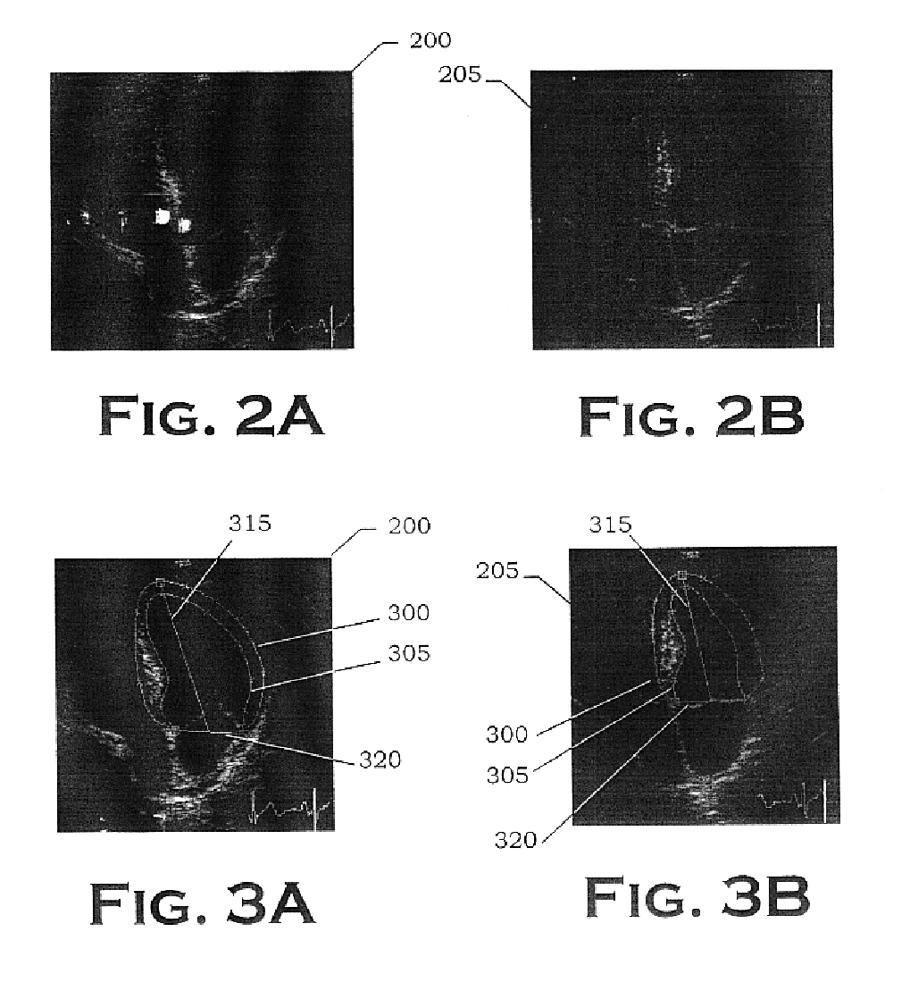
This example comprises a fully automated computer-based algorithm that identifies the epicardial and endocardial borders of the LV for echocardiographic image sequences acquired from the apical 4-chamber view during systole. Since the fractional area computation requires area estimates at both ED and ES, the first and last borders in the sequence are of particular interest. Difficulties encountered with this view include a complex shape, poor image information along the LW, poor image information at the apex, and the fact that the LV must be distinguished from the other 3 chambers present in the image. The dynamic motion and changing shape of the MVA create additional problems since it may move upward through the image by as much as 18 mm during systole and the MV may (or may not) be open (or partially open) for some image frames during the cycle. D...
example 2
5.2 Example 2
Developing Images Manifesting a Predetermined Pathology Using an Apical Two-chamber View
A similar system has been developed which approximates the epicardial and endocardial borders for echocardiographic image sequences acquired from the apical two-chamber view. In the apical two-chamber view, the epicardial border and the endocardial border may be comparable in brightness on the endocardial slightly more prominent and the epicardial along the inferior / posterior wall. However, the epicardial border is determined first for control. The reason for interest in the MV plane for making automated measurements is that this anatomical structure usually forms a prominent feature in this type of image and thus provides a good starting point for placement of the epicardial border. If the image has been acquired by the sonographer with proper viewing angle (Weyman, 1984), gain settings, and depth settings, then the portion of the MVA closest to the inferior wall will be in the lowe...
example 3
5.2 Example 3
Developing Images Manifesting a Predetermined Pathology Using a Parasternal, Short-axis View
Major features for the parasternal short axis view are the posterior epicardial-pericardial interface with the lung, and the right ventricular surface of the anterior septum. These interfaces are found with large elliptical arc filters. The model for the LV in the short axis view is an ellipse. For the apical four-chamber view, the major feature is the cavity of the LV followed by the interventricular septum, the MVA, and finally the EA. Once the MVA and EA positions are located, the model used to control the search for specific endocardial and epicardial points is a composite of expert drawn epicardial and endocardial borders from a large database.
From among the standard views to choose from in echocardiography, there are a number of reasons for considering the parasternal short-axis view. The first is that the borders typically have an oval (and thus easily described) shape. A ...
PUM
 Abstract
Description
Claims
Application Information