High resolution, multispectral, wide field of view retinal imager

a retinal imager and multi-spectral technology, applied in the field of high resolution, multi-spectral, wide field of view retinal imager, can solve the problems of spatial resolution, inability to detect biochemical and cellular-scale features, and inability to detect them with current funduscopic instruments, so as to improve the level of opthmological care, increase the resolution and field of view of retinal imaging, and reduce the bandwidth

Inactive Publication Date: 2004-05-18
KESTREL CORP
View PDF3 Cites 76 Cited by
  • Summary
  • Abstract
  • Description
  • Claims
  • Application Information

AI Technical Summary

Benefits of technology

It is an object of the present invention to provide, in association with any commercially available fundus imager, an improved, low bandwidth adaptive optics system and an optimized depth sensitive deconvolution technique to increase retinal imaging resolution and field of view, to thereby enable a clinical device to improve the level of opthmological healthcare.
It is another object of the present invention to provide a deconvolution technique which takes into account the reflectance of difficult colors from the various layers of the retina to provide a high spatial resolution, multi-spectral image over a wide field of view.
It is yet another object of the present invention to provide a retinal imaging system which uses a scanning (or dithered) reference spot to mitigate the speckle problems associated with the instrumentation disclosed by Liang et al. and which allows wavefront estimates and images of the retina to be taken with one exposure instead of multiple exposures.

Problems solved by technology

Certain biochemical and cellular-scale features, which may be present in the early stages of many retinal diseases (e.g., ARMD), cannot be detected today with current funduscopic instruments because of the losses in spatial resolution introduced by the ocular medium of the eye and the lack of selectable spectral data.
Additionally, the presence of aberrations within the eye limits the effective input pupil size of a standard fundus retinal imager to about 2 mm.
This limit leads to a decrease in the contrast of the small image details due to diffraction effects.
1. Sensitively to speckle modulation within the eye;
2. The deformable mirror can only provide limited correction;
3. It is a panchromatic instrument, not multispectral;
4. It operates with a limited field of view, on the order of 2-5 degrees; and
5. Several renditions of the S-H output are required to estimate the wavefront.
Further, while it is claimed that it is useful in determining aberrations beyond defocus and astigmatism and providing improved imaging inside of the eye, there is no discussion of its use as a clinical instrument to be used in the diagnosis of the major causes of vision loss and blindness.
Finally, the Liang et al. instrument is a laboratory device composed of very expensive one-of-a-kind components.

Method used


Image

  • High resolution, multispectral, wide field of view retinal imager
  • High resolution, multispectral, wide field of view retinal imager
  • High resolution, multispectral, wide field of view retinal imager

Examples


Embodiment Construction

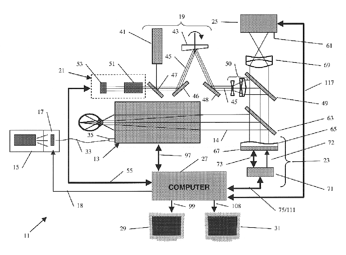
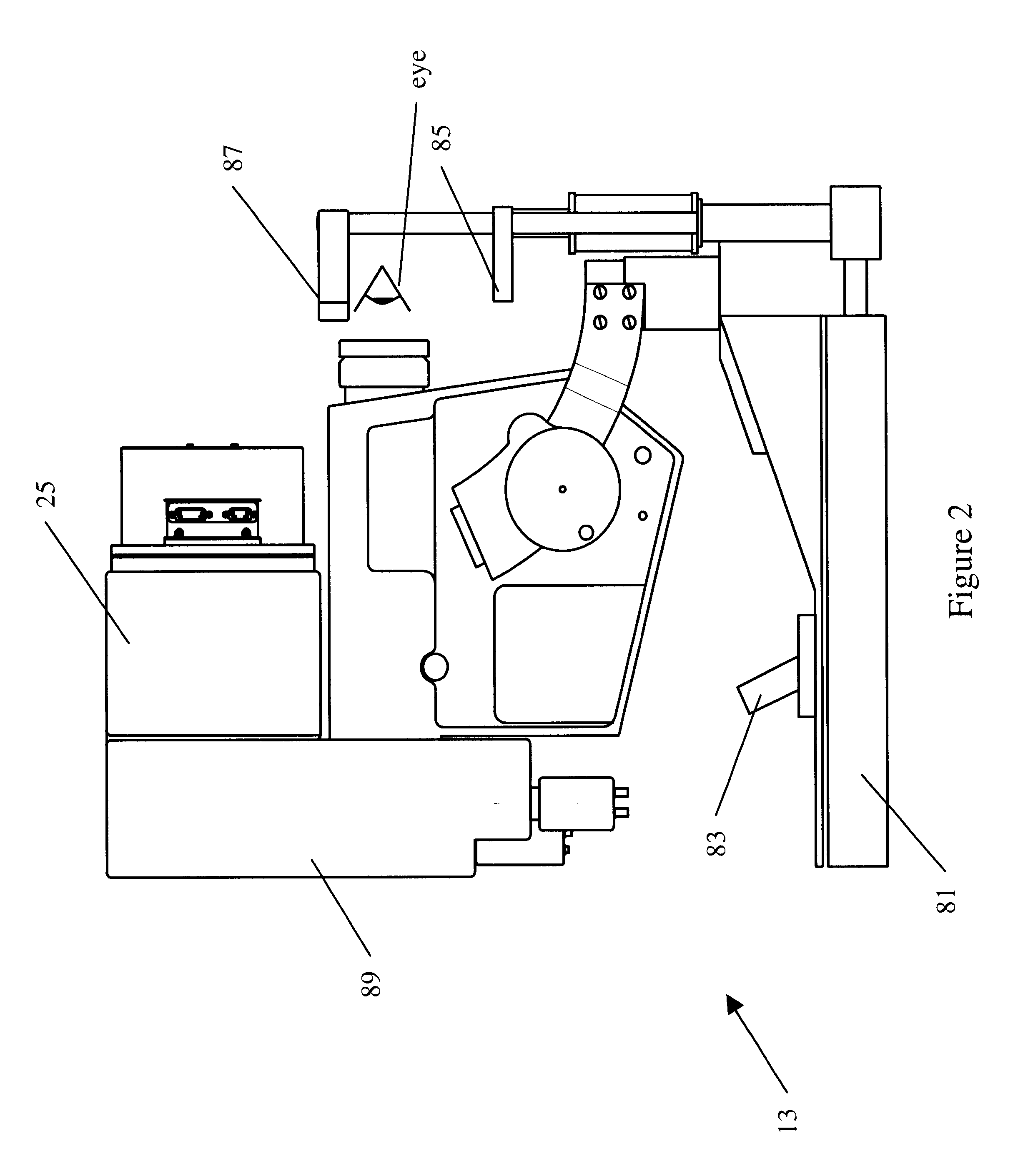
With reference to FIG. 1, improved retinal imaging system 11 includes fundus retinal imager 13, light source 15, filter assembly 17, dithered reference source generator 19, wavefront sensor 21, deformable mirror assembly 23, large format, high resolution detector 25, computer 27 and monitors 29 and 31.

In the present embodiment, fundus retinal imager 13 is a JST ZOMZ, Model KFG3, which provides collimated output 14. However, as those skilled in the art will appreciate other fundus imagers such as the Zeiss FF4 or FF5, the Topcon TRC-50 series or the Canon CF-60 series or CR5-45 can also be used. Light source 15 and filter assembly 17 are connected to fundus retinal imager 13 via fiber optic cable 33 and the standard fiber optic port 35 provided on fundus retinal imager 13. Source 15 includes an illumination lamp such as a tungston, xenon or metal halide lamp (not shown). Filter assembly 17, which is controlled by computer 27 via control line 18, includes up to 10 filters for use in c...


PUM


Abstract

An ophthalmic instrument (for obtaining high resolution, wide field of area multi-spectral retinal images) including a fundus retinal imager, (which includes optics for illuminating and imaging the retina of the eye); apparatus for generating a reference beam coupled to the fundus optics to form a reference area on the retina; a wavefront sensor optically coupled to the fundus optics for measuring the wavefront produced by optical aberrations within the eye and the imager optics; wavefront compensation optics coupled to the fundus optics for correcting large, low order aberrations in the wavefront; a high resolution detector optically coupled to the imager optics and the wavefront compensation optics; and a computer (which is connected to the wavefront sensor, the wavefront compensation optics, and the high resolution camera) including an algorithm for correcting, small, high order aberrations on the wavefront and residual low order aberrations.

Description

The present invention is directed to an improved fundus retinal imaging system which provides high resolution multispectral retinal images over a wide field of view to permit early diagnosis of various pathologies such as diabetic retinopathy, ARMD (age related malocular degeneration) and glaucoma. More specifically, the present invention relates to a conventional fundus retinal imager combined with, inter alia, a multispectral source, a dithered reference, a wavefront sensor, a deformable mirror, a high resolution camera and deconvoluting software to produce wide field, high resolution, multispectral images of the retina.The ability to resolve fine details on retinal images can play a key role in the early diagnosis of vision loss. Certain biochemical and cellular-scale features, which may be present in the early stages of many retinal diseases (e.g., ARMD), cannot be detected today with current funduscopic instruments because of the losses in spatial resolution introduced by the o...

Claims


Application Information

Patent Timeline
Patent Type & Authority Patents(United States)
IPC IPC(8): A61B3/12
CPCA61B3/1015A61B3/1225
Inventor KUDRYASHOV, ALEXISLARICHEV, ANDREY VIKTOROCVICHOTTEN, L. JOHN
Owner KESTREL CORP
Why Eureka
Try Eureka