
Methods and systems using field-based light scattering spectroscopy

a field-based light scattering and spectroscopy technology, applied in the field of optical imaging techniques, can solve the problems of based lss only providing a two-dimensional image, and affecting the quality of tissue spectroscopic information, so as to achieve the effect of preventing the interference of internal sample reflections

Inactive Publication Date: 2005-01-25
MASSACHUSETTS INST OF TECH
View PDF12 Cites 42 Cited by
  • Summary
  • Abstract
  • Description
  • Claims
  • Application Information

AI Technical Summary

Benefits of technology

The present invention relates to systems and methods employing field-based light scattering spectroscopy to provide three-dimensional images and for determining physical characteristics of biological tissue. The three-dimensional images provide information regarding the characteristics of cellular or other structures including, for example, size, shape, texture of the cell nuclei as well as cellular dynamics which can include motility and chaos. Field-based LSS systems incorporate spectroscopic information into a low coherence interferometer system. This field-based type of LSS can be differentiated from intensity-based LSS as it is able to isolate small regions of a biological sample for study, and yields information regarding the phase, as well as the amplitude, of the scattered field. Because field-based LSS measures phase of the wavefront, and wavefront uniformity, as well as providing a high degree of spatial resolution, field-based LSS can measure the shape and spatial distribution of cell nuclei along with size and refractive index of the nuclei. To ascertain the shape of cell nuclei, measurements of the scattered light's amplitude wavefront, phase wavefront, and wavefront non-uniformity are combined. Spatial distribution of cell nuclei is ascertained by three dimensional profiling which allows for imaging of cell nuclei density over the probed region.

Problems solved by technology

Generally, obtaining spectroscopic information from tissue in vivo is difficult as light scattered from surrounding tissues often obscures the desired optical signal.
However, intensity based LSS only provides a two-dimensional image.
Due to the limited coherence length of the source (typically 10-15 μm), light returning from the interferometer reference arm and light backscattered by internal sample reflections interferes constructively or destructively only when the interferometer arm optical path lengths are matched to within the source coherence length.
However, OCT does not provide a functional image as the resultant resolution does not provide an image of the cell nuclei, whose size, shape and other characteristics can reveal abnormal cell activity.

Method used


Image

  • Methods and systems using field-based light scattering spectroscopy
  • Methods and systems using field-based light scattering spectroscopy
  • Methods and systems using field-based light scattering spectroscopy

Examples


Embodiment Construction

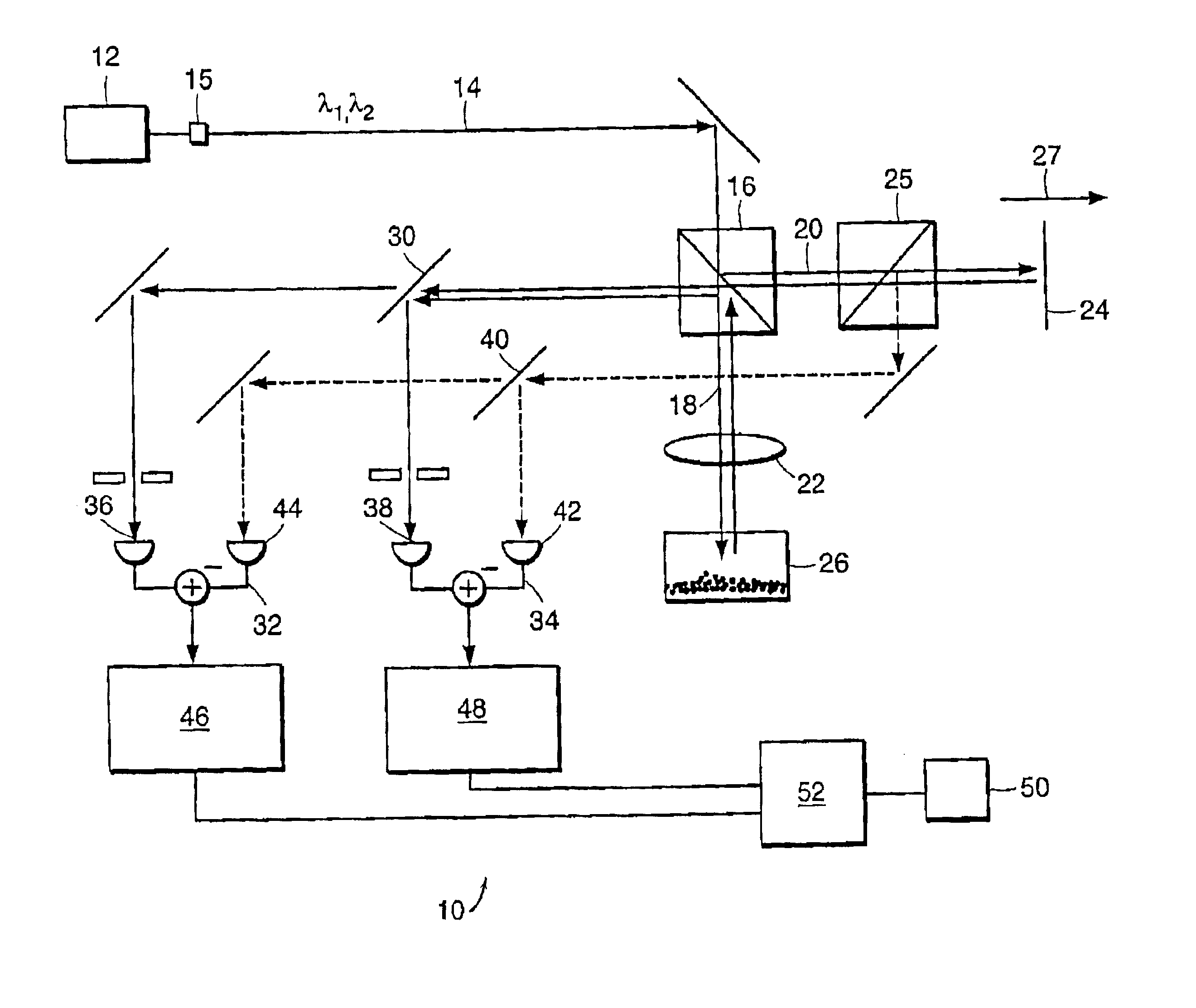
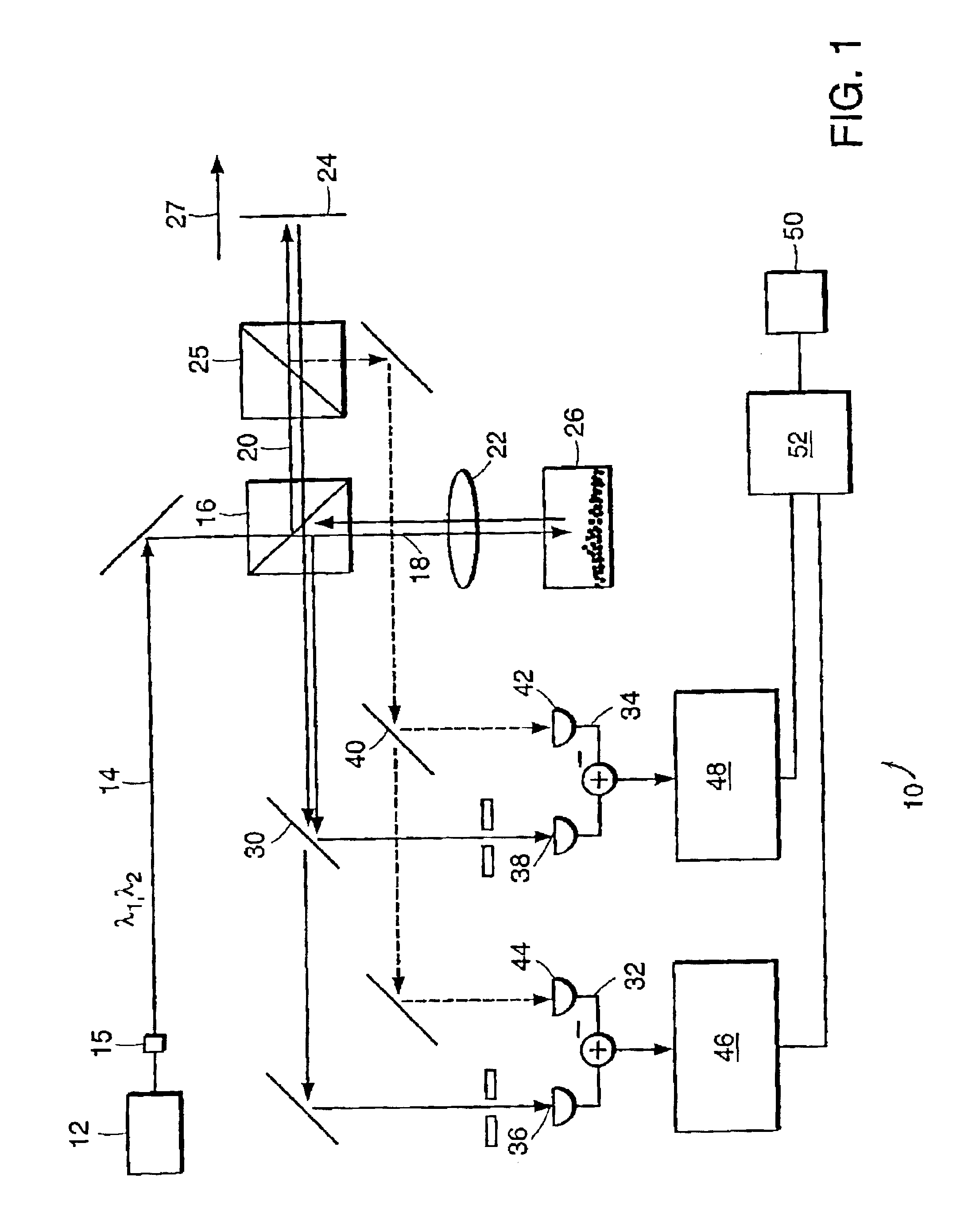
Referring to FIG. 1, a preferred embodiment of a field based LSS system 10 is illustrated, including a Michelson interferometer with two low-coherence light sources. In this particular embodiment, a Coherent MIRA Ti:sapphire laser 12 operating in femtosecond mode (150 fs) produces 800 nm light. The measured coherence length is about 30 μm. A portion of this light is handled by converter 15 such that it is split off and up-converted to 400 nm by means of a CSK Optronics LBO second harmonic generation crystal. Note that two or more separate light sources can be used to provide the two or more wavelengths used for particular measurements. The converted light is then recombined with the original beam. There is preferably substantial overlap (i.e., greater than 50% and preferably greater than 80%) between the two wavelength components. Reduced overlap will increase the scan time to illuminate a given surface are or volume. The combined beam 14 is then divided by a beam-splitter 16 into a...


PUM

PropertyMeasurementUnit
sizesaaaaaaaaaa
coherence lengthaaaaaaaaaa

Abstract

The present invention relates to systems and methods of field-based light scattering spectroscopy. These systems and methods provide for the diagnosis of tissue by measuring the size and distribution of cellular characteristics. Field based measurements provide phase information resulting from the interaction of scatterers within the material and the incident wavefront. These measurements can be used to provide three dimensional images of tissue.

Description

BACKGROUND OF THE INVENTIONOptical imaging techniques have been shown to be useful diagnostic tools for a number of biomedical applications. Increasingly, the structure of biological tissue is being studied using the interaction of the tissue with light. For example, using light scattering spectroscopy (LSS) the size and index of refraction of scatterers comparable in size to the wavelength of light are profiled by measuring the periodicity of the spectra of reflected light. In standard pathological analysis, sensitivity to variations in the size and index of refraction of cell nuclei can give indications of abnormal cell activity. For example, pre-cancerous epithelial cells exhibit nuclear enlargement.Generally, obtaining spectroscopic information from tissue in vivo is difficult as light scattered from surrounding tissues often obscures the desired optical signal. This can be overcome through a combination of spectroscopy with a biomedical imaging techniques which provide the abil...

Claims


Application Information

Patent Timeline
Patent Type & Authority Patents(United States)
IPC IPC(8): A61B5/00G01N21/47G01B9/02G01N21/17G01N21/27G01N21/45
CPCA61B5/0059G01N21/4795G01N21/45A61B5/0066G01N21/47
Inventor YANG, CHANGHUEIWAX, ADAM P.PERELMAN, LEV T.DASARI, RAMACHANDRA R.FELD, MICHAEL S.
Owner MASSACHUSETTS INST OF TECH
Learn More