Methods for functional kidney imaging using small dendrimer contrast agents

a technology of dendrimer and contrast agent, applied in the field of mri methods, can solve the problems of loss of mri signal through saturation effect, limited cell penetration, and selection of dendrimer-based contrast agent suitable for imaging a particular tissue or tissue function, and achieve the effect of abnormal kidney function

Inactive Publication Date: 2005-02-08
US DEPT OF HEALTH & HUMAN SERVICES
View PDF11 Cites 14 Cited by
  • Summary
  • Abstract
  • Description
  • Claims
  • Application Information

AI Technical Summary

Benefits of technology

[0012]Contrast agents and methods useful for imaging the internal structure and function of the mammalian kidney are disclosed. Contrast agents prepared from small DAB and PAMAM dendrimers are disclosed to provide visualization of the proximal straight tubules of the kidney. In one disclosed method, an image-enhancing amount of contrast agent prepared from a DAB-G2D, D

Problems solved by technology

Faster scans with higher resolution require more rapid RF pulsing, and can lead to loss of the MRI signal through saturation effects.
Because many paramagnetic metal ions, including gadolinium (III), are toxic, they are often administered in a sequestered form, for example, as metal chelates.
However, metal chelates, because of their small size and relative hydrophillicity, tend to be cleared rapidly from blood, giving rise to limited cell penetration (but good tolerability).
Unfortunately, selection of a dendrimer-based contrast agent that is suitable for imaging a particular tissue or tissue function (by virtue of a combination of distribution to the tissue and image enhancement of particular features of the tissue) is complicated by the current und

Method used


Image

  • Methods for functional kidney imaging using small dendrimer contrast agents
  • Methods for functional kidney imaging using small dendrimer contrast agents
  • Methods for functional kidney imaging using small dendrimer contrast agents

Examples


example 1

Preparation of Dendrimer Conjugates

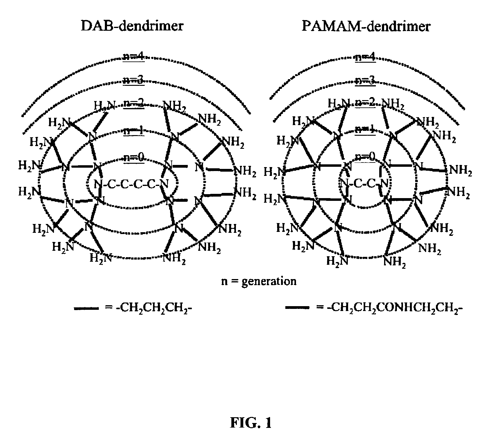
[0059]Dendrimers were concentrated to 10 mg / ml, and diafiltrated against 0.1 M sodium phosphate buffer at pH 9 and reacted at 40° C. with a 16, 32, or 64-fold molar, excess of 2-(p-isothiocyanatobenzyl)-6-methyl-diethylenetriaminepentaacetic acid (1B4M) for the generation-2, -3, and -4 PAMAM and DAB dendrimers, respectively. The reaction solutions were maintained at pH 9 with 1 M NaOH over the reaction time of 48 hr. Additional 1B4M equal to the initial amount was added as a solid after 24 hours to each reaction. The resulting preparation was purified by diafiltration using a Centricon 30 (Amicon Co., Beverly, Mass.) for the generation-4 dendrimers and a Centricon 10 (Amicon Co.) for generation-2 and -3 dendrimers. Over 98% of the amine groups on the dendrimers reacted with the 1B4M as determined by a 153Gd labeling assay of the products.

[0060]Radioactive dendrimer conjugates used for biodistribution studies were prepared as follows. 153GdCl3 conta...

example 2

Biodistribution and Whole Body Retention of 153Gd-Labeled Dendrimer-1B4M-Gd Conjugates

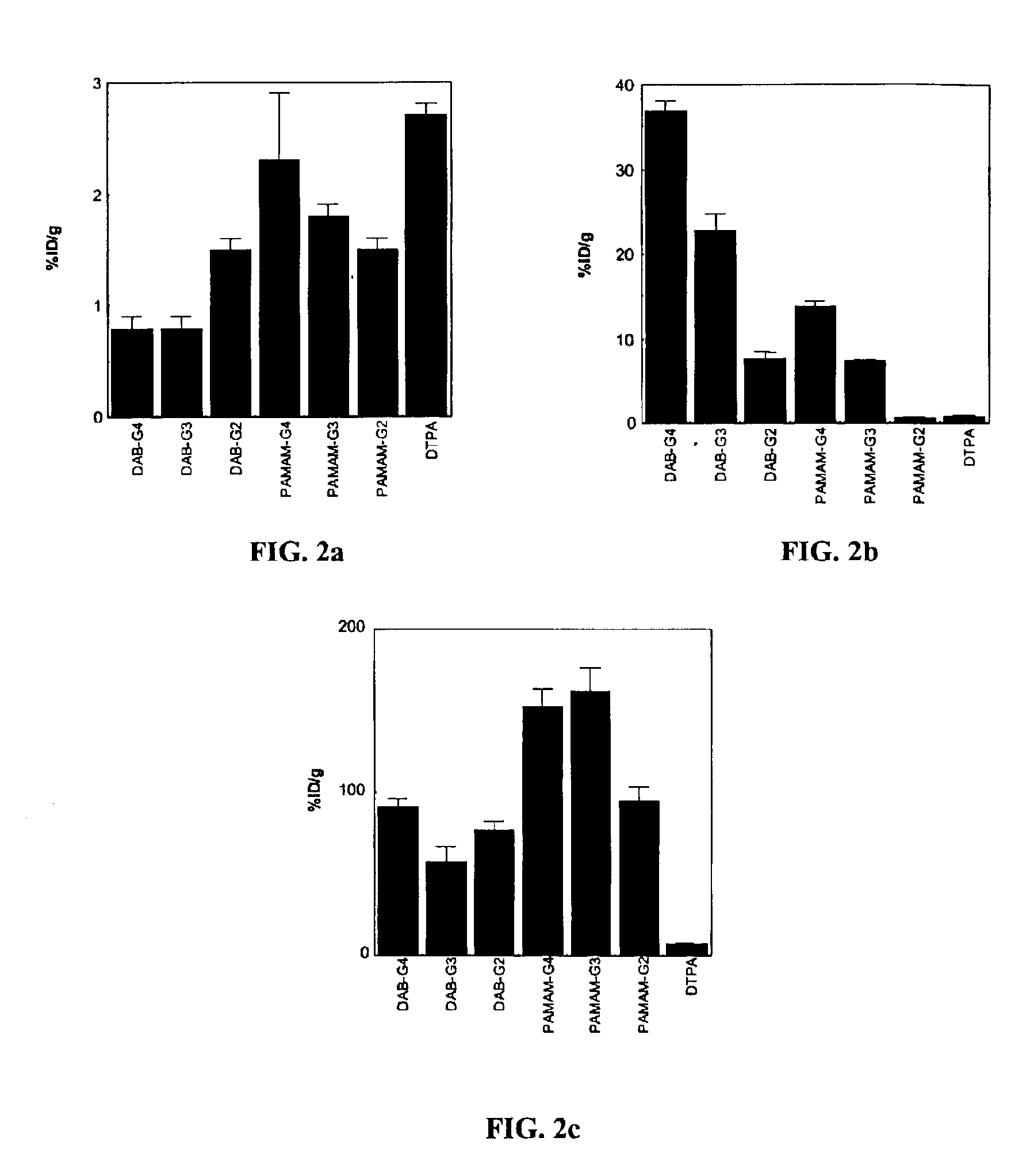
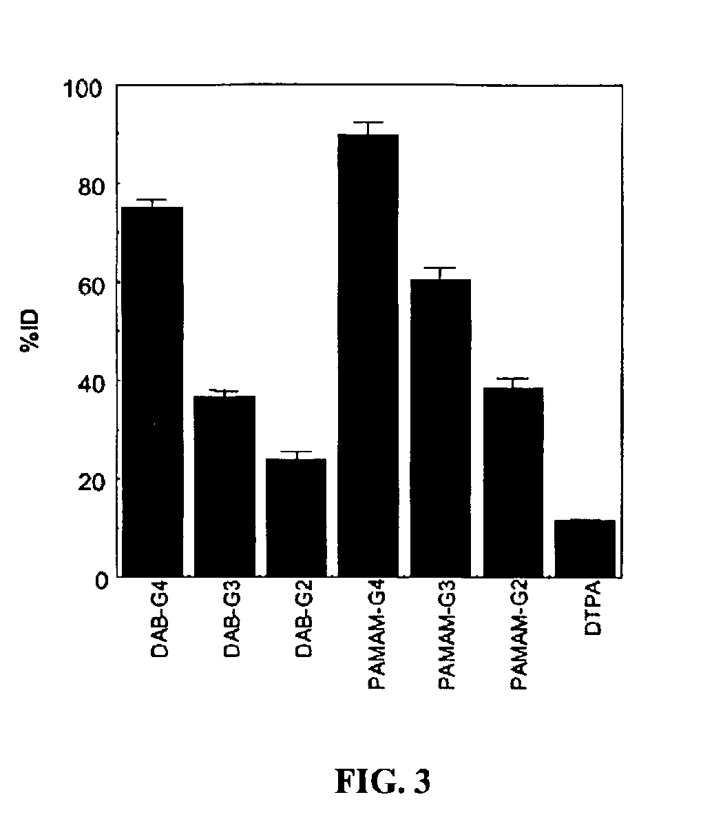
[0063]Seven groups of nude mice (n=4 in each group) were injected with 37 kBq (1 μCi) / 200 μl of 153Gd-labeled dendrimer-1B4M-Gd conjugates or 153Gd-DTPA. The injected samples were added to non-radioactive preparations and the total gadolinium dose was adjusted to 0.02 mmol Gd / kg, 20% of the dose of Gd-[DTPA]-dimeglumine routinely used clinically. The mice were sacrificed 15 min after the injection of the 153Gd-labeled preparations and biodistribution studies were performed. The data were expressed as the percentage of the injected dose per gram (% ID / g) of tissue. The carcasses of the mice were also counted to calculate the whole body retention (% ID).

[0064]The 153Gd-labeled DAB-based agents accumulated significantly greater in the liver (FIG. 2b) and less in the kidney (FIG. 2c) than 153Gd-labeled PAMAM-based agents (p153Gd-labeled DAB-based agents remaining in the blood (FIG. 2a) increased as the...

example 3

Contrast-Enhanced Dynamic 3D-Micro-MRI of Mice

[0065]To evaluate the whole body pharmacokinetics of the contrast agents, seven groups of 8-week-old female nude mice (n=4 in each group) (NCI, Frederick, Md.) were used to obtain contrast-enhanced dynamic 3D-micro-MR images. Briefly, either 0.03 mmol Gd / kg (30% of clinical dose) of dendrimer-1B4M-Gd conjugates or 0.1 mmol Gd / kg of Gd-[DTPA]-dimeglumine (Magnevist, Schering, Berlin, Germany) were intravenously injected into the left tail vein. All images were obtained using the high-resolution wrist coil (GE) with a custom mouse holder using a clinical-grade 1.5-tesla superconductive magnet unit (Signa LX, General Electric Medical System, Milwaukee, Wis.). The mice were anesthetized with 1.15 mg of sodium pentobarbital (Dainabot, Osaka, Japan) and placed in the center of the coils. The fast spoiled gradient echo technique (FSPGR; TR / TE 19.4 / 4.2; flip angle 60°; scan time 1′40″; phase encoding steps 256×256; 3 number of excitations; slab ...


PUM

PropertyMeasurementUnit
Substance countaaaaaaaaaa

Abstract

Small dendrimer-based MRI contrast agents are disclosed to accumulate in renal tubules. The accumulation enables visualization of renal structure and function, permitting assessment of structural and functional damage to the kidneys. In a disclosed embodiment, six, small dendrimer-based MRI contrast agents were synthesized, and their pharmacokinetics, whole body retention, and renal MRI images were evaluated in mice. Surprisingly, despite having unequal renal clearance properties, all of the dendrimer agents clearly visualized the renal anatomy and proximal straight tubules of the mice better than Gd-[DTPA]-dimeglumine. Dendrimer conjugate contrast agents prepared from PAMAM-G2D, DAB-G3D, and DAB-G2D dendrimers were excreted rapidly and may be acceptable for use in clinical applications.

Description

FIELD[0002]Methods of imaging kidney tissue are disclosed. More specifically, the disclosure relates to MRI methods that provide images that may be used to assess renal structure and function, and to detect renal injury.BACKGROUND[0003]MRI is a technique that allows whole body in vivo imaging in three dimensions at high resolution. In MRI, a static magnetic field is applied to the object of interest while simultaneously or subsequently applying pulses of radio frequency (RF) to change the distribution of the magnetic moments of protons in the object. The change in distribution of the magnetic moments of protons in the object from their equilibrium (normal) distribution to a non-equilibrium distribution and back to the normal distribution (via relaxation processes) constitute the MRI signal.[0004]The longitudinal relaxation time, T1, is defined as the time constant of the exponential recovery of proton spins to their equilibrium distribution along an applied magnetic field after a di...

Claims


Application Information

Patent Timeline
IPC IPC(8): A61K49/18A61K49/06A61K49/12A61K51/12A61K49/08
CPCA61K49/124B82Y5/00A61K51/065
Inventor BRECHBIEL, MARTIN W.STAR, ROBERT A.KOBAYASHI, HISATAKA
Owner US DEPT OF HEALTH & HUMAN SERVICES
Why Eureka
Try Eureka