
Filtered backprojection algorithms for compton cameras in nuclear medicine

Inactive Publication Date: 2006-03-21
GUNTER DONALD LEE
View PDF6 Cites 16 Cited by
  • Summary
  • Abstract
  • Description
  • Claims
  • Application Information

AI Technical Summary

Benefits of technology

[0010]With another aspect of the invention, an idealized mathematical model expresses the observed Compton camera data in terms of an integral over the source distribution. An exact analytic inversion is then found for this idealized model. The new

Problems solved by technology

It is generally acknowledged, however, that one of the major limitations of SPECT is the loss of counts due to the collimator in the gamma-ray camera.
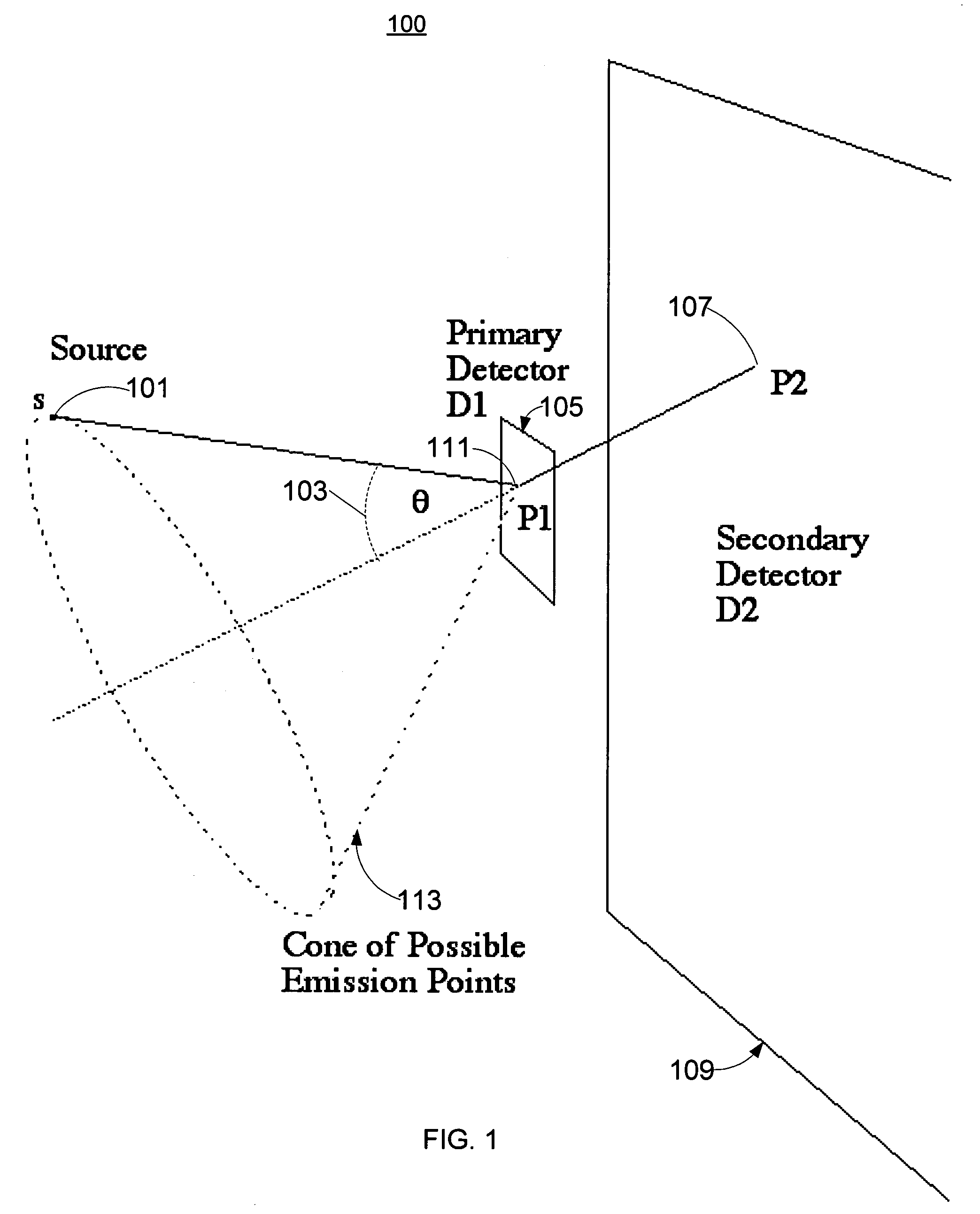
In medical applications, however, the patient is located at a finite distance, so that the interpretation of the angular information provided by Compton events is complicated by huge parallax effects.
Among the most significant of these problems is the 3D reconstruction of the radiopharmaceutical distribution from the data provided by a Compton camera.
The reconstruction of 3D images from Compton camera data poses a formidable mathematical problem.
This geometric difference completely changes the nature of the reconstruction problem.
However, the reason for preferring iterative algorithms was not the accuracy of the mathematical model.
An algorithm for Compton cameras developed by Cree and Bones restricts the acceptable data as much as would a standard collimator—and is, therefore, unsuitable for practical applications.
This rebinning procedure, which is the major drawback of the technique, overcomes the basic problem of Compton camera reconstruction; namely, the cone of incident gamma-ray directions.
Although Parra and Tomitani and Hirasawa use a slightly different mathematical model than Basko et al, they face the same problem; namely, the additional degree of freedom introduced by the acceptance cone of the Compton camera.
Not only were simulations and sophisticated statistical analysis used for camera design, but the reconstruction problem was attacked using a variety of techniques.
Unfortunately, simple backprojection onto the appropriate cones (without filtering) does not produce accurate reconstruction of the source distribution.
The current art also provides analytic-inversion techniques that may be inherently faster to execute but are typically deficient with respect to relative immunity to statistical noise and the desired accuracy of reconstructing the source distribution.

Method used


Image

  • Filtered backprojection algorithms for compton cameras in nuclear medicine
  • Filtered backprojection algorithms for compton cameras in nuclear medicine
  • Filtered backprojection algorithms for compton cameras in nuclear medicine

Examples


Embodiment Construction

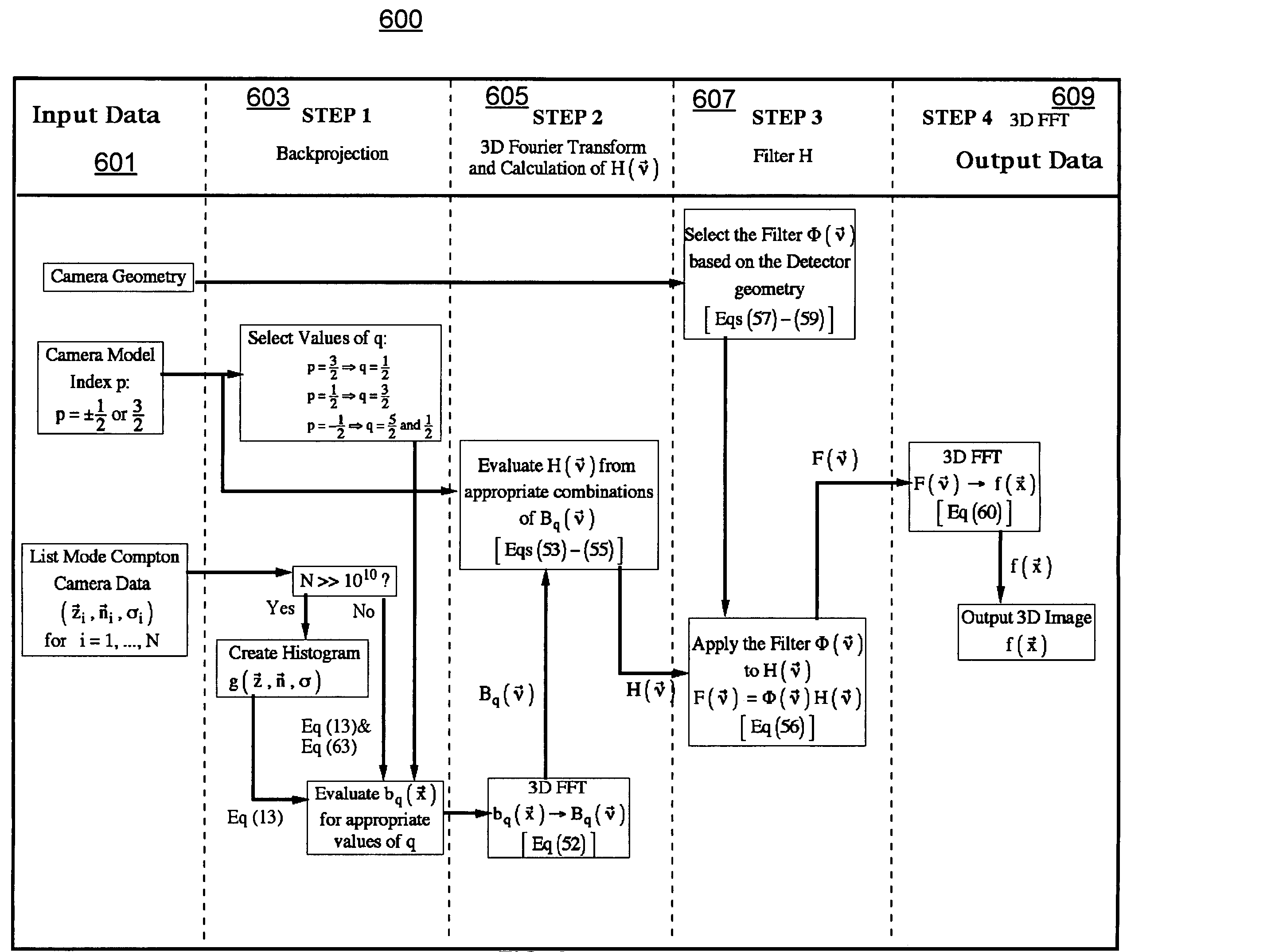
[0028]The following terminology and notation is referenced in the following discussion.[0029]BP backprojection operator[0030]FBP filtered backprojection[0031]FT Fourier transformation[0032]LHS left hand side of an equation[0033]RHS right hand side of an equation[0034]voxel The smallest distinguishable box-shaped part of a three-dimensional space. A particular voxel will be identified by the x, y and z coordinates of one of its eight corners, or perhaps its centre. The term is generally used in three dimensional modeling and sometimes generalized to higher dimensions.[0035]δ( ) Dirac delta function. The function is defined by the relation f(0)=∫dx f(x)δ(x) for all integrable functions f(x). An alternative definition is δ⁡(x)=limλ→0⁢12⁢⁢π⁢λ⁢⁢exp⁡(-x2λ2).[0036]{right arrow over (e)}x, {right arrow over (e)}y, {right arrow over (e)}z unit vectors corresponding to the x, y, and z coordinates, respectively.

[0037]A mathematical model is proposed for Compton cameras. This model describes th...


PUM


Abstract

The present invention provides methods and apparatuses for constructing observed data from a Compton camera in order to provide a three-dimensional image of a radiopharmaceutical source distribution within a patient. No intermediate two-dimensional images are formed. Observed data may be analyzed by a processor in order to construct a three-dimensional image representing the source distribution. An idealized mathematical model may express the observed Compton camera data in terms of an integral over the source distribution. An exact analytic inversion is then found for this idealized model. The new analytic solutions arise from a generalization of the integral equation used to model the Compton camera. The kernel of the integral equation is modified by the introduction of an index p that describes the effect of source distance from the camera.

Description

[0001]This application claims priority to provisional U.S. Application. No. 60 / 458,131, filed Mar. 27, 2003.FIELD OF THE INVENTION[0002]The present invention relates to nuclear medicine for reconstructing a source distribution from Compton camera data in the application of nuclear medicine. In particular, the source distribution corresponds to a radiopharmaceutical distribution within a patient.BACKGROUND OF THE INVENTION[0003]In nuclear medicine, a radiopharmaceutical is injected into a patient and accumulates in tissues of physiological interest (e.g., cancer tumors or heart muscle). The gamma-ray emissions from this radiopharmaceutical are then observed and the local density of radiopharmaceutical within the patient reconstructed. Two clinical imaging techniques, single photon emission computed tomography (SPECT) and positron emission tomography (PET), are based on this simple idea and are currently available commercially. Both these imaging techniques use filtered backprojection...

Claims


Application Information

Patent Timeline
IPC IPC(8): G01T1/20A61K
CPCG06F15/16G06T11/006G06T2211/421
Inventor GUNTER, DONALD LEE
Owner GUNTER DONALD LEE
Learn More