Polynucleotides encoding GFRα3

a polynucleotide and coding technology, applied in the field of identifying and isolating novel dna, can solve the problems of different malignancies and pathological disorders, and achieve the effects of facilitating identification and characterization, high throughput, and facilitating trapping and detection

Inactive Publication Date: 2006-04-11
GENENTECH INC
View PDF8 Cites 27 Cited by
  • Summary
  • Abstract
  • Description
  • Claims
  • Application Information

AI Technical Summary

Benefits of technology

[0017]In view of the surprising finding herein that the α-subunit receptor can dimerize upon ligand binding, and further that such dimerization can activate a kinase domain fused the α-subunit receptor, a method is provided herein to measure ligand-induced α-subunit receptor activation, i.e. homo-dimerizaiton or homo- oligomerization. In one embodiment is provided a sensitive, reliable assay that measures agonist- or ligand-induced α-subunit receptor activation, i.e., homo-dimerization or homo-oligomerization, preferably by measuring receptor protein tyrosine kinase (rPTK) autophosphorylation of a polypeptide fusion comprising the ligand binding domain of an α-subunit receptor and the intracellular catalytic domain of a receptor protein tyrosine kinase. The construct can further optionally comprise a flag epitope to facilitate trapping and detection of the activated (e.g., dimerized, phosphorylated) α-subunit receptor. The assay is desirably useful for qualitatively and quantitatively measuring α-subunit receptor activation as well as facilitating identification and characterization of potential agonists and antagonists for a selected α-subunit receptor. It is a further object of the invention to provide an assay which enables ligand-receptor interactions to be studied for any selected α-subunit receptor, and preferably a GFRα subunit receptor.
[0018]This assay must have a capacity for high throughput, that is, the ability to reliably evaluate large numbers of samples in a relatively short period of time (e.g., in one day). The assay ideally does not use radioactive materials and is also amenable to automation.
[0019]In at least one embodiment of the invention is provided a generic assay which enables a α-subunit receptor of interest to be studied, regardless of whether or not a receptor-specific capture agent having the desired characteristics is available. Furthermore, it is an object of the invention to provide an assay which substantially represents the ligand-binding activity of the α-subunit receptor in situ. This is desirable insofar as it reduces the possibility that altered interactions between the receptor and the ligand may occur as a consequence of the receptor not being membrane bound. In one embodiment of the assay is provided a method for measuring ligand binding by detecting serine-threonine kinase phosphorylation, phosphorylation of intracellular kinases and phosphatase activity of a catalytic domain fused to the α-subunit receptor. Accordingly, the invention provides an assay for measuring activation or ligand binding of an α-subunit receptor construct chimera by detecting its homo-dimerization or homo-oligomerization by in turn measuring kinase or phosphatase activity (i.e., by autophosphorylation) of catalytic domain that is fused to the ligand- binding domain of an α-subunit receptor of interest.
[0024](b) An analyte is then added to the wells having the adhering cells, such that the receptor construct is exposed to (or contacted with) the analyte. This assay enables identification of agonist and antagonist ligands for the α-subunit receptor of interest. In order to detect the presence of an antagonist ligand which blocks binding and / or activation of the receptor by an agonist ligand, the adhering cells are exposed to the suspected antagonist ligand first and then to the agonist ligand (or to a mixture of the agonist and antagonist) so that competitive inhibition of receptor binding and activation can be measured. Also, the assay can identify an antagonist which binds to the agonist ligand and thereby reduces or eliminates its ability to bind to, and activate, the kinase domain. To detect such an antagonist, the suspected antagonist and the agonist for the receptor are incubated together and the adhering cells are then exposed to this mixture of ligands.

Problems solved by technology

The aberrant expression or uncontrolled regulation of any one of these receptor tyrosine kinases can result in different malignancies and pathological disorders.

Method used


Image

  • Polynucleotides encoding GFRα3
  • Polynucleotides encoding GFRα3
  • Polynucleotides encoding GFRα3

Examples


example 1

Cloning of Mouse GFRα3

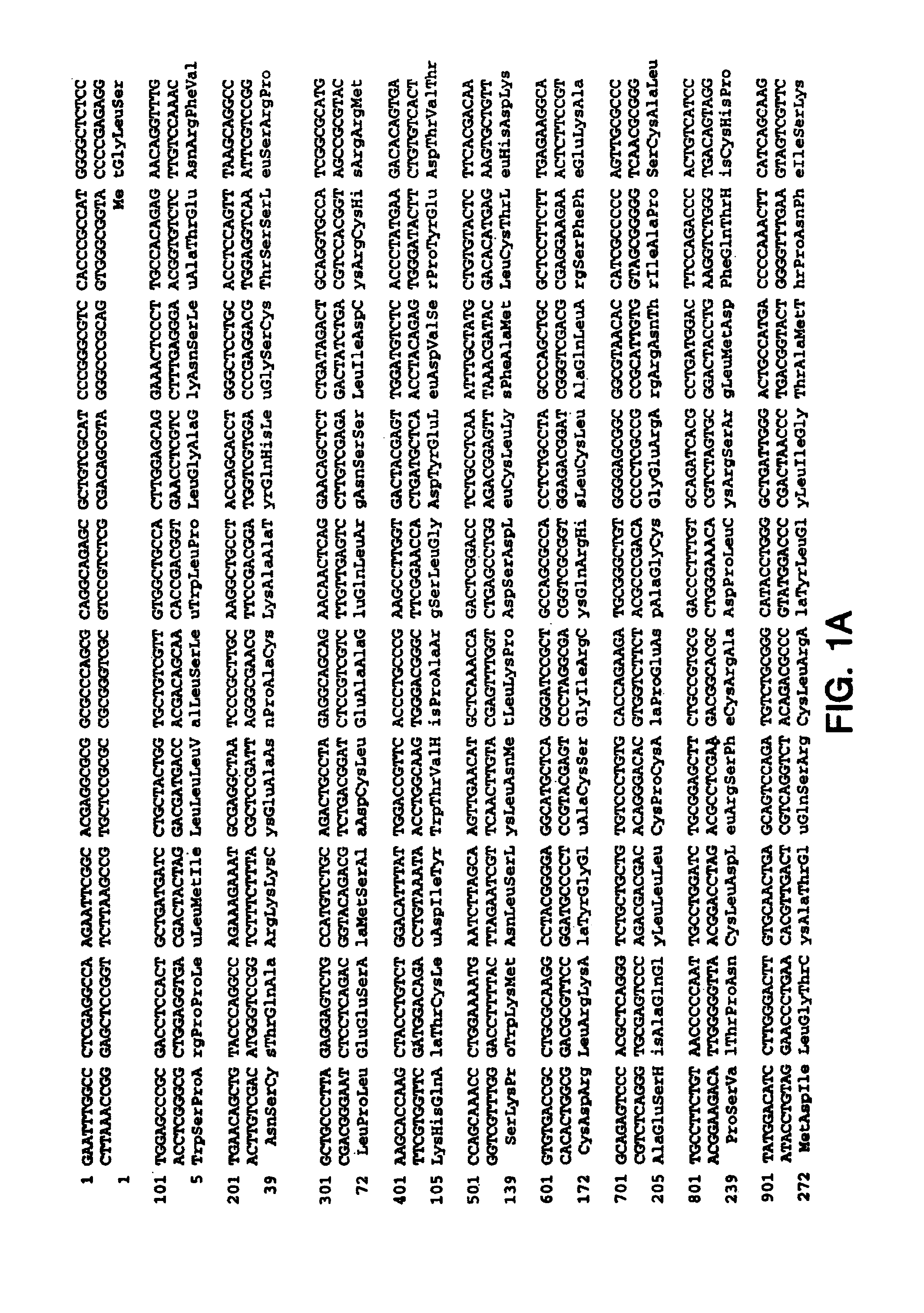
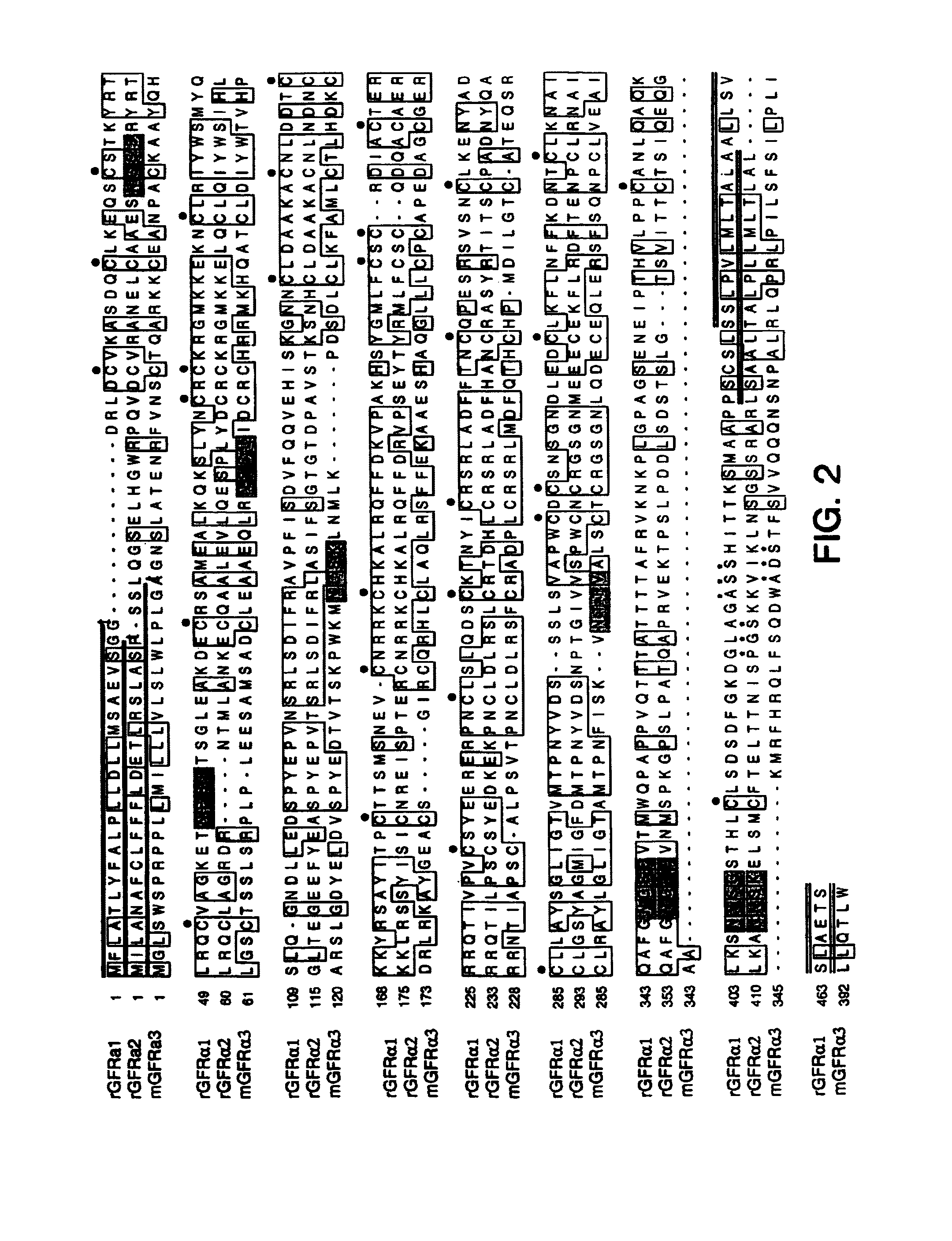
[0277]Using sequences from the neurturin receptor, now known as GFRα2 (“glial-cell-line-derived neurotrophic factor family receptor alpha”), a novel, potential member of the GFRα family was identified as a mouse EST in a public gene database (Accession Numbers W99197 (SEQ ID NO: 1), AA041935 (SEQ ID NO: 2) and AA050083 (SEQ ID NO: 3)). A DNA fragment corresponding to this potentially new receptor was obtained by Marathon PCR using mouse E15 cDNA (Clontech, Inc. USA) as template and PCR primers derived from the mouse EST. The PCR product was then used to screen a lambda gt10 mouse E15 library (Clontech, Inc. USA) to obtain a full length clone. The nucleotide sequence of the full length mouse cDNA is provided as SEQ ID NO: 4 (FIGS. 1A–1B). The protein sequence (SEQ ID NO: 5; see FIGS. 1A–1B) encoded by the isolated DNA was designated GFRα3, since it was determined to be a novel protein with sequence identity to GFRα 1 (formerly the GDNF Receptor alpha) and GFRα2 ...

example 2

Isolation of cDNA Clones Encoding Human GFRα3

[0279]To identify rapidly whether a human homolog of this new receptor might exist, an expressed sequence tag (EST) DNA database (a proprietary EST database, LIFESEQ™, Incyte Pharmaceuticals, Palo Alto, Calif.) was searched and an EST (INC3574209) was identified having the sequence:

[0280]

GCGCTGNNTGNCNGNANGNGGGGGCGGGAGGTGCCGGTCGAGGGAGCCCCGCTCTCAGAG(SEQ ID NO: 10).CTCCAGGGGAGGAGCGANGGGAGCGCGGAGCCCGGCCGCCTACAGCTCGCCATGGTGCGCCCCCTGAACCCGCGACCGCTGCCGCCCGTAGNNNNNNNNNNNNNNNNNNNNNNNNNNNNNNNNNNNNNGCCTCTCGCAGCCGGAGACCCCCTTCCCACAGAAAGCCGACTCATGAACAGCTGTCTCCAGGCCAGGAGGAAGTGCCAGGCTGATCCCACCTGC

This sequence had 61% identity to the murine GFRα3.

[0281]To clone the corresponding full length cDNA, a panel of cDNA libraries were screened with primers:

[0282]

newa3.F5′ GCC TCT CGC AGC CGG AGA CC 3′(SEQ ID NO: 11)newa3.R5′ CAG GTG GGA TCA GCC TGG CAC 3′(SEQ ID NO: 12)

DNA from the libraries was screened by PCR amplification, as per Ausubel et al., Current Protoc...

example 3

Use of GFRα3 as a Hybridization Probe

[0290]The following method describes use of a nucleotide sequence encoding GFRα3 as a hybridization probe.

[0291]DNA comprising the coding sequence of GFRα3 (shown in SEQ ID NO: 4, SEQ ID NO: 14 or SEQ ID NO: 16), or a fragment thereof, is employed as a probe to screen for homologous DNAs (such as those encoding naturally-occurring GFRα3 or variants of GFRα3) in human tissue cDNA libraries, human tissue genomic libraries, RNA isolated from tissues, tissue preparations in situ, or chromosome preparations (such as for chromosome mapping).

[0292]Hybridization and washing of filters containing either library DNAs is performed under the following high stringency conditions. Hybridization of radiolabeled GFRα3-derived probe to the filters is performed in a solution of 50% formamide, 5×SSC, 0.1% SDS, 0.1% sodium pyrophosphate, 50 mM sodium phosphate, pH 6.8, 2× Denhardt's solution, and 10% dextran sulfate at 42° C. for 20 hours. Washing of the filters is ...


PUM

PropertyMeasurementUnit
molecular weightaaaaaaaaaa
timeaaaaaaaaaa

Abstract

The present invention relates to nucleotide sequences, including expressed sequence tags (ESTs), oligonucleotide probes, polypeptides, vectors and host cells expressing, and immunoadhesions and antibodies to mammalian GFRα3, a novel α-subunit receptor of the GDNF (i.e. GFR) receptor family. It further relates to an assay for measuring activation of an α-subunit receptor by detecting tyrosine kinase receptor activation (i.e., autophosphorylation) or other activities related to ligand-induced α-subunit receptor homo-dimerization or homo-oligomerization.

Description

[0001]This is a non-provisional application of co-pending application(s) provisional application Nos. 60 / 079,124 filed 23 Mar. 1998, and 60 / 081,569 filed 13 Apr. 1998 the entire disclosure of which is hereby incorporated by reference and to which application(s) priority is claimed under 35 USC §119.TECHNICAL FIELD[0002]The present invention relates generally to the identification and isolation of novel DNA and to the recombinant production of novel polypeptides which are characterized by the presence of GFRα3 sequences, an α-subunit receptor. It further relates to an assay for measuring ligand-induced activation of an α-subunit receptor by detecting autophosphorylation of a kinase domain of an α-receptor-receptor protein tyrosine kinase (rPTK) fusion using a kinase receptor activation, enzyme-linked immunosorbent assay (KIRA ELISA) or by other means to detect α-subunit homodimerization.INTRODUCTIONBACKGROUND[0003]Neurotrophic factors such as insulin-like growth factors, nerve growth...

Claims


Application Information

Patent Timeline
Patent Type & Authority Patents(United States)
IPC IPC(8): C12N15/12C12N15/79C12N15/85
CPCC07K14/71
Inventor DE SAUVAGE, FREDERIC J.KLEIN, ROBERT D.PHILLIPS, HEIDI S.ROSENTHAL, ARNON
Owner GENENTECH INC
Why Eureka
Try Eureka