Suturing instruments and methods of use

a technology of sutures and instruments, applied in the field of sutures for approximation, ligation and fixation of tissue using a suture, can solve the problems of severe limitation, limited mulhollan's instrument, time-consuming suturing of body tissues, etc., and achieves accurate placement, good suturing effect, and good suturing

Inactive Publication Date: 2006-04-25
BOSTON SCI SCIMED INC
View PDF94 Cites 177 Cited by
  • Summary
  • Abstract
  • Description
  • Claims
  • Application Information

AI Technical Summary

Benefits of technology

[0052]The method of use of this device is as follows: an incision is made bilaterally in the anterior vaginal wall, just lateral to the bladder neck. The space of Retzius is dissected by sweeping the surgeon's finger along the inferior aspect of the pubic ramus, from the symphysis pubis out as far laterally as can be reached, preferably as far as the obturator fossa. The Cooper's ligament may be palpated, and using the symphysis as the medial landmark, and the obturator fossa as the lateral landmark, the instrument is guided by palpation up onto the superior aspect of the pubic ramus. A notch in the face of the head of the device is provided to allow the instrument to be accurately placed for delivery of the suture through Cooper's ligament.
[0053]After proper positioning of the instrument is verified, the needle driver button is pushed, driving the needle carrier and needle suture combination through the ligament and into the needle catch. The shouldered needle is captured in the needle catch, and the needle carrier is retracted back into the device head. The entire device is withdrawn through the colpotomy, trailing the suture. A second pass through the ligament is optional. The free ends of the suture are then passed through the vaginal mucosa, and tied with a pulley stitch to allow for proper positioning of the bladder neck. The procedure is repeated on the contralateral side, both pulley st...

Problems solved by technology

Suturing of body tissues is a time consuming aspect of most surgical procedures.
A limitation of these types of devices is that they are particularly adapted for use in open surgical procedures where there is room for the surgeon to manipulate the instrument.
A limitation of these designs is that they also are adapted primarily for open surgery, in that they require exposure of the tissues to be sutured in order that the tissue may be grasped or pinched between the jaws of the instrument.
This is a severe limitation in the case of endoscopic surgery.
Mulhollan's instrument is limited in that the arc through which the needle must be driven is perpendicular to the axis of the device.
An improper or non-existent closure can lead to a herniation of the bowel and/or bowel obstruction.
Many patients are obese and present considerable fat in this region.
Because the abdominal wall may be several inches thick, it is extremely difficult, tedious and time consuming to approximate the fascial tissues with a suture.
Often times, following removal of a large trocar, the puncture site needs to be enlarged to accomplish this, thus negating some of the advantages of endoscopic surgery previously discussed.
One of the problems with the procedure described above is that it is normally done only in conjunction with other scheduled abdominal surgical procedures such as a hysterectomy.
This is because, as described earlier, an open surgical approach requiring a large abdominal incision must be used, and it is not very common for a patient to elect to have a major abdominal surgical procedure just for the treatment of incontinence.
There are three difficulties associated with the laparoscopic approach; access, suture placement, and knot tying.
Because of their size, the needles used in these procedures are generally not able to be held in the jaws of the needle driver while being introduced through the operative trocar.
This is a difficult and time-consuming aspect of the current endoscopic technique for suturing.
It may be seen that a limitation of this type of needle driver is that the needle may only be driven or rotated in a plane perpendicular to the axis of rotation, such axis being described by the elongate shaft and the position of the surgical trocar.
Thus the current endoscopic needle drivers will not allow the surgeon to swing the needle in an arc paralle...

Method used


Image

  • Suturing instruments and methods of use
  • Suturing instruments and methods of use
  • Suturing instruments and methods of use

Examples


Embodiment Construction

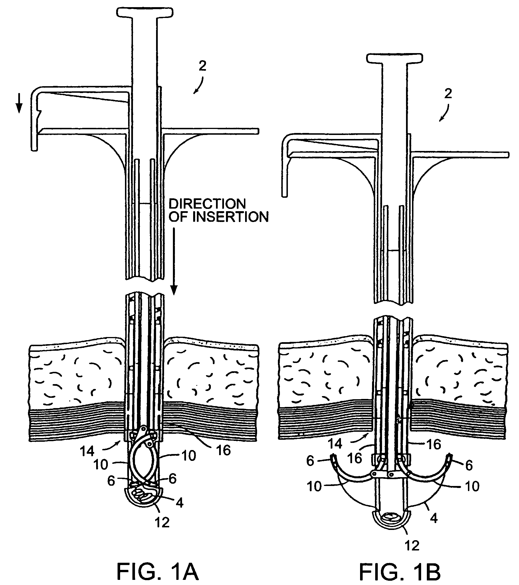
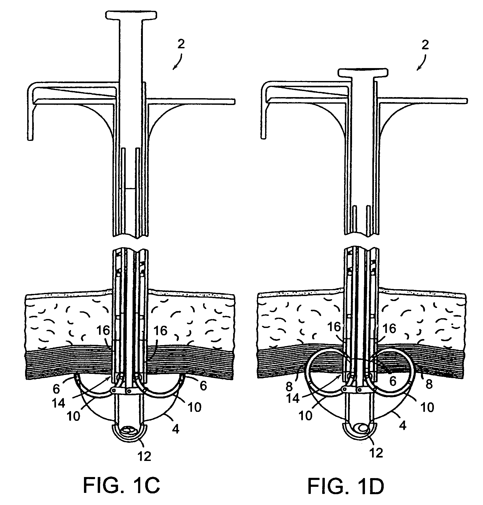
[0083]Although the principles of the present invention are applicable to any device suitable for use in surgical procedures, whether performed on humans or animals, particular utility is effected in human abdominal surgery performed using endoscopic techniques for closure of the wounds created during the introduction of trocars into the abdominal cavity, and particularly the puncture wounds created thereof, as well as closure or approximation of the wounds created either during the resection of benign or malignant lesions, or during the performance of other therapeutic procedures on an organ or organs within a body cavity.

[0084]FIGS. 1A through 1H illustrate the general structure and operation of a first embodiment of the present invention. FIGS. 1A and 1B show a device 2, according to the present invention, which incorporates a length of standard suture material 4 with a needle 6 on each end. The needles 6 are held by a needle carrier 8 (FIG. 1D) and loaded into two guiding tracks ...


PUM


Abstract

A method and device for the placement of sutures and for the purpose of approximating tissue. The invention relates to devices for approximation, ligation and fixation of tissue using a suture, to various constituent parts comprising said devices, and particularly to the placement of sutures into certain difficult to access ligamental structures, to the approximation of tissue separated by means of an endosurgical trocar being inserted into a body cavity, and to approximation, ligation, and fixation of body tissue using both traditional open surgical and endosurgical techniques and instruments. The invention provides for the loading of suture material including needles into the device, introduction and placement of the device into the body cavity, with the distal end having deployable needle guides, extending the needle guides either simultaneously or individually to the periphery of the wound, engaging the wound with the needle guides, driving the needles and suture material through the tissue to be approximated into a catch mechanism, retracting the needle guides and withdrawing the device, leaving a loop of suture material in the margin of tissue. The suture may then be tied to approximate the wound and excess suture material cut off. The invention also provides for the placement of sutures for the endoscopic approximation, fixation, and ligation of tissues within a body cavity including the driving and retrieval of needle and suture combinations, and facilitating the tying of knots.

Description

RELATED APPLICATIONS[0001]This patent application is a continuation-in-part of patent application Ser. No. 09 / 002,875 filed Jan. 5, 1998, by inventors Norman S. Gordon, Robert P. Cooper and Richard L. Quick, and entitled “Needle Guidance System for Endoscopic Suture Device”; which is a continuation of patent application Ser. No. 08 / 554,743 filed Nov. 7, 1995, by inventors Norman S. Gordon, Robert P. Cooper and Richard L. Quick, and entitled “Needle Guidance System for Endoscopic Suture Device”; which is a continuation-in-part of patent application Ser. No. 08 / 311,967, filed Sep. 26, 1994, by inventors Norman S. Gordon and Robert P. Cooper, and entitled “Endoscopic Suture System”; which is a continuation-in-part of patent application Ser. No. 08 / 205,042, filed Mar. 2, 1994, by inventors Norman S. Gordon, Robert P. Cooper and Gordon C. Gunn, and entitled “Endoscopic Suture System”; which is a continuation-in-part of patent application Ser. No. 08 / 057,699, filed May 4, 1993, by invento...

Claims


Application Information

Patent Timeline
IPC IPC(8): A61B17/04A61B17/00A61B17/06A61B17/28
CPCA61B17/0469A61B17/0625A61B17/0057A61B17/0482A61B17/06066A61B17/2909A61B2017/2901A61B2017/00663A61B2017/047A61B2017/0472A61B2017/0474A61B2017/0479A61B2017/06057A61B2017/00637
Inventor GORDON, NORMAN S.COOPER, ROBERT P.QUICK, RICHARD L.
Owner BOSTON SCI SCIMED INC
Why Eureka
Try Eureka