Four-dimensional helical tomographic scanner

Inactive Publication Date: 2006-05-09
KONINKLIJKE PHILIPS ELECTRONICS NV
View PDF14 Cites 55 Cited by
  • Summary
  • Abstract
  • Description
  • Claims
  • Application Information

AI Technical Summary

Benefits of technology

[0015]One advantage of the present invention resides in increased reconstruction accuracy compared with conventional cardiac gating employing circular or low-pitch spiral orbits. The improved accuracy is particularly apparent for large volumes of interest.
[0016]Another advantage of the present invention resides in improved temporal resolution at the voxel level.
[0017]Yet another advantage of the present invent

Problems solved by technology

In cardiac imaging, problems arise due to a limited temporal resolution of computed tomography, which is controlled by the rotation rate of the x-ray source.
However, data combined from adjacent cardiac cycles may not readily form a complete data set due to angular redundancies.
Another source of error with cardiac gating is inaccuracy in associating the electrocardiographic data with the cardiac cycle.
Particularly in cases of heart arrhythmia where the cardiac cycle period is sometimes variable over a few neighbor

Method used


Image

  • Four-dimensional helical tomographic scanner
  • Four-dimensional helical tomographic scanner
  • Four-dimensional helical tomographic scanner

Examples


Example

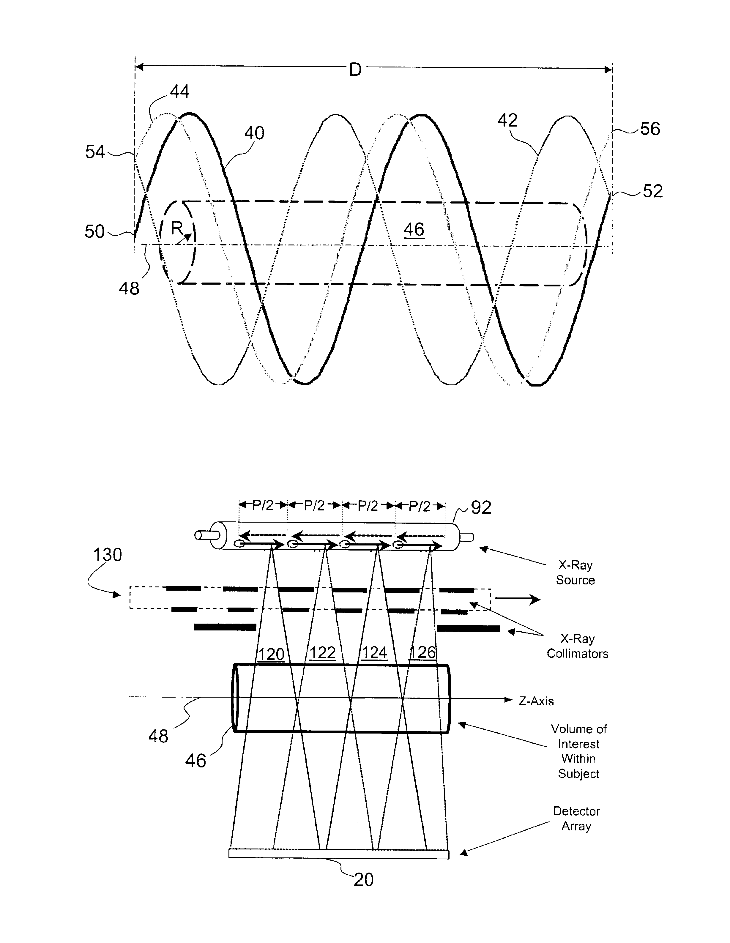
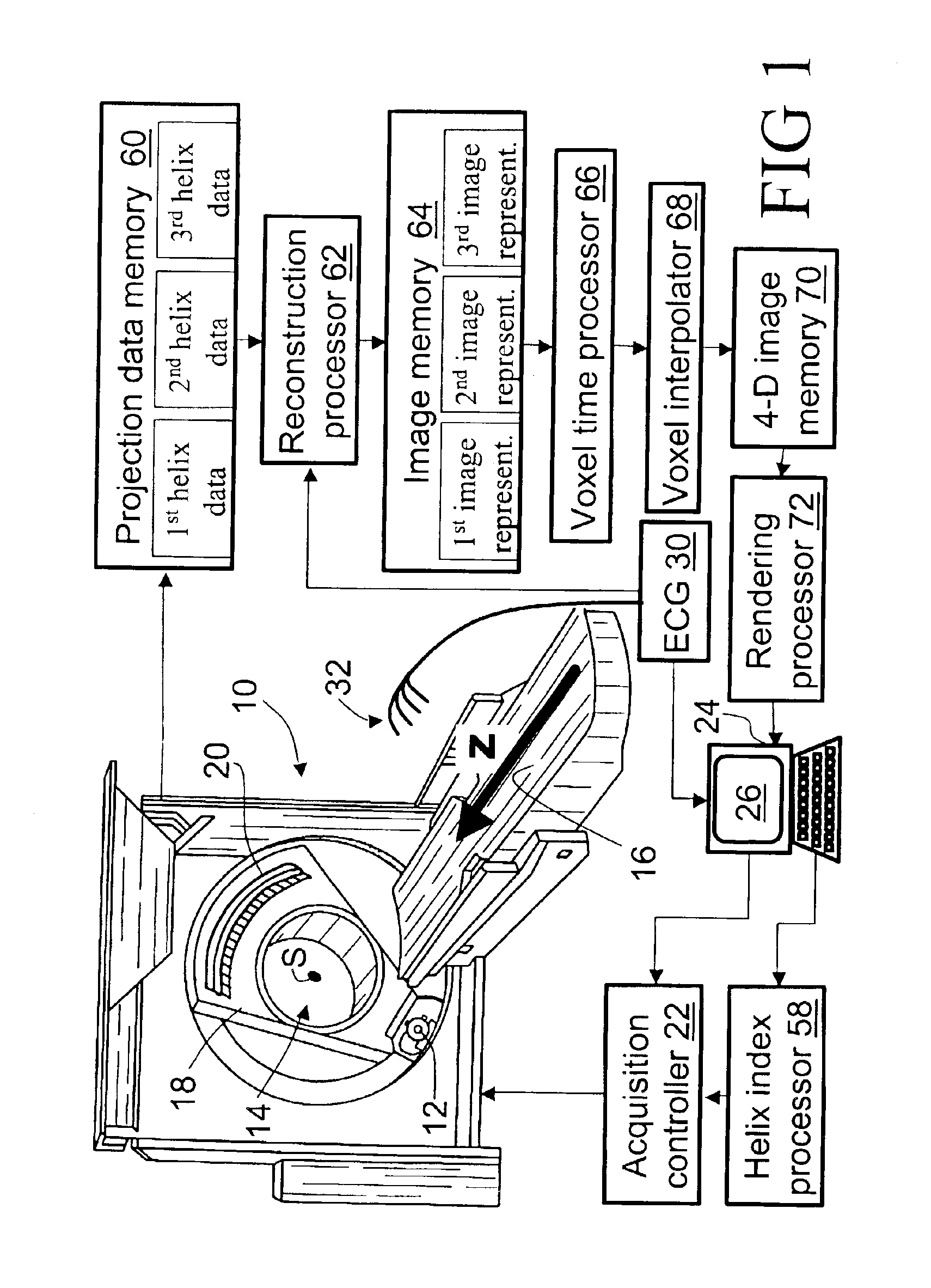
[0035]With reference to FIG. 1, a computed tomography (CT) imaging scanner 10 includes an x-ray source 12 that produces a fan-shaped, cone-shaped, wedge-shaped, or otherwise-shaped x-ray beam directed into an examination region 14 which contains an imaging subject (not shown) arranged on a subject support 16. For cardiac imaging, a patient is positioned with the subject heart substantially centered within the examination region 14. The subject support 16 is linearly movable in a Z-direction while the x-ray source 12 is mounted on a rotating gantry 18 that rotates around the Z-axis.

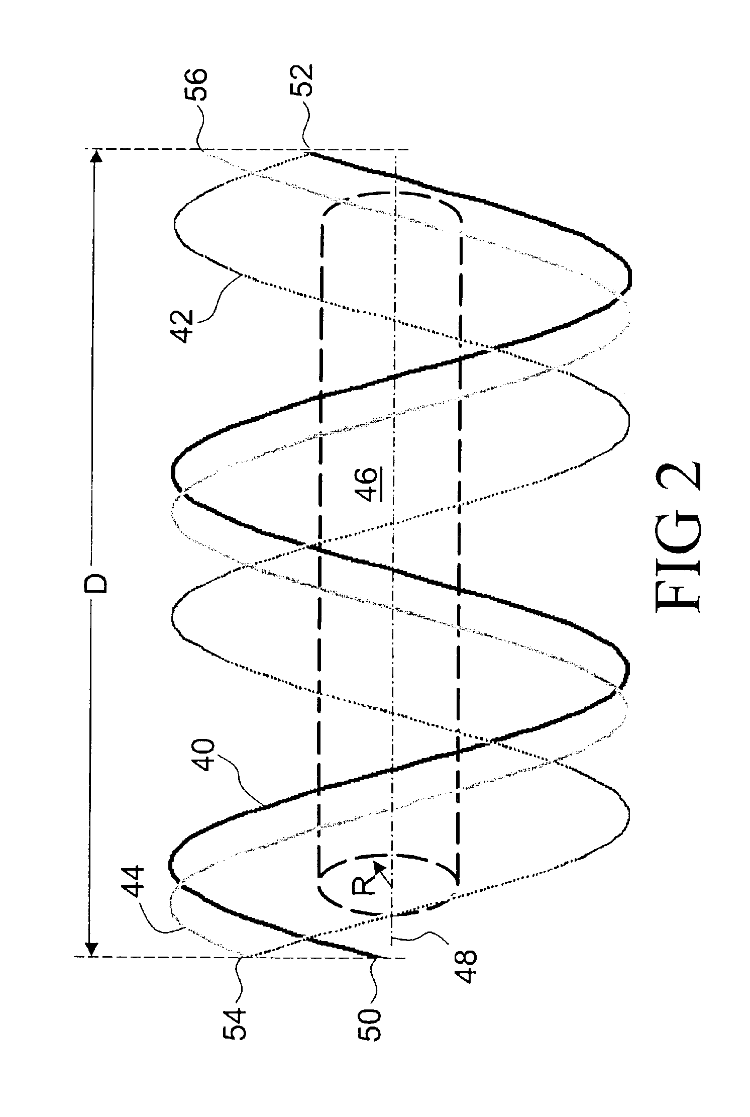
[0036]In a mechanical computed tomography imaging embodiment, the rotating gantry 18 rotates simultaneously with linear advancement of the subject support 16 to produce a generally helical trajectory of the x-ray source 12 about the examination region 14. In an electronic operating embodiment, the subject support 16 remains stationary and the x-ray source 12 electronically sweeps the x-ray beam axially acr...


PUM


Abstract

A computed tomography imaging scanner (10) acquires helical cone beam projection data for a volume of interest (46) using at least two source trajectory helices. A reconstruction processor (62) reconstructs the projection data for each helix to generate a corresponding time skewed image representation. A voxel time processor (66) computes an acquisition time for each voxel in each time skewed image representation. A voxel interpolator (68) computes an interpolated voxel value for each voxel based on values of the voxel in the time skewed image representations and corresponding voxel acquisition times. In an electronic embodiment, the computed tomography scanner (10) includes an x-ray source (12) with an axially oriented cylindrical anode (92), an electron source (961, 962) irradiating the cylindrical anode (92) to produce an x-ray beam (120, 122, 124, 126), and an electron beam deflector (98, 100) that deflects the electron beam along the anode (92) to axially sweep the x-ray beam.

Description

BACKGROUND OF THE INVENTION[0001]The present invention relates to the medical imaging arts. It particularly relates to high-speed and time-dependent helical or multi-slice volumetric cardiac computed tomography (CT) imaging, and will be described with particular reference thereto. However, the invention will also find application in volumetric computed tomographic imaging of other dynamically moving organs, in high resolution contrast agent intake and blood perfusion studies, and the like.[0002]Cardiac computed tomography imaging typically employs an x-ray source that generates a fan-beam, wedge-beam, cone-beam or otherwise-shaped beam of x-rays that traverse an examination region within which a patient's heart is disposed. The cardiac tissue, coronary arteries, and blood interacts with and absorbs a portion of the traversing x-rays. Typically, a contrast agent is administered to the patient to improve blood contrast. A one- or two-dimensional radiation detector arranged opposite th...

Claims


Application Information

Patent Timeline
IPC IPC(8): A61B6/00A61B6/03G06T11/00H01J35/30
CPCA61B6/032A61B6/4085A61B6/5288A61B6/541H01J35/30A61B6/027G06T11/008A61B6/4488A61B6/507G06T2211/412A61B6/4021
Inventor HEUSCHER, DOMINIC J.
Owner KONINKLIJKE PHILIPS ELECTRONICS NV
Why Eureka
Try Eureka