Antibody methods for selectively inhibiting VEGF

a technology of antibody and vegf, applied in the field of antibody, angiogenesis and tumor treatment, can solve the problems of significant drawbacks of immunotoxin therapy applied to solid tumors, significant distance and interstitial pressure within the tumor, and significant limitations of this type of therapy. the effect of binding and binding of control antibodies

Inactive Publication Date: 2006-06-06
BOARD OF RGT THE UNIV OF TEXAS SYST
View PDF73 Cites 183 Cited by
  • Summary
  • Abstract
  • Description
  • Claims
  • Application Information

AI Technical Summary

Benefits of technology

[0018]A particular advantage of the present invention is that the antibodies provided inhibit VEGF binding only to VEGFR2, and not VEGFR1. This contrasts with the leading antibodies in the prior art, including A4.6.1, which inhibit VEGF binding to both VEGFR2 and VEGFR1. As VEGFR1 has important biological roles unconnected to angiogenesis, particularly in macrophage migration and chemotaxis, and osteoclast and chondroclast function, the present ability to inhibit only VEGFR2-mediated angiogenesis is a distinct advantage. This translates into notable clinical benefits in that macrophages are still able to mediate host anti-tumor responses and that bone metabolism, e.g., in the treatment of pediatric cancers, is not adversely affected.
[0019]A further advantage is that, as binding of VEGF to VEGFR1 is maintained in the presence of the antibodies of the invention, they can be used to specifically deliver attached therapeutic agents to tumor vasculature by virtue of binding to VEGF that is bound to VEGFR1, which is upregulated on tumor endothelium. In the context of immunoconjugates, therefore, the present invention provides agents that have both anti-angiogenic and tumor destructive properties within the same molecule.
[0020]Yet a further advantage exists in the ability of the compositions provided to neutralize the survival signal of VEGF, which is mediated through VEGFR2. The naked and conjugated antibodies of the invention thus form synergistic combinations with other therapies and / or attached agents, particularly those methods and agents that fail to achieve maximal effectiveness in vivo due to the ability of VEGF to counteract their destructive properties.
[0021]The present invention thus provides antibodies that specifically block VEGF binding to the VEGFR2 receptor, or that block VEGF binding to essentially only the VEGFR2 receptor. Such antibodies significantly inhibit VEGF binding to the VEGFR2 receptor (KDR / Flk-1) without significantly inhibiting VEGF binding to the VEGFR1 receptor (Flt-1). The antibodies thus inhibit VEGF binding to the VEGFR2 receptor (KDR / Flk-1), do not substantially inhibit VEGF binding to the VEGFR1 receptor (Flt-1), exhibit anti-angiogenic and anti-tumor effects in vivo and do not significantly inhibit macrophage chemotaxis, osteoclast or chondroclast functions
[0022]The antibodies of the invention are thus succinctly termed “VEGFR2-blocking, non-VEGFR1-blocking, anti-VEGF antibodies”. Even more succinctly, they are termed “VEGFR2-blocking, anti-VEGF antibodies”, which is used for simplicity in reference to all compositions, uses and methods of the invention. A “VEGFR2-blocking, anti-VEGF antibody” is an antibody against VEGF that blocks VEGF binding to the VEGFR2 receptor. It will be clear that such antibodies are not antibodies against the VEGFR2 receptor itself.
[0023]Prior to the present invention, there was no motivation to generate anti-VEGF antibodies that specifically block VEGF binding to the VEGFR2 receptor, but not the VEGFR1, neither were any advantages of such antibodies envisioned. Importantly, as blocking antibodies need to physically prevent the interaction of a growth factor and its receptor(s), and as receptor binding sites on growth factors are limited in size, there was nothing to suggest that such specific VEGFR2-blocking, anti-VEGF antibodies could be developed.

Problems solved by technology

Tumor cell resistance to chemotherapeutic agents represents a significant problem in clinical oncology.
However, in common with chemotherapeutic approaches, immunotoxin therapy also suffers from significant drawbacks when applied to solid tumors.
Both the physical diffusion distances and the interstitial pressure within the tumor are significant limitations to this type of therapy.

Method used


Image

  • Antibody methods for selectively inhibiting VEGF
  • Antibody methods for selectively inhibiting VEGF
  • Antibody methods for selectively inhibiting VEGF

Examples


example i

Generation and Unique Characteristics of Anti-VEGF Antibody 2C3

A. Materials and Methods

[0859]1. Immunogens

[0860]Peptides corresponding to the N-terminal 26 amino acids of human VEGF (huVEGF; SEQ ID NO:10) and the N-terminal 25 amino acids of guinea pig VEGF (gpVEGF; SEQ ID NO:11) were synthesized by the Biopolymers Facility of the Howard Hughes Medical Institute at UT Southwestern Medical Center at Dallas. The peptides had the following sequences (N to C):

[0861]

APMAEGGGQNHHEVVKFMDVYQRSYC;SEQ ID NO:10;andAPMAEGEQKPREVVKIFMDVYKRSYC;SEQ ID NO:11.

[0862]Peptides were conjugated via the C-terminal cysteine to thyroglobulin using succinimidyl 4-(N-maleimidomethyl) cyclohexane-1-carboxylate (SMCC) linker (Pierce, Rockford, Ill.). Control conjugates were also prepared that consisted of L-cysteine linked to thyroglobulin. Conjugates were separated from free peptide or linker by size exclusion chromatography.

[0863]Recombinant human VEGF was also separately used as an immunogen (obtained from D...

example ii

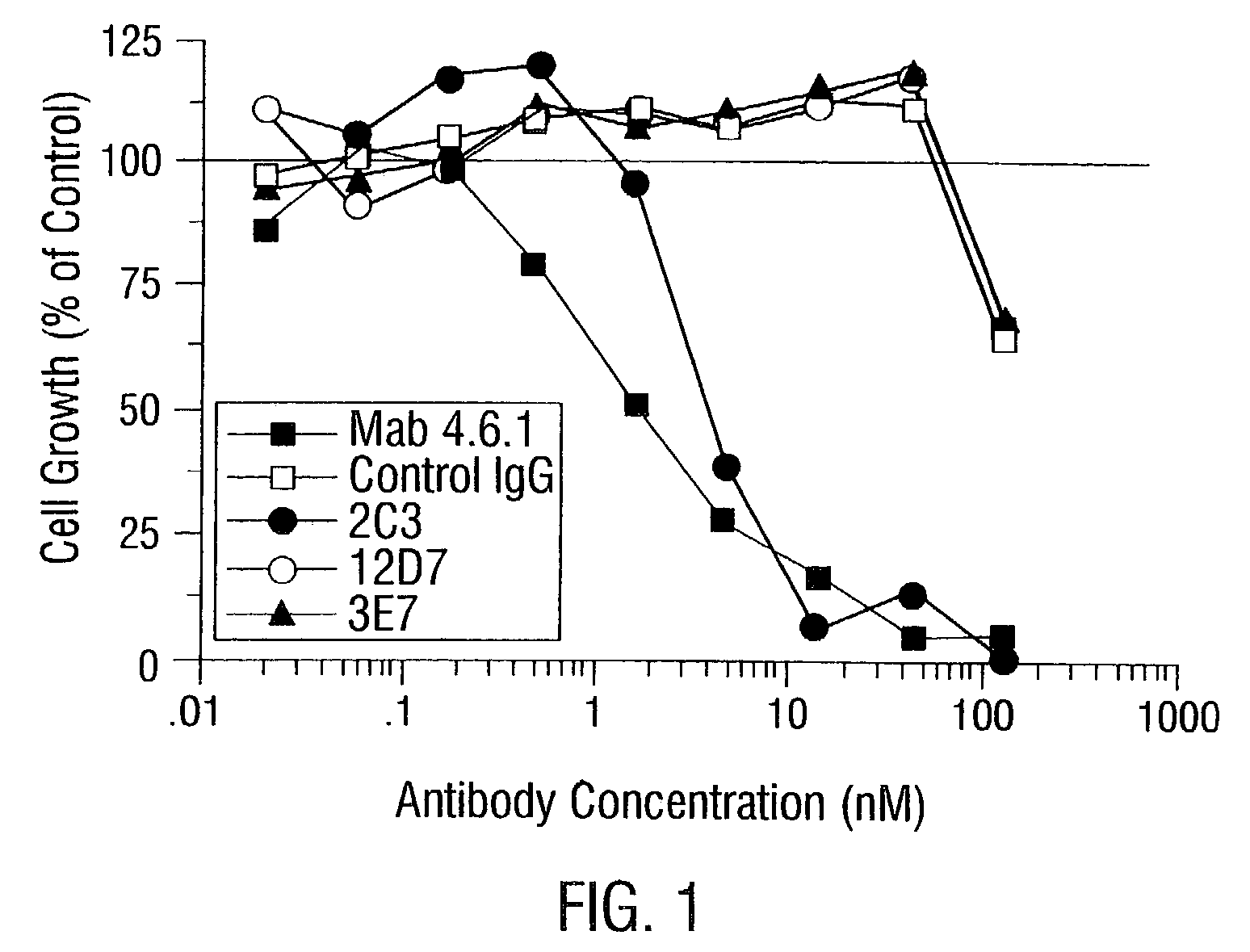
2C3 Inhibits Endothelial Cell Migration

A. Materials and Methods

[0912]Endothelial Cell Growth Assay

[0913]Adult bovine aortic endothelial (ABAE) cells were plated into 96-well tissue culture plates at 1500 cells / well and grown in the presence of 0.5 nM human VEGF with the addition of the various sample and control antibodies. Control wells received media with or without VEGF.

[0914]After 4 days of incubation, the cells were quantified by an MTS (3-(4,5-dimethylthiazol-2-yl)-5-(3-carboxylmethoxyphenyl)-2-(4-sulfophenyl)-2H-tetrazolium, inner salt) conversion assay, where MTS conversion to a formazan is proportional to cell number and can be followed by absorbance at 490 nM (Cell Titer 96 AQueous One Solution Cell Proliferation Assay, Promega, Madison, Wis.). The assay was performed according to the manufacturer's instructions.

[0915]Cell growth was estimated by subtracting the MTS conversion in cultures to which VEGF was not added. Results were expressed as a percentage of the MTS conver...

example iii

2C3 Specifically Localizes to Tumors in Vivo

A. Materials and Methods

[0921]In Vivo Localization to Human Tumor Xenografts

[0922]Tumors were grown subcutaneously in immunocompromised mice (NCI-H358 NSCLC in nu / nu mice and HT29 colon adenocarcinoma in SCID mice) until the tumor volume was approximately 1 cm3. 100 μg of unlabeled antibody for studies using SCID mice, or 100 μg of biotinylated antibody for studies using nude mice, was injected intravenously via a tail vein. Twenty four hours later, the mice were anesthetized, perfused with PBS, and tumor and organs including heart, lungs, liver, kidneys, intestines and spleen were collected and snap frozen in liquid nitrogen.

[0923]The tumor and organs from each mouse were sectioned on a cryostat and stained for antibody immunohistochemically as above, with the exception that sections from the nude mice were developed using peroxidase labeled streptavidin-biotin complex (Dako, Carpinteria, Calif.) and the sections from the SCID mice were d...


PUM


Abstract

Disclosed are antibodies that specifically inhibit VEGF binding to only one (VEGFR2) of the two VEGF receptors. The antibodies effectively inhibit angiogenesis and induce tumor regression, and yet have improved safety due to their specificity. The present invention thus provides new antibody-based compositions, methods and combined protocols for treating cancer and other angiogenic diseases. Advantageous immunoconjugate and prodrug compositions and methods using the new VEGF-specific antibodies are also provided.

Description

[0001]The present application is a continuation of U.S. patent application Ser. No. 09 / 561,499, filed Apr. 28, 2000, which issued as U.S. Pat. No. 6,524,583 on Feb. 25, 2003, which claims priority to U.S. provisional patent application Ser. No. 60 / 131,432, filed Apr. 28, 1999, the entire text and drawings of which applications are specifically incorporated by reference herein without disclaimer.[0002]The U.S. Government owns rights in the present invention pursuant to grant numbers 1RO1 CA74951, 5RO CA54168 and T32 GM07062 from the National Institutes of Health.BACKGROUND OF THE INVENTION[0003]1. Field of the Invention[0004]The present invention relates generally to the fields of antibodies, angiogenesis and tumor treatment. More particularly, it provides anti-VEGF antibodies that specifically inhibit VEGF binding to only one (VEGFR2) of the two VEGF receptors. Such antibodies inhibit angiogenesis and induce tumor regression, and yet have improved safety due to their specific blocki...

Claims


Application Information

Patent Timeline
Patent Type & Authority Patents(United States)
IPC IPC(8): A61K39/395C07K16/00C12P21/08A61K45/00C12N15/02A61K47/48A61P9/10A61P27/02A61P35/00A61P43/00C07K5/103C07K14/52C07K16/22C07K16/24C07K16/30C12N5/02C12N5/10
CPCA61K39/3955A61K47/4843A61K47/48438A61K47/48546A61K47/48561C07K5/101C07K14/52C07K16/22C07K16/24A61K47/48415Y10S530/865A61K2039/505C07K2317/73C07K2319/00C07K2319/30Y10S530/864Y10S530/866Y10S435/81Y10S530/809A61K2300/00A61K47/6811A61K47/6815A61K47/6817A61K47/6845A61K47/6849A61P1/04A61P15/00A61P17/00A61P17/02A61P17/06A61P19/02A61P19/08A61P27/02A61P27/06A61P27/10A61P27/14A61P29/00A61P31/04A61P31/18A61P33/00A61P33/02A61P35/00A61P35/02A61P37/02A61P37/06A61P43/00A61P7/06A61P7/10A61P9/00A61P9/10A61K39/395
Inventor THORPE, PHILIP E.BREKKEN, ROLF A.
Owner BOARD OF RGT THE UNIV OF TEXAS SYST
Why Eureka
Try Eureka