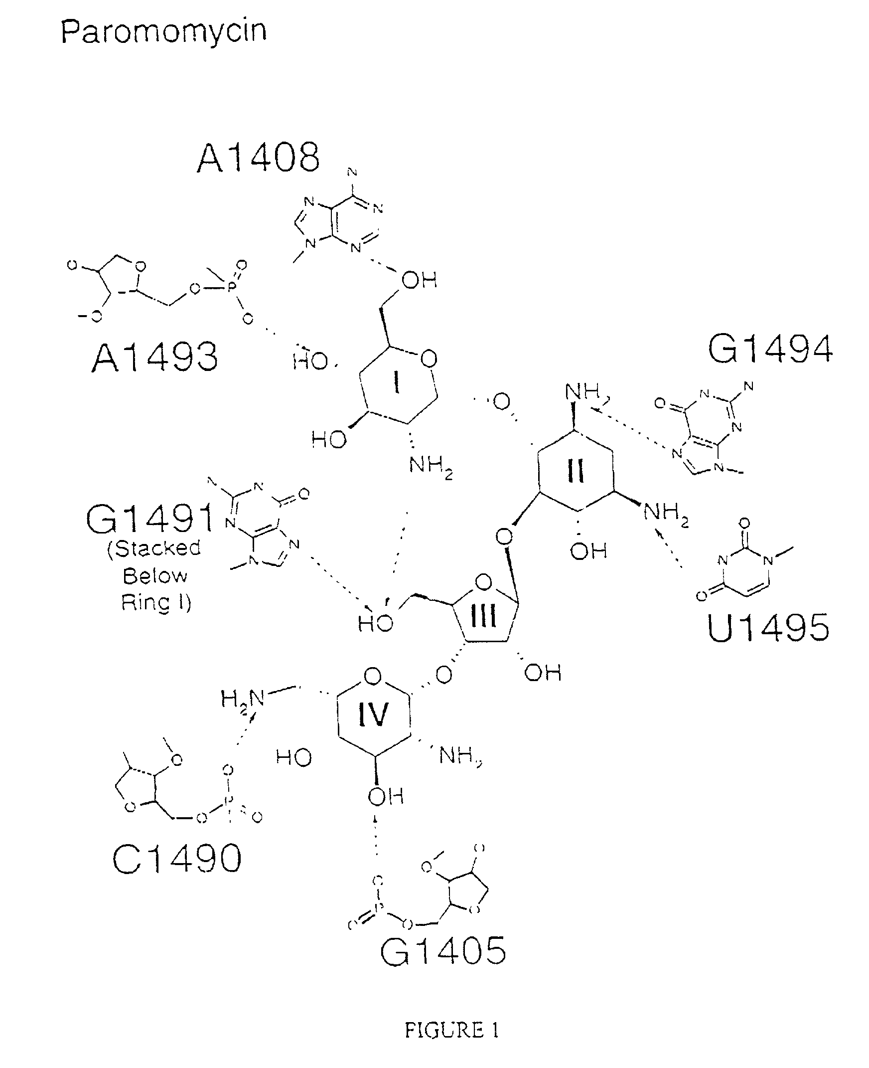
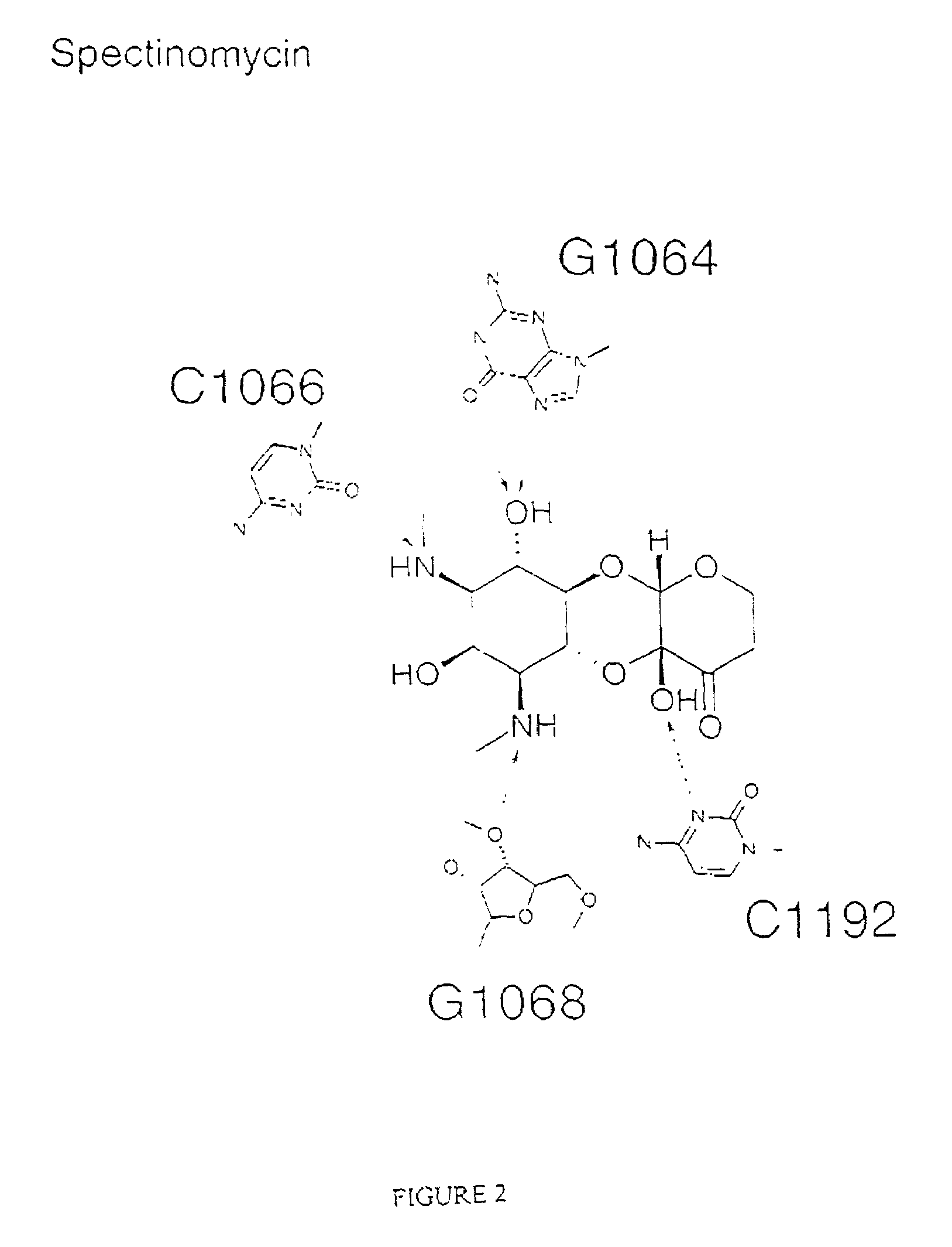
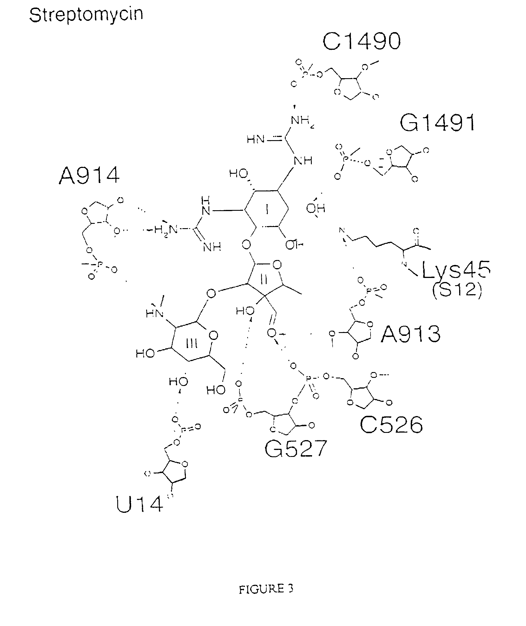
Crystal structure of antibiotics bound to the 30S ribosome and its use

a crystal structure and ribosome technology, applied in the field of crystal structure of the prokaryotic 30s ribosome subunit, can solve the problems of resistance to current antibiotics, few new antibiotics available, and poorly understood molecular details of the process, so as to improve the resolution of the crystal structure

Inactive Publication Date: 2006-07-18
UK RES & INNOVATION LTD
View PDF0 Cites 9 Cited by
  • Summary
  • Abstract
  • Description
  • Claims
  • Application Information

AI Technical Summary

Benefits of technology

[0012]The refined atomic resolution models of the 30S presented here allows the interpretation of a vast amount of biochemical data on its function in precise structural terms. The structure will also serve as a basis for the interpretation in molecular terms of lower resolution models of various functional states by electron-microscopy or X-ray crystallography. The 30S structure will help produce testable models for various aspects of ribosome function.
[0054]30S crystals do not contain the S1 subunit protein. Selective removal of the S1 subunit prior to crystallization is shown to improve the resolution of the crystals of the 30S subunit according to the invention described herein. Although the atomic co-ordinates, provided herein, allow those of skill in the art to bypass the need to undertake the crystallization of the 30S, this crystallization method nonetheless forms a further aspect of the invention.

Problems solved by technology

Despite several decades of work, the molecular details of the process are poorly understood, and the elucidation of the mechanism of translation is one of the fundamental problems in molecular biology today.
However, resistance to current antibiotics is currently a major problem in the field of medicine.
There are presently very few new antibiotics available which can be used to treat the highly resistant strains of bacteria such as MRSA (methicilin resistant Staphylococcus aureus) which are becoming increasingly widespread.

Method used


Image

  • Crystal structure of antibiotics bound to the 30S ribosome and its use
  • Crystal structure of antibiotics bound to the 30S ribosome and its use
  • Crystal structure of antibiotics bound to the 30S ribosome and its use

Examples


example 1

Generation of Crystals of the 30S Ribosomal Subunit Bound to Antibiotics

Crystallization of the 30S Ribosomal Subunit

[0281]Crystals of T. thermophilis 30S ribosomal subunits were obtained by an optimization of the procedure reported by Trakhanov et al. (Trakhanov, S. D. et al FEBS Lett. 220, 319±322 (1987)) with respect to pH and concentrations of Mg2+ ions and 2-methyl-2,4-pentanediol (MPD) as described in Wimberly et al. Nature 407, 327–339 (2000). The final conditions were 250 mM KCl, 75 mM NH4Cl, 25 mM MgCl2 and 6 mM 2-mercaptoethanol in 0.1 M potassium caco-dylate or 0.1 M MES (2-N-morpholino-ethanesulphonic acid) at pH 6.5 with 13±17% MPD as the precipitant. 30S crystals completely lacked ribosomal protein S1. Selective removal of the S1 subunit prior to crystallization improved both the crystal size and reproducibility in crystal growth. Typical procedures used to remove the S1 subunit include the use of poly-U sepharose chromatography followed by extensive salt washing as des...

example 2

Computer Based Method of Rational Drug Design

[0283]This example provides a computer based method of rational drug design. To obtain structural information about the protein / drug interaction to allow rational drug design, it is first necessary to prepare a crystal of the complex, the complex being the drug bound to the 30S ribosomal subunit. A crystal of the complex can be produced using two different methods. In a first method, the actual complex itself is crystallized using the prescribed conditions set forth for the native 30S ribosomal subunit. Once crystals of a suitable size have grown, x-ray diffraction data are collected and analyzed as described by Greer et al., J. of Medicinal Chemistry, Vol. 37, (1994), 1035–1054. This process usually involves the measurement of many tens of thousands of diffracted x-rays over a period of one to several days depending on the crystal form and the resolution of the data required. According to the method, crystals are bombarded with x-rays. T...

example 3

An Example of a Computer Based Method for Identifying a Potential Inhibitor of the 30S Ribosome

[0284]This example discloses a computer based method to identify a potential inhibitor of the 30S ribosome. The structure of an antibiotic bound to the 30S defines “crystallographically known sites”. A number of molecular docking procedures can be used to design novel binders that also bind to these sites. For example, the programs MCSS and Ludi can be used to dock small fragments that can then be linked together to form a novel ligand. Molecular docking procedures, as used herein, refer to the use of computer programs, as defined herein, to identify potential ligands that are predicted to stably interact with a defined site on the 30S ribosome subunit. Alternatively, libraries of compounds as described herein are converted into 3D format using programs such as Concord (Tripos), CeriusII (Accelrys) etc. The 3D structures of the ligands are then docked against a site on the ribosome. This s...


PUM

PropertyMeasurementUnit
wavelengthsaaaaaaaaaa
pHaaaaaaaaaa
pHaaaaaaaaaa

Abstract

The invention provides high resolution X-ray crystal structures of the 30S ribosome, obtained from Thermus thermophilus 30S subunit, having a tetragonal space group P41212 to which are bound an antibiotic selected from the group paromomycin, streptomycin, spectinomycin, tetracycline, pactamycin and hygromycin B. An advantageous feature of the structure is that it diffracts at about 3 Å resolution. The invention also provides a crystal of 30S having the three dimensional atomic coordinates of the 30S ribosome, the coordinates being provided in any one of tables 1 to 4. The data may be used for the rational design and modelling of inhibitors for the 30S ribosome, which have potential use as antibiotics.

Description

CROSS REFERENCE TO RELATED APPLICATIONS[0001]This application claims priority from United Kingdom (U.K.) applications 0017376.5 filed Jul. 14, 2000, and 0022943.5 filed Sep. 19, 2000, 0029872.9 filed Dec. 7, 2000, 0029870.3 filed Dec. 7, 2000, 0029871.1 filed Dec. 7, 2000, and 01 10885.1 filed May 3, 2001, the contents of which are incorporated herein by reference. This application is a continuation-in-part of U.S. Ser. No. 09 / 905,212, filed Jul. 13, 2001.[0002]This invention was made in part with U.S. Government support under NIH grant GM 44973 awarded by the PHS. The U.S. Government has certain rights in the invention.FIELD OF THE INVENTION[0003]The present invention relates to the provision of a high resolution crystal structure of the prokaryotic 30S ribosome subunit, particularly the 30S ribosome subunit bound to various antibiotics, and the use of this structure in drug discovery.BACKGROUND OF THE INVENTION[0004]The wealth of information made available through efforts in struc...

Claims


Application Information

Patent Timeline
Patent Type & Authority Patents(United States)
IPC IPC(8): G06F19/00C12Q1/00G01N31/00G01N33/48G06G7/48G16B15/30C07K14/195C12Q1/18G01N33/68
CPCC07K14/195G06F19/16G01N33/6803C12Q1/18C07K2299/00G16B15/00G16B15/30
Inventor RAMAKRISHNAN, VENKATRAMANBRODERSEN, DITLEV EGESKOVCARTER, ANDREW PHILIP
Owner UK RES & INNOVATION LTD
Why Eureka
Try Eureka