Display of receptors and analysis of binding interactions and drug libraries
a technology of receptors and binding interactions, applied in the field of non-cellular display of 7transmembrane receptors, can solve the problems of not having the ability to assess a library of ligands, the assay cannot allow quantification or elucidation of actual binding events, and the steps are not optimal for real-time kinetic analysis of rapidly equilibrating systems, etc., to achieve rapid screening of large combinatorial drug libraries and solubility of drugs
- Summary
- Abstract
- Description
- Claims
- Application Information
AI Technical Summary
Benefits of technology
Problems solved by technology
Method used
Image
Examples
example 1
[0058]The hexahistidine tag was incorporated into a C-terminal oligonucleotide. This oligonucleotide was used in conjunction with an N-terminal oligonucleotide and pfu polymerase for PCR amplification of the FPR. Automated dideoxy sequencing was performed to confirm the sequence. The receptor-tagged constructs were transfected into U937 cells by electroporation and selected with G418. The transfected cells were identified by fluorescent peptide binding and sorted by flow cytometry. In typical preparations, the receptor density was determined using fluorescent peptide and flow cytometric analysis to be ˜300,000 per cell.
[0059]U937 C-His FPR cells were harvested, centrifuged at 200×g for 5 minutes and resuspended in cavitation buffer at a density of 107 cells / ml at 4° C. (10 nM PIPES, 100 mM KCl, 3 mM NaCl, 3.5 mM MgCl2, 600 μg / ml ATP, 50 μM PMSF, 20 μg / ml chymostatin, and 0.05% DFP). The cell suspension was placed in a nitrogen bomb and pressurized to 450 psi using N2 gas for 20 minu...
example 2
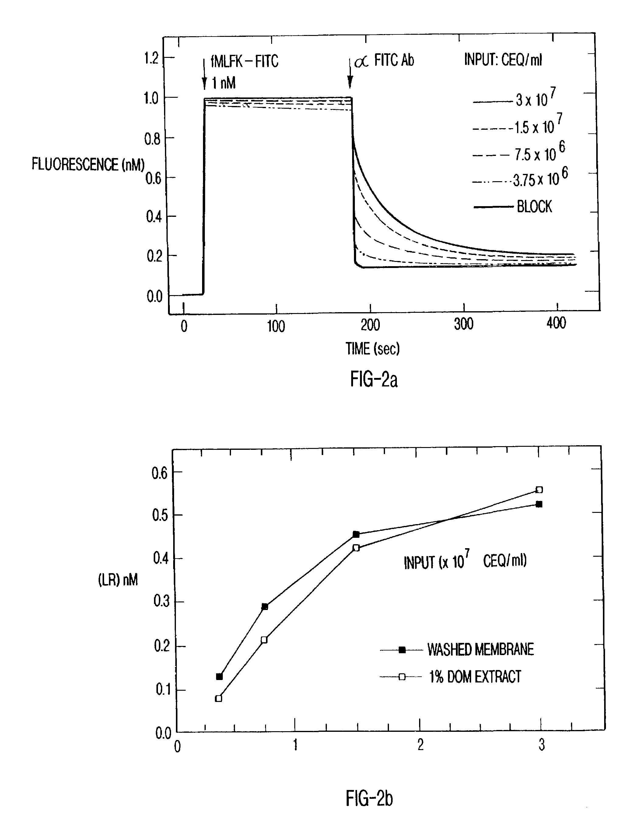
[0061]Membranes from transfected cells expressing C-terminal His-tagged N-formyl peptide receptors were prepared as above. Membranes were centrifuged at 135,000×g for 30 minutes and resuspended to 6×10⁸ CEQ / ml in BB containing a broad protease inhibitor cocktail (Calbiochem, La Jolla, Calif.) and 1% n-dodecyl β-D-maltoside (DOM). Preparations were incubated 60 minutes at 4° C. with agitation. The insoluble fraction was separated by centrifugation at 87,750×g for 30 minutes. The supernatant was removed, and this extract was used for experimentation.
[0062]To affinity-couple formyl peptide receptors to a particulate substrate, Ni2+-nitriloacetate coated silica particles (Ni-NTA, Qiagen, Santa Clarita, Calif.) were added to U937 C-His FPR membrane extracts at 10 mg / ml and incubated at 4° C. for 30 minutes with gentle mixing. This concentration of silica produced 1.15×10⁶ silica particles / ml as measured using a hemocytometer. Silica particles ranged from approximately 2–20 μm in diameter...
example 3
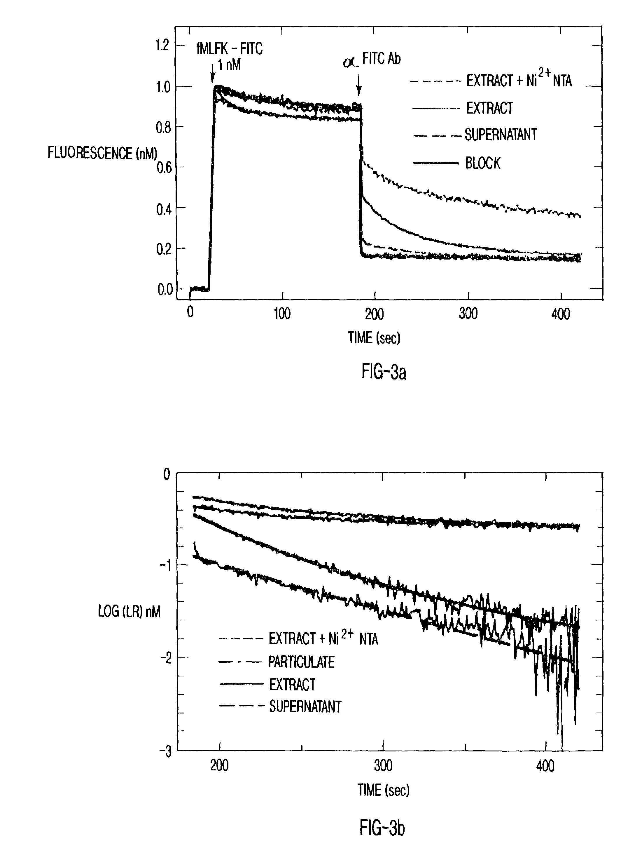
[0064]The solubilized receptors were displayed on silica particles in a format compatible with flow cytometry. FIG. 3a shows the results of a receptor recovery assay in which the particles were incubated with solubilized receptors. The uptake of C-His FPR onto Ni2+-NTA silica particles was demonstrated by the depletion of receptor from FPR extracts. The experiments were performed with 1 nM fMLFK-FITC, 1.5×10⁷ cell equivalents / ml of membrane and 20 mg silica particles / ml. The spectroscopic analysis used the antibody to fluorescein to examine ligand binding. The binding curves are depicted from top to bottom: receptors present on silica particles, receptors present in the membrane extract, receptors present in the supernatant after silica particles have been removed from the extract, control sample in which a blocking peptide (10−5 M tboc-phe-leu-phe-leu-phe) (SEQ ID NO. 1) inhibits the specific binding. In the presence of the particles, receptors were quantitatively sedimented out of...
PUM
| Property | Measurement | Unit |
|---|---|---|
| pH | aaaaa | aaaaa |
| diameter | aaaaa | aaaaa |
| concentration | aaaaa | aaaaa |
Abstract
Description
Claims
Application Information
 - R&D
- Intellectual Property
- Life Sciences
- Materials
- Tech Scout
- Unparalleled Data Quality
- Higher Quality Content
- 60% Fewer Hallucinations
Browse by: Latest US Patents, China's latest patents, Technical Efficacy Thesaurus, Application Domain, Technology Topic, Popular Technical Reports.
© 2025 PatSnap. All rights reserved.Legal|Privacy policy|Modern Slavery Act Transparency Statement|Sitemap|About US| Contact US: help@patsnap.com


