
Method of assaying cellular adhesion with a coated biochip

a biochip and cell adhesion technology, applied in the field of biological assay and biological assay equipment, can solve the problems of difficult monitoring and recording, difficult observation of biological transmigration through the micropore, and incongruous geometries

Inactive Publication Date: 2006-10-17
TRINITY COLLEGE DUBLIN
View PDF30 Cites 23 Cited by
  • Summary
  • Abstract
  • Description
  • Claims
  • Application Information

AI Technical Summary

Benefits of technology

[0023]It is envisaged that more than one cell type may be held in suspension as this often happens in practice and indeed in many instances, it may be advantageous to deliver a reagent liquid and a sample liquid through the microchannel to form multilaminar flow and then if the reagent liquid comprises a chemoattractant suitable for one of the types of the cell, it will be possible to effectively separate that particular type of cell from the sample.
[0043]It is envisaged that assemblies comprising a plurality of biochips as described above will be formed on the one base sheet and will preferably have various common feeder microchannels having ports therein. The advantage of a whole lot of biochips all on the one sheet is that they can be readily easily examined by the one microscope.

Problems solved by technology

One of the major disadvantages of this and the previous adhesion assays is the geometrical design (microscope slides and multiple well chambers), which does not at all resemble the in vivo situation.
The main disadvantage of the assays described in those specifications is that the biological process of transmigration through the micropores is difficult to observe due to the geometrical configuration of the apparatus involved.
This obviously leads to difficulties due to optical aberrations.
In effect, the study of the cells morphology changes while transmigrating across the membrane and their subsequent cytoskeletal changes reverting to their former state is a process which is difficult to monitor and record due to limitations with current techniques.
In addition, although it is possible to alter such an experiments parameters following the initiation of the experiment, such as the introduction of a second chemoattractant, recombinant or cell-derived, at some specified time after commencing the experiment, it is not possible to distinguish separate effects from each said chemoattractant.
In addition to cell biology studies, the pharmaceutical industry has major problems in the drug screening process and while high throughput screening (HTS) has been extremely successful in the elimination of the large majority of unsuitable drugs, it has not progressed beyond that and usually, after a successful HTS assay, a pharmaceutical company may still have 7,000 possible drugs requiring assessment.
This again, unfortunately, is still a static test.
There are other disadvantages such as the transport and subsequent reaction of the drug following its injection into the animal.
Probably the most important disadvantage is that it does not in any way test, in a real situation, drug efficacy.
Finally, there are no techniques at the present moment for performing assays to test the interaction of a large number of chosen compounds with living cells while the cells or compounds mimic the in vivo situation of continuous flow.

Method used


Image

  • Method of assaying cellular adhesion with a coated biochip
  • Method of assaying cellular adhesion with a coated biochip
  • Method of assaying cellular adhesion with a coated biochip

Examples


Embodiment Construction

[0071]In the drawings, there are described many micro-fabricated biochips having a plurality of ports which it would be very confusing to identify by different reference numerals in the drawings of each microchip or biochip. Thus, all the ports in the drawings are identified by the reference numerals 1 to 40. Accordingly, in certain circumstances, an outlet port will be identified by the reference numeral 4 and in another embodiment, it may be an inlet port. However, for clarity in viewing the drawings, this scheme of identification has been adopted.

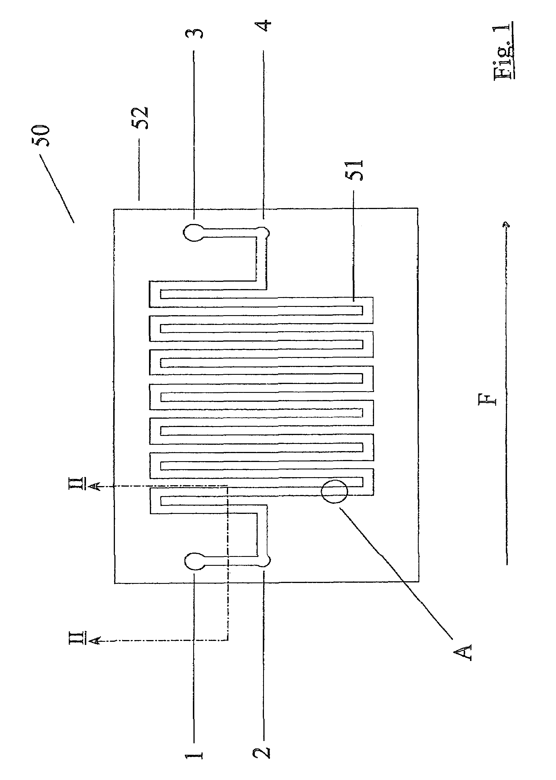
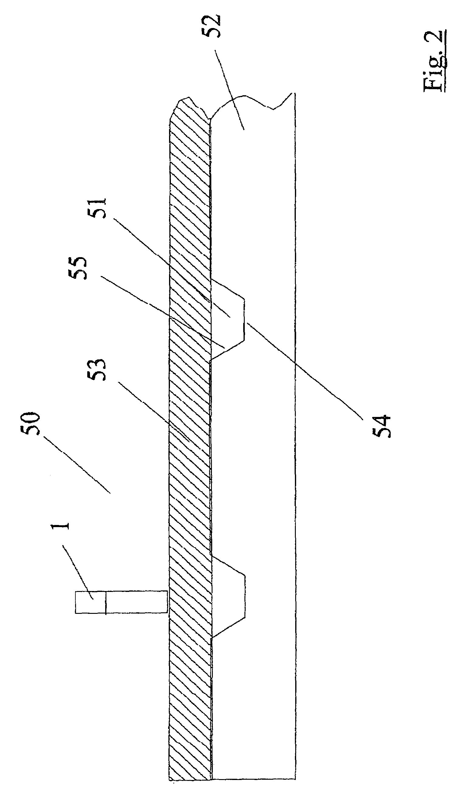
[0072]Referring to FIGS. 1 and 2, there is illustrated a biochip, indicated generally by the reference numeral 50 comprising a microchannel 51 formed in a base sheet 52. The microchannel 51 comprises a top wall 53 formed from plastics film and has a planar bottom wall 54 and tapering side walls 55 which taper outwardly away from the bottom wall 54. A fluid inlet port 1 is illustrated in FIG. 2. The biochips 50 are fabricated using standa...


PUM


Abstract

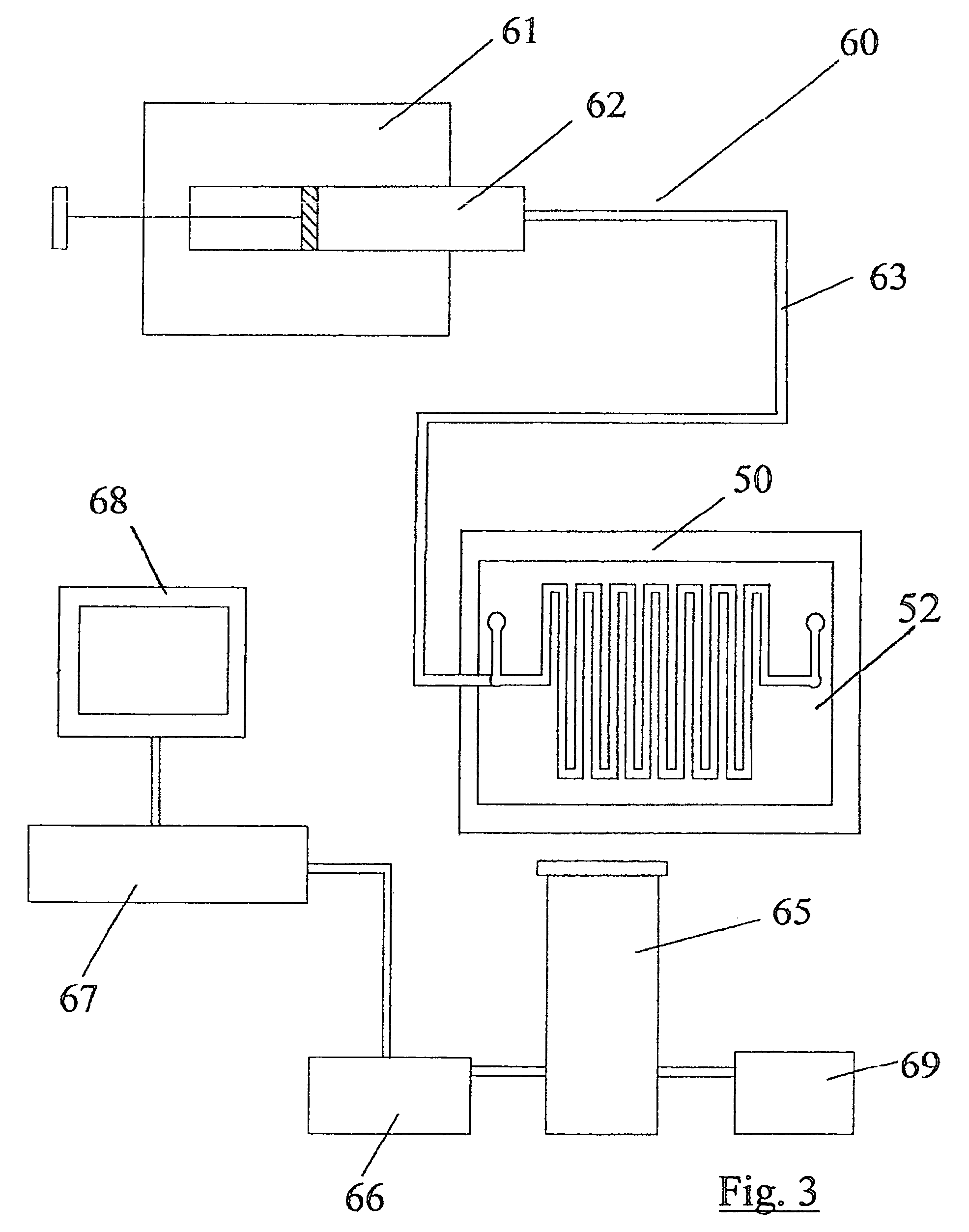
Biological assays using various constructions of biochips are disclosed to mirror in vivo situations. The biochip 50 comprises a microchannel 51 having a liquid outlet port 1, bubble release port 2 and a liquid outlet port 3 with an associated bubble release port 4. A multiplicity of tests can be performed often by coating the bore of the microchannel 50 which various adhesion mediating proteins or the use of chemoattractants. The assay assembly 60 comprises a syringe pump feeding the biochip 50. An inverted microscope 65, digital camera 66 and recorder 67 are provided. A sample liquid containing cells in suspension is injected slowly through the biochip and the effect of the assay recorded over a long period.

Description

[0001]This application is a continuation of application Ser. No. 09 / 750,348, filed on Dec. 29, 2000, now U.S. Pat. No. 6,770,434, the entire contents of which are hereby incorporated by reference and for which priority is claimed under 35 U.S.C. § 120.BACKGROUND OF THE INVENTION[0002]1. Field of the Invention[0003]The present invention relates to a biological assay and a biological assay apparatus.[0004]Biochemical, microbiological, chemical and many other assays are being performed every day in laboratories. While a considerable amount of attention has naturally been placed on such biological cell assaying for humans, this is also be becoming more important in the field of animal welfare and indeed plant production generally.[0005]A rapidly advancing research area in biology is the study of cell receptor-ligand interactions resulting in cell-substratum and cell-cell adhesion followed by subsequent cell migration. The pre-requisite to transendothelial migration of certain cell lines...

Claims


Application Information

Patent Timeline
IPC IPC(8): C12Q1/00B01L3/00G01N33/50
CPCB01L3/5027B01L3/502761G01N33/5005B01L3/5635B01L2200/027B01L2200/0636B01L2200/0647B01L2300/0636B01L2300/0816B01L2300/0864B01L2300/0867B01L2300/0887B01L2300/163B01L2400/0487
Inventor SHVETS, IGORKASHANIN, DMITRIYKELLEHER, DERMOTWILLIAMS, VIVIENNEVOLKOV, YURI
Owner TRINITY COLLEGE DUBLIN
Learn More