
Apparatus and method for electroporation of biological samples

a biological sample and apparatus technology, applied in the field of apparatus and method for electroporation of biological samples, can solve the problems of damage to cells and cell components, insufficient surface area of electrodes, and large heat buildup in prior art flow cells, and achieve the effect of simple and efficien

Inactive Publication Date: 2006-11-28
MAXCYTE
View PDF94 Cites 119 Cited by
  • Summary
  • Abstract
  • Description
  • Claims
  • Application Information

AI Technical Summary

Benefits of technology

[0180]Another advantage of the present invention is the speed at which a large population of cells can be transfected. For example, a population of lymphocytes can be transfected by electroporation by electroporating the sample in less than 5 hours, preferably less than 4 hours and most preferable in less than 3 hours and most preferably in less than 2 hours. The time of electroporation is the time that the sample is processed by the flow electroporation process.
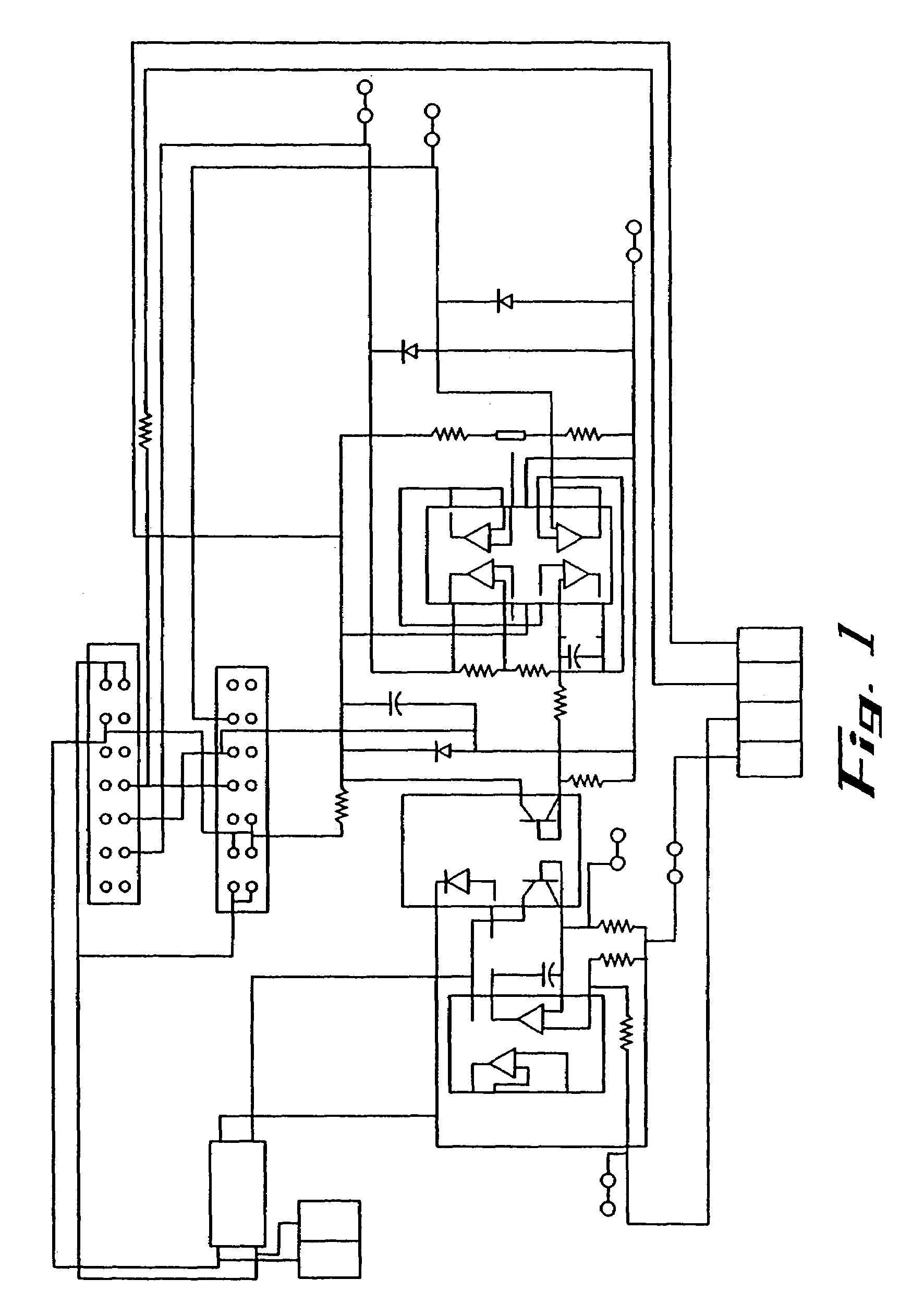
[0181]The flow electroporation device of the present invention is, in one embodiment, comprised of the following:
[0184]Monitor (displays graphical user interface [GUI] and enables user interaction); and
[0185]Disposable Set (component in which flow electroporation of cell suspension occurs.
[0186]The present invention uses flow electroporation to overcome the practical limitations in respect to the number of cells that can be electroporated, the time in which they can be electroporated and the volume of solution in which they are suspended that attend to static or batch electroporation methods. With this method, a cell suspension is passed across parallel bar electrodes that are contained in a flow cell that is preferably disposable. It is to be understood that different configurations of flow cells can be used in the present invention. For example, see below. During this passage, the cells are subjected to electrical pulses with predetermined characteristics. For example, the specific settings for preparation of IHP-loaded red blood cells are: voltage, 750V; pulse width, 650 μsec; time between pulses, 100 μsec; 2 biphasic pulses in a burst; time between bursts, 12 sec; flow rate, 0.05 mL / sec. The molecule of interest (e.g., IHP) then diffuses into the cell following concentration and / or electrical gradients. The present invention is optionally capable of subjecting the cells to a range of electric field strengths. Generally speaking, field strengths useful in the present invention are greater than approximately 1 kV / cm; preferably, approximately 1 kV / cm to 3.5 kV / cm.
[0187]The process is initiated by attaching the flow cell with solutions and cell suspensions in the containers with the necessary fluids and samples. The flow cell snaps into place and is secured by closing a hinged panel. Priming solution (saline) and cell suspension are introduced by providing the required commands to the electroporation system, which controls operation of the pump and pinch valves. As the cells transit the flow path between electrodes, electric pulses of the chosen voltage, duration, and frequency are applied. Product and waste fluids are collected in the designated containers.

Problems solved by technology

The major problem with this flow cell as well as other prior art flow cells is that the surface area of the electrode is not sufficient to dissipate heat as the cells are being electroporated.
Thus, the heat buildup in the prior art flow cells is very large as the cells are being electroporated.
This heat build up can cause damage to cells and cell components and decrease the efficiency of the electroporation process.

Method used


Image

  • Apparatus and method for electroporation of biological samples
  • Apparatus and method for electroporation of biological samples
  • Apparatus and method for electroporation of biological samples

Examples


example 1

Electroporation of Red Blood Cells with IHP

[0293]It has been determined that one of the most important parameters to control is production of heat. Red blood cells are extremely sensitive to heat and this was found to be an important parameter in the inefficiency of prior art electroporation methods. The following example was performed using the apparatus described herein.

[0294]60 mL of blood is collected from a human donor into a collection set containing 15 to 20 m of CPDA-1 (21 CFR 640.4) buffer 26.3 g trisodium citrate 3.27 g citric acid: 31.9 g dextrose: 2.22 g monobasic sod phosphate: 0.275 g adenine:, H2O to 1000 mL. The volume per 100 ml of blood is 14 mL. It is to be understood that larger volumes of blood can be treated with the present invention. The blood is transferred to two 50 ml centrifuge tubes and then is centrifuged for 10 minutes at 2000 g. The plasma is collected from above the cell pellets and transferred into a separate sterile 50 ml tubes and stored. The buff...

example 2

To Electroporate Non-stimulated Lymphocyte

[0298]Primary lymphocytes were suspended in B&K buffer (125 mM KCl, 15 mM NaCl, 1.2 mM MgCl2, 3 mM glucose, 25 mM Hepes, pH 7.4) cell concentration was set from 1×10⁷ cells / mL to 6×10⁸ cells / mL together with DNA plasmid from 50 μg / mL to 1 mg / mL. DNA concentration was 50 ug / mL–1000 ug / mL. Electroporation was performed at 2.3 kV / cm, 400 μs, 4 pulses for small volume experiments (15 μl) or 2.2 kV / cm, 1.6 ms, 1 pulse for large volume experiments (0.5 ml–2 ml) was performed under static conditions at room temperature. Following electroporation, cells were incubated in B&K buffer for 20 minutes at 37° C. for small volume experiments, or diluted by 10×volume of culture medium (RPMI-1640+10% fetal bovine serum+1% Pen-strep+2 mM L-glutamine) for large volume experiments. Cells were not diluted for post pulse incubation. Cells were cultured in culture medium for various periods (up to 72 hours) and the transfection efficiency was analyzed.

[0299]Primar...

example 3

Electroporation of Stimulated Lymphocytes

[0300]Primary lymphocytes (5×10⁶ cells / mL) were stimulated with PHA-P (up to 10 μg / mL) and 10 U / mL hr IL-2 for 48 hours in culture medium (RPMI-1640+10% Fetal bovine serum+1% Pen-strap+2 mM L-glutamine). Large cell aggregates were collected by incubating the cells in a 15 ml centrifuge tube for 10 min in the incubator and re-suspended in B&K buffer with 200 μg / mL of plasmid DNA. Following electroporation under static conditions at 1.8 kV / cm, 400 μs, 4 pulses, cells were incubated for 20 min in B&K buffer at 37° C. for small volume experiments or diluted with 10×volume of culture medium containing 20 U / mL hr IL-2. Cells were cultured with culture medium containing 20 U / mL IL-2 for various periods (up to 20 days) and the transfection efficiency was analyzed.

[0301]This is the first demonstration of gene transfer of large amount of (greater than 1×10⁸ cells) stimulated-lymphocytes by a non-viral method.


PUM

PropertyMeasurementUnit
distanceaaaaaaaaaa
distanceaaaaaaaaaa
distanceaaaaaaaaaa

Abstract

The present invention relates to methods and apparatus for the encapsulation of biologically-active substances in various cell populations. More particularly, the present invention relates to a method and apparatus for the encapsulation of biologically-active substances in various cell populations in blood by electroporation to achieve therapeutically desirable changes in the physical characteristics of the various cell populations in blood.

Description

CROSS REFERENCE TO RELATED APPLICATIONS[0001]This application claims benefit of provisional patent application Ser. No. 60 / 354,571, filed Feb. 5, 2002 and provisional patent application Ser. No. 60 / 314,241, filed Aug. 22, 2001.FIELD OF THE INVENTION[0002]The present invention relates to methods and apparatus for the introduction of biologically active substances into various types of living cells or cell particles. More particularly, the present invention relates to a method and apparatus for the introduction of biologically active substances into various cell or cell particle populations suspended in a fluid by electroporation to achieve therapeutic results or to modify cells being used in research to increase their experimental utility. Blood represents one example of cells suspended in a fluid. Other examples include cells being grown in culture outside the body, commonly known as “cell culture” or “tissue culture.”BACKGROUND OF THE INVENTIONRed Blood Cells[0003]In the vascular s...

Claims


Application Information

Patent Timeline
Patent Type & Authority Patents(United States)
IPC IPC(8): C12N15/87A61K9/50A61K48/00A61N1/30A61N1/32C12N15/867
CPCA61K9/5068A61K48/0091A61N1/0412A61N1/327C12M35/02C12N15/87C12N2740/10051C12N2840/203
Inventor DZEKUNOV, SERGEY M.LEE, HYUNG J.LI, LINHONGSINGH, VININDERLIU, LINDAHOLADAY, JOHN W.
Owner MAXCYTE
Learn More