Hedgehog signaling promotes the formation of three dimensional cartilage matrices
a three-dimensional cartilage and hedgehog signaling technology, applied in the direction of prosthesis, drug composition, peptide, etc., can solve the problems of reducing the effect of cartilage replacement, affecting the quality of life of sufferers, so as to improve the treatment of the range of cartilage related conditions, reduce the impact, and achieve the effect of reducing the impa
- Summary
- Abstract
- Description
- Claims
- Application Information
AI Technical Summary
Benefits of technology
Problems solved by technology
Method used
Image
Examples
example 1
Isolation of Human Mesenchymal Stem Cells
[0242]Mesenchymal stem cells were derived from human volunteers under general anesthesia. Bone marrow was harvested from the iliac crest by aspiration. Approximately 5 mL of bone marrow was suspended in essential medium containing 20% fetal bovine serum (FBS), antibiotics, and glutamine (Sigma). Under these conditions, mesenchymal stem cells adhere to the culture flask while hematopoietic stem cells do not adhere. The non-adherent cells, as well as any debris, is easily separated from the adherent mesenchymal stem cells.
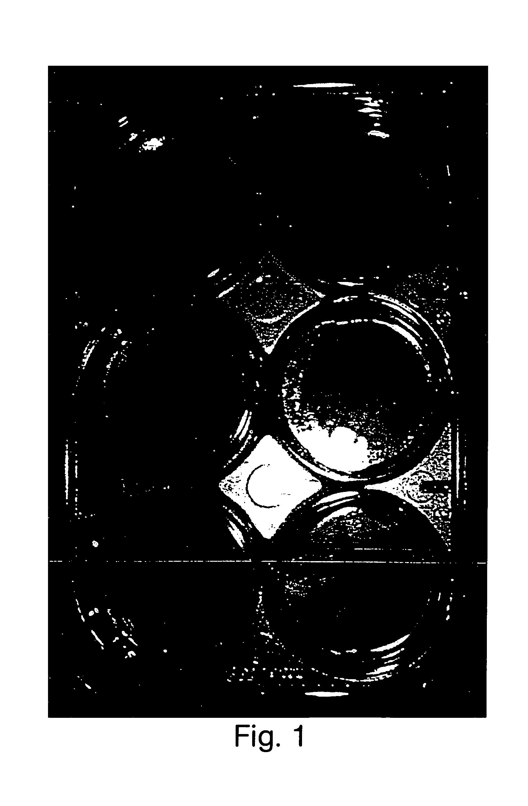
[0243]Mesenchymal stem cells were plated at a density of 1×10⁶ / mL in a six well plate. Following adhesion of the cells to the plate, cells were treated with either medium containing 20% FBS, with medium supplemented with TGFβ-1, or with medium supplemented with sonic hedgehog.
example 2
Change in Mesenchymal Stem Cell Behavior Following Factor Treatment
[0244]Following adhesion of cells to the plate, cells were treated with either medium containing 20% FBS (negative control), medium containing 1 ng / mL TGFβ-1, or medium containing 100 ng / mL of the lipid modified N-terminal fragment of sonic hedgehog. In all cases, the medium was changes twice per week throughout the duration of the experiment.
[0245]Cells treated with 20% FBS attached and proliferated in culture. Within three weeks, the cells proliferated to form a monolayer of stromal cells over the entire culture well. No areas of differentiation were observed in the well.
[0246]Cells treated with TGFβ-1 appeared morphologically distinct after a week in culture. The cells appeared larger, and later molecular characterization of these cells revealed a correlation between this change in cell size and matrix production (data not shown).
[0247]Cells treated with sonic hedgehog proliferated efficiently during the first two...
example 3
Molecular Characterization of Cell Behavior Following Factor Treatment
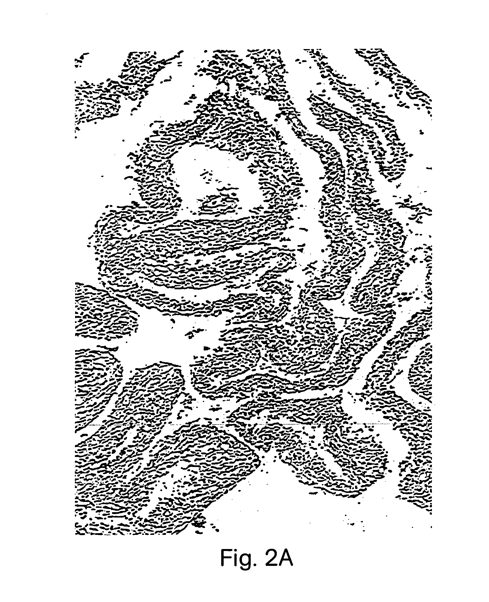
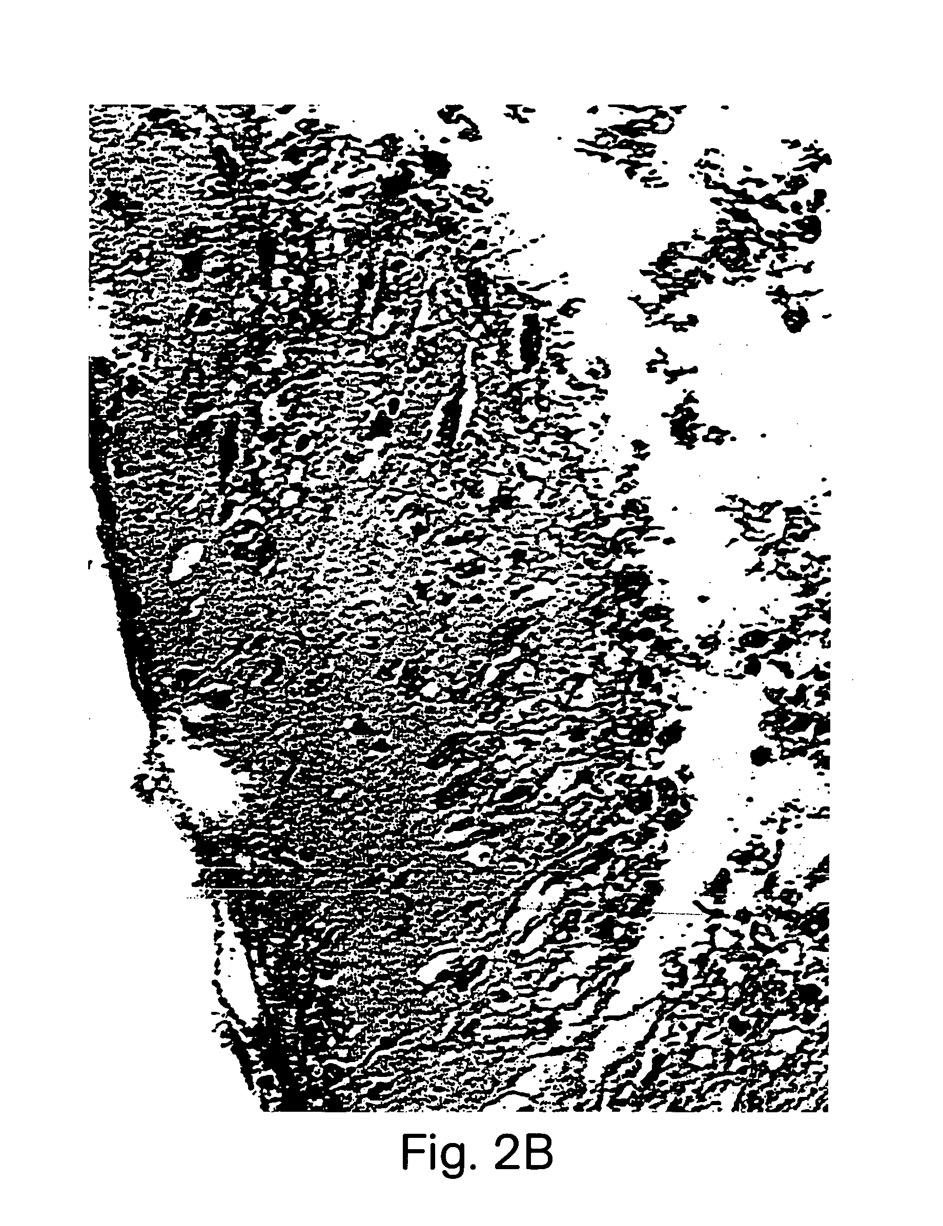
[0248]Mesenchymal stem cell cultures were established, as described in detail above. Cells were cultured in the presence of 20% FBS, 1 ng / mL TGFβ-1, or 100 ng / mL lipid modified sonic hedgehog, and the medium was changed twice per week. After 21 days in culture, tissue was harvested, and fixed overnight in formaline solution. Fixed tissue was washed, dehydrated, embedded in paraffin, and 4 μm sections were cut using a microtome. Sections were mounted on adhesive slides, and processed for histology and immunohistochemistry.
[0249]a. Hematoxyline-Eosine Staining
[0250]Mounted paraffin sections were stained with hematoxyline-eosine, and viewed under a light microscope. FIG. 2 shows that tissue harvested following 21 days of treatment with sonic hedgehog is highly organized, contains several folded layers, and produces abundant matrix.
[0251]b. Alcian Blue staining
[0252]Mounted paraffin sections were stained with Alcian b...
PUM
| Property | Measurement | Unit |
|---|---|---|
| Fraction | aaaaa | aaaaa |
| Shape | aaaaa | aaaaa |
Abstract
Description
Claims
Application Information
 - R&D
- Intellectual Property
- Life Sciences
- Materials
- Tech Scout
- Unparalleled Data Quality
- Higher Quality Content
- 60% Fewer Hallucinations
Browse by: Latest US Patents, China's latest patents, Technical Efficacy Thesaurus, Application Domain, Technology Topic, Popular Technical Reports.
© 2025 PatSnap. All rights reserved.Legal|Privacy policy|Modern Slavery Act Transparency Statement|Sitemap|About US| Contact US: help@patsnap.com


