Methods of using an intravascular balloon catheter in combination with an angioscope

a balloon catheter and angioscope technology, applied in the field of intravascular devices, can solve the problems of limited angiography to view the subject vessel, jeopardizing the health of the patient, and not being able to accurately identify the pathology of abnormal deposits within the vessel

Inactive Publication Date: 2007-01-30
BOSTON SCI SCIMED INC
View PDF96 Cites 329 Cited by
  • Summary
  • Abstract
  • Description
  • Claims
  • Application Information

AI Technical Summary

Problems solved by technology

Angiography is limited to viewing the subject vessel in a monochromatic two-dimensional plane with no depth of field.
However, this method inherently involves a certain amount of human error and since each view must be taken in sequence, critical time is wasted which may jeopardize the health of the patient.
In addition, since angiography is limited to monochromatic views, it is not able to accurately identify the pathology of abnormal deposits within the vessel.
However, utilizing an occluding balloon deprives portions of the heart of essential oxygenated blood and often results in ischemia and patient discomfort

Method used


Image

  • Methods of using an intravascular balloon catheter in combination with an angioscope
  • Methods of using an intravascular balloon catheter in combination with an angioscope
  • Methods of using an intravascular balloon catheter in combination with an angioscope

Examples


Embodiment Construction

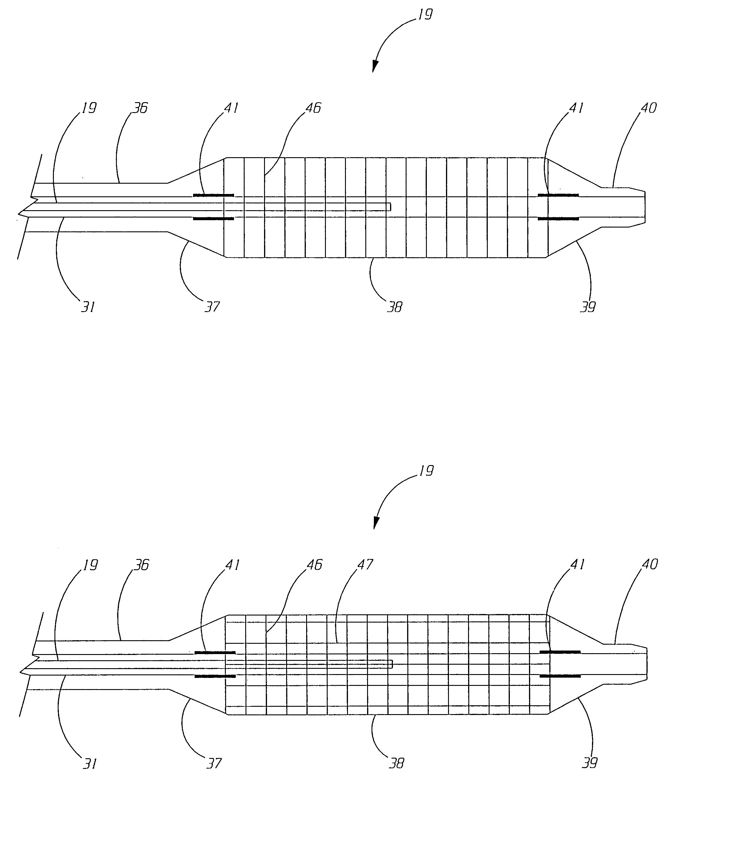
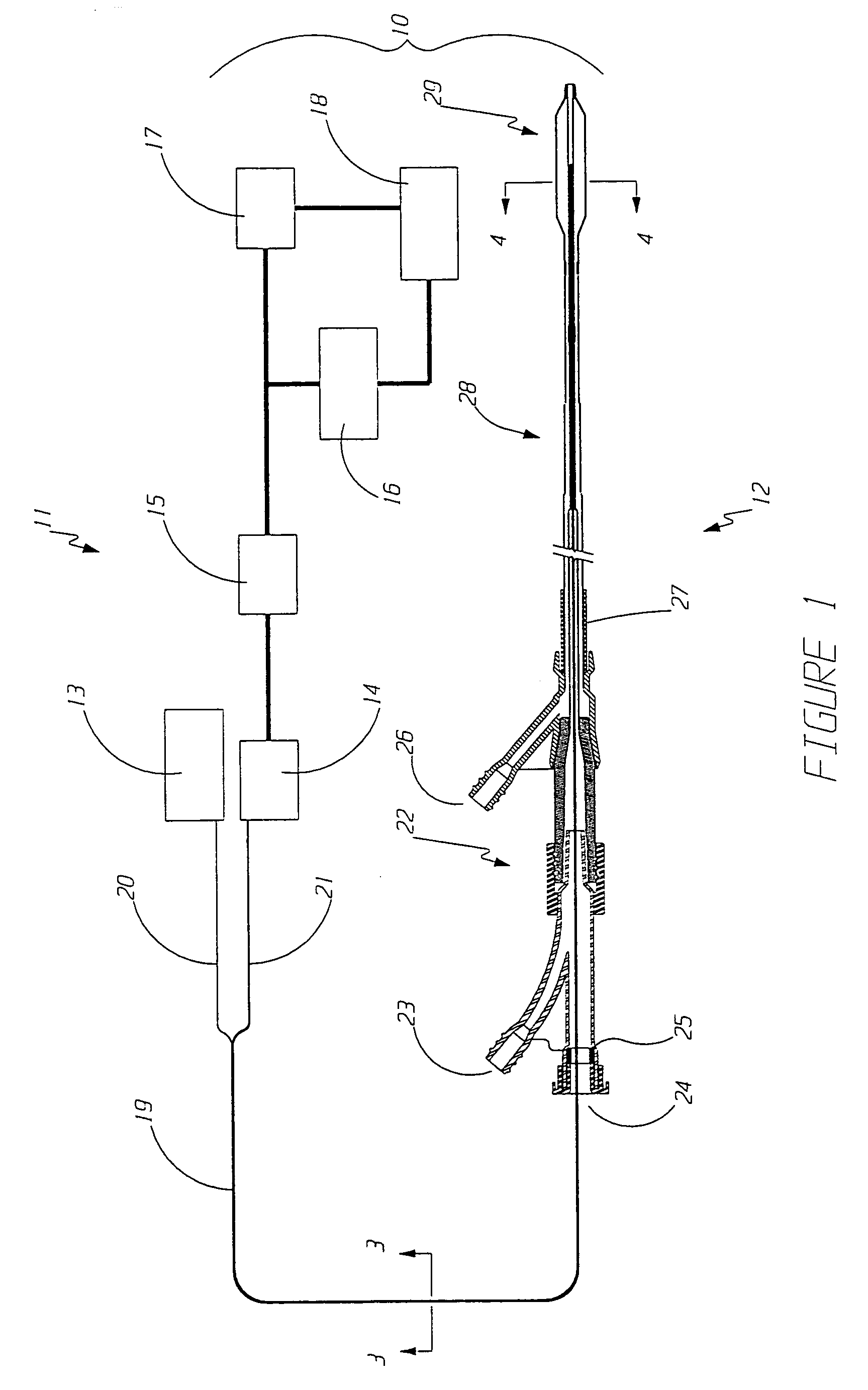
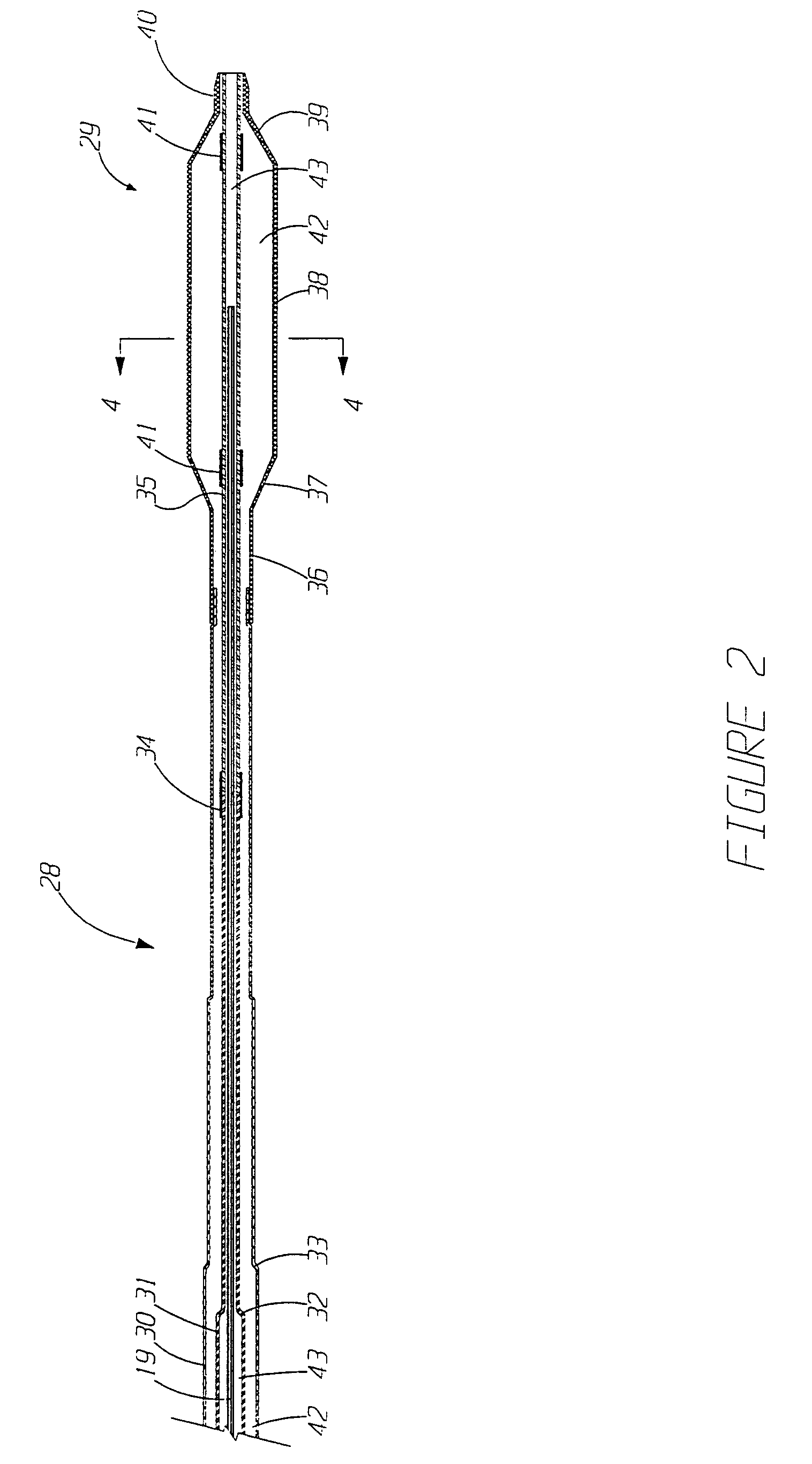
[0023]Referring to FIG. 1, a medical system 10 in accordance with the present invention includes an angioscopy system 11 and a balloon catheter 12. The angioscopy system 11 includes an angioscope 19 which includes a plurality of illumination fibers 20 and an imaging bundle 21 with an objective lens at its distal end (not shown). The proximal end of the illumination fibers 20 are connected to a light source 13. The proximal end of the imaging bundle 21 is connected to a series of image processing subsystems, including focusing optics 14, camera 15, computer 16, video cassette recorder (VCR) 17, and video monitor 18. The light source and the image processing subsystems can be arranged as well known in the art such as shown in U.S. Pat. No. 4,331,132 to Mukasa, which is herein incorporated by reference. The computer 16 can be used to digitally enhance the image and / or quantitatively process the image in order to determine dimensional aspects of the objects being viewed. The dimensional...


PUM


Abstract

A method of performing a medical procedure is disclosed which involves the use of a balloon catheter in combination with an angioscope to monitor an intravascular stent before, during or after deploying the stent. Specifically, the method may include the steps of (1) delivering an intravascular stent into the vasculature of a patient, (2) positioning a balloon catheter in the vasculature such that the inflatable balloon is adjacent the stent, (3) inserting an angioscope into the balloon catheter such that the distal end of the angioscope is adjacent an optically-transparent tube traversing the interior of the balloon, and (4) visually monitoring the stent before, during or after deploying the intravascular stent. The optically-transparent tube may define a guide wire lumen for use with a guide wire and the angioscope may be inserted into the guide wire lumen. The intravascular stent may be a balloon-expandable stent, a self-expanding stent, or a stent made of a photo-responsive polymer.

Description

CROSS-REFERENCE TO RELATED CASES[0001]This is a continuation of application Ser. No. 08 / 432,318, filed May 1, 1995 now abandoned, which is a continuation-in-part of application Ser. No. 08 / 200,122, filed Feb. 22, 1994, now issued U.S. Pat. No. 5,411,016, dated May 2, 1995.FIELD OF THE INVENTION[0002]The present invention generally relates to intravascular devices and methods of use thereof. More specifically, the present invention relates to methods of using balloon angioplasty devices in combination with an angioscope.BACKGROUND OF THE INVENTION[0003]A wide variety of devices and techniques have been developed to diagnose and treat vascular diseases. Coronary artery disease (CAD) is a vascular disease in which blood flow to the heart muscle is restricted by abnormal deposits in the coronary arteries. The abnormal deposits deprive portions of the heart muscle of essential oxygenated blood. The wide spread impact of coronary artery disease has stimulated the development of diverse ty...

Claims


Application Information

Patent Timeline
IPC IPC(8): A61M25/098A61B1/04A61B5/00A61B19/00A61M29/02
CPCA61B1/00082A61B1/00135A61B5/0084A61M25/104A61B1/042A61M2025/0008
Inventor KUME, STEWART M.
Owner BOSTON SCI SCIMED INC
Why Eureka
Try Eureka