Methods for detecting papillomavirus DNA in blood plasma and serum

Inactive Publication Date: 2007-02-27
PENN STATE RES FOUND
View PDF25 Cites 22 Cited by
  • Summary
  • Abstract
  • Description
  • Claims
  • Application Information

AI Technical Summary

Benefits of technology

[0031]One advantageous application of this invention is for identifying individuals having epithelial malignancies and premalignancies. Another advantageous application of this invention is for selecting individuals for HPV-or disease-directed therapies, including biotherapies, chemotherapies, anti-viral therapies, radiation therapies, and surgical therapies. Yet another advantageous application of the methods of this invention is to provide a marker for determining whether an adequate therapeutic effect has been achieved by a therapy, or whether additional or more advanced therapy is required, and to assess prognosis in these patients. A further advantageous application of this invention is to permit identification or analysis, either quantitatively or qualitatively, of papillomavirus nucleic acid in plasma or serum

Problems solved by technology

Unfortunately, the Pap smear does not detect all instances of cervical dysplasia or premalignancy.
In an additional 20–30% of cases, the Pap smear may be insufficient for interpretation due to the presence of inflammatory cells.
However, HPV infection cannot be detected in all cases of cervical dysplasia/pre-malignancy or even of cervical carcinoma.
Unfortunately, this method identifies both individuals with prior HPV exposure but without persistent infection and those with persistent infection, but does not distinguish between ongoing, persistent, and chronic HPV infections.
Serologic testing for HPV antibodies thus does not correlate well with the presence of cervical dysplasia, because it does not permit discrimination between previous HPV exposure and persistent HPV infection (Olsen et al., 1996, Int. J. Cancer 68: 415–419).
This method is disadvantageous because the examiner may miss an infected region of the cervix, or the infected lesion may not be superficial and readily accessible, so that the test provides a false-negati

Method used


Image

  • Methods for detecting papillomavirus DNA in blood plasma and serum

Examples


example 1

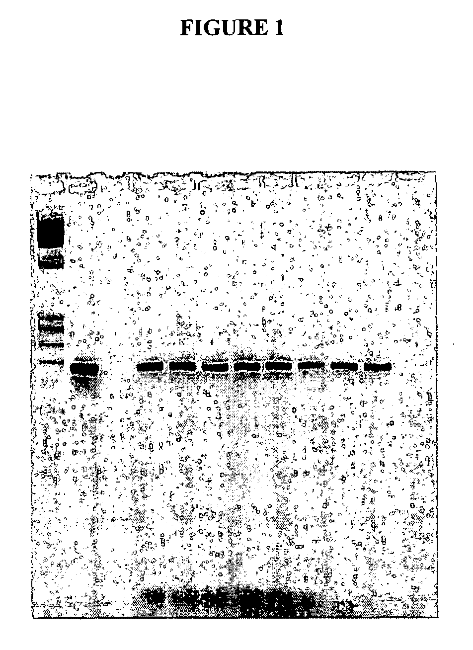
[0046]Three cottontail rabbits were infected at time zero with the cottontail rabbit papilloma virus (CRPV), a form of the virus specific to the squamous epithelium of the cottontail rabbit. The cottontail rabbit is used extensively as a model for the human version of papilloma virus infection and progression to dysplasia and carcinoma. Blood was drawn and serum separated prior to infection and approximately every month thereafter. The serum was stored frozen at −80°C. Each of the animals developed pre-malignant lesions (papillomas) approximately 5 weeks after the initial inoculation, and cancer approximately 4–13 months after time zero.

[0047]DNA was extracted from the serum by a method described in U.S. patent application Ser. No. 08 / 818,058. One tenth of the resulting extract was utilized in a polymerase chain reaction, also as described except that primers to the E8 region of the CRPV genome were used at 25 picomoles each per reaction. The oligonucleotide primer sequences were as...

example 2

[0051]A young, sexually active woman visits her gynecologist for a routine examination. A Pap smear is obtained as standard practice, and blood for the HPV nucleic acid assay is also obtained. The Pap smear is reviewed by a cytotechnologist, who finds no abnormalities. The blood test is performed by isolating nucleic acids, both DNA and RNA, from the plasma or serum component. A broad spectrum assay for several dozen HPV types is performed by the PCR method, with detection by the hybrid capture method commonly used for testing of tissue samples for HPV. A positive result is obtained, and a portion of the PCR product is genotyped by digestion with a number of restriction endonucleases. The patient is found to have two types of virus, high risk type 18 and low risk type 4. The sample is used to quantify the mRNA for these two types and a value of 500 genomes / microliter of blood is obtained for the type 18.

[0052]In retrospect, the “normal” Pap smear may be attributable to lab error, sa...


PUM


Abstract

This invention relates to the detection of extracellular papillomavirus DNA in blood plasma or serum from a human or animal. In particular, the invention relates to the detection, identification, evaluation, or monitoring of neoplastic, premalignant or malignant disease associated with a papillomavirus. The invention thereby provides methods for the identification of individuals at risk for, or having, cervical dysplasia, cervical intraepithelial neoplasia, or cervical cancer.

Description

[0001]This application is a divisional of U.S. Ser. No. 09 / 653,644, filed Aug. 31, 2000, now U.S. Pat. No. 6,511,805, issued Jan. 28, 2003, which is a continuation-in-part of U.S. patent application Ser. No. 09 / 456,222, filed Dec. 7, 1999, now U.S. Pat. No. 6,630,301, issued Oct. 27, 2003, which is a continuation-in-part of U.S. patent application Ser. No. 09 / 049,234, filed Mar. 27, 1998, now abandoned, which is a continuation-in-part of U.S. patent application Ser. No. 08 / 818,058, filed Mar. 14, 1997, now U.S. Pat. No. 6,156,504, which is a continuation-in-part of U.S. Provisional Application, Ser. No. 60 / 028,180, filed Oct. 15, 1996, which is a continuation-in-part of U.S. Provisional Application, Ser. No. 60 / 026,252, filed Sep. 17, 1996, which is a continuation-in-part of U.S. Provisional Application, Ser. No. 60 / 013,497, filed Mar. 15, 1996, the entire disclosure of each of the foregoing is hereby incorporated by reference.BACKGROUND OF THE INVENTION[0002]1. Field of the Inventi...

Claims


Application Information

Patent Timeline
IPC IPC(8): C12Q1/68C12P19/34C07K14/47C07K14/82C12Q1/70
CPCC07K14/47C07K14/4746C07K14/82C12Q1/686C12Q1/6865C12Q1/6883C12Q1/6886C12Q1/708C12Q2600/16C12Q2600/156C12Q2600/112C07K2319/00C12Q1/683C12Q2535/125
Inventor GOCKE, CHRISTOPHER D.CHRISTENSEN, NEIL
Owner PENN STATE RES FOUND
Why Eureka
Try Eureka