X-ray tomography apparatus and operating method for generating multiple energy images

a tomography and energy image technology, applied in tomography, instruments, applications, etc., can solve the problems of low energy difference between the achievable energy difference between the x-ray radiation used for generating the high-energy image and the low-energy image, and the inability to completely separate bony tissue and soft tissue in the calculated images

Active Publication Date: 2007-04-24
SIEMENS HEALTHCARE GMBH
View PDF5 Cites 27 Cited by
  • Summary
  • Abstract
  • Description
  • Claims
  • Application Information

AI Technical Summary

Benefits of technology

[0010]An object of the present invention is to provide a tomography apparatus and a method for operating a tomography apparatus that enable, in a simple manner, generation of multiple energy images with essentially the same x-ray dose of an x-ray beam.
[0013]The generation of the projections ensues according to the invention with essentially equal x-ray doses, such that high-energy projections and low-energy projections can be acquired with an essentially identical image quality given simultaneous low radiation exposure of a subject, i.e. a patient.
[0015]An exposure time that can be adjusted quickly and in a simple manner in an alternating fashion is advantageously provided as a further control variable. The adjustment of the exposure time is achieved with a substantially constant current.
[0026]Starting from the set first voltage value and the set first exposure time of the high-energy projections, a direct adaptation of the voltage to the second voltage value and to the second exposure time is in the subsequent low-energy projections. In reverse, starting from the set second voltage value and a set second exposure time a direct adaptation of the voltage to the first voltage value and to the first exposure time likewise is used in the following low-energy projections. Given such an adjustment, at least given a change of the projections, the voltage must only be adjusted by the difference between the first voltage and the second voltage, such that in a simpler operation of the high-voltage generator is achieved.

Problems solved by technology

Such detectors are very expensive to produce due to the use of two separate detector arrays.
Moreover, with such a detector the achievable energy difference of the x-ray radiation used for generation of the high-energy image and of the low-energy image is very slight, such that bony tissue and soft tissue cannot always be completely separated in the calculated images.

Method used


Image

  • X-ray tomography apparatus and operating method for generating multiple energy images
  • X-ray tomography apparatus and operating method for generating multiple energy images
  • X-ray tomography apparatus and operating method for generating multiple energy images

Examples


Embodiment Construction

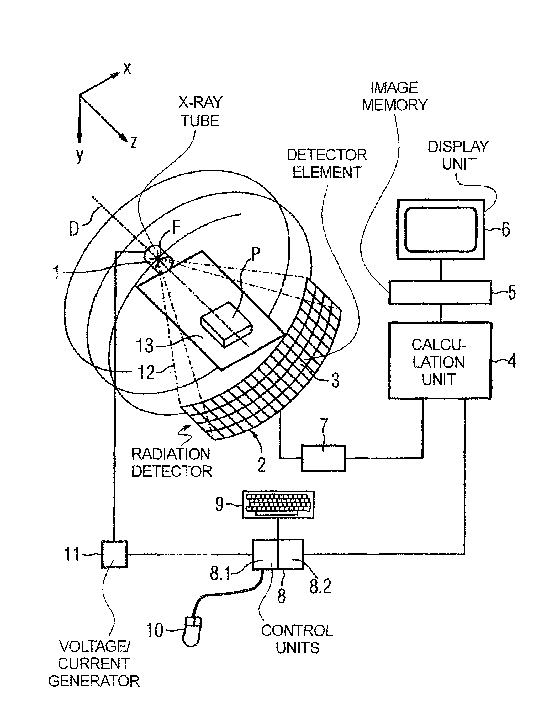
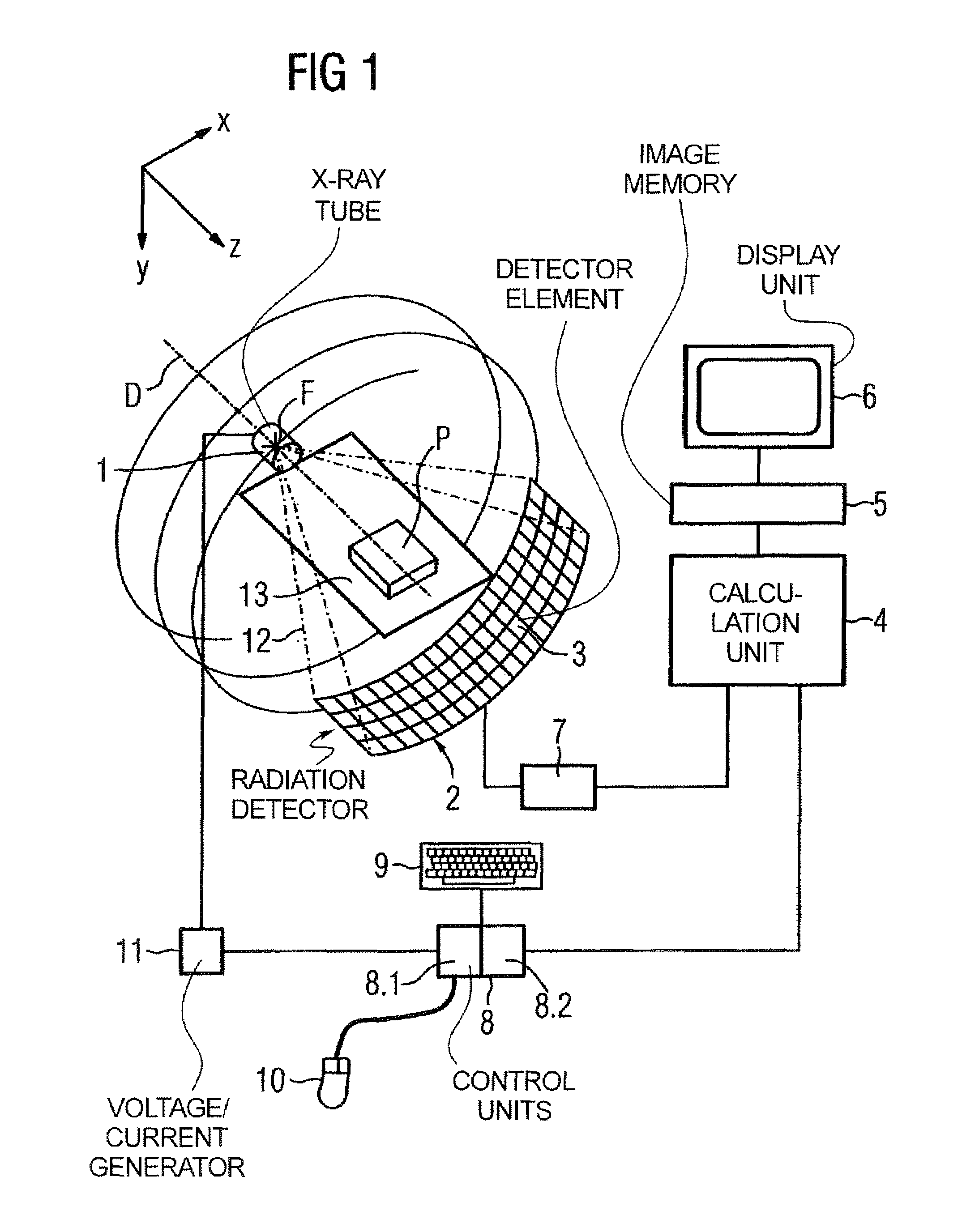
[0047]An inventive tomography apparatus, here in the form of a computed tomography apparatus, is shown in FIG. 1. The computed tomography apparatus includes: an x-ray radiator in the form of an x-ray tube 1, a radiation detector 2 composed of detector elements 3 in columns and rows in a detector array, an adjustment device 8 having a first control unit 8.1 for alternating adjustment of a voltage and a second control unit 8.2 for alternating adjustment of a further control variable; a calculation unit 4 for preparation of the acquired projections and for calculation of diverse result images (for example a low-energy image and a high-energy image in the form of slice images or topograms); an image memory 5; and a display unit 6.

[0048]The x-ray tube 1 and the radiation detector 2 are part of an acquisition system and are mounted opposite one another on a rotary frame (not shown) such that x-rays emanating from a focus F of the x-ray tube 1 and limited by edge rays 12 strike the radiati...


PUM


Abstract

In a tomography apparatus and a method for operating a tomography apparatus for generation of multiple energy images, in which high-energy projections and low-energy projections are acquired by alternating adjustment of a voltage and a further control variable (namely the current or the exposure time), given a set first voltage value and a set first control value of the further control variable, and given a set second voltage value and a set second control value of the further control variable, x-rays generated by an x-ray radiator exhibit essentially the same x-ray dose or photon flow.

Description

BACKGROUND OF THE INVENTION[0001]1. Field of the Invention[0002]The present invention concerns an x-ray tomography apparatus for generation of multiple energy images and a method for generation of multiple energy images with such a tomography apparatus.[0003]2. Description of the Prior Art[0004]“Multiple energy images,” are x-ray images that are generated with different x-ray energies. For example, by a weighted subtraction on the basis of a low-energy image and a high-energy image it is possible to calculate images in which essentially only substances with a specific absorption characteristic are still visible. This fact is in particular used in diagnostic medicine for visualization of soft tissue separate from bony tissue. Multiple energy images can be generated in different manners.[0005]In FujiFilm Co., Technical review no. 12, “Upright image reader that supports energy subtraction processing software”, an x-ray apparatus is described that enables the simultaneous generation of ...

Claims


Application Information

Patent Timeline
Patent Type & Authority Patents(United States)
IPC IPC(8): A61B6/00H05G1/44
CPCA61B6/032A61B6/405A61B6/482G01N23/046H05G1/46A61B6/542G01N2223/612G01N2223/419
Inventor POPESCU, STEFAN
Owner SIEMENS HEALTHCARE GMBH
Why Eureka
Try Eureka