Apparatus for handling biopsy specimens, and method for using it

a biopsy specimen and apparatus technology, applied in the field of apparatus for handling biopsy specimens, can solve the problems of difficult processing of frozen specimens, time-consuming and labor-intensive capillaries, and difficult production of 200 m-thick specimens with a diameter of 1.2 to 3 mm, so as to improve the reliability and reproducibility of the preparation process, improve the quality of snapshots of specimens, and increase the speed of preparation operation

Active Publication Date: 2007-05-15
LEICA MICROSYSTEMS GMBH
View PDF0 Cites 9 Cited by
  • Summary
  • Abstract
  • Description
  • Claims
  • Application Information

AI Technical Summary

Benefits of technology

[0008]It was accordingly the object of the present invention greatly to increase the speed of the preparation operation (time requirement between excision and onset of freezing less than 30 seconds), and thereby to improve the quality of the snapshots of the specimens being examined. A further object of the invention was to improve the reliability and reproducibility of the preparation process as compared to manual preparation. Lastly, the further object presented was to decrease the manual and motor demands on implementing personnel, i.e. to facilitate working procedures for preparation and thereby to give even technical personnel of average training and skill the capability of producing perfect specimens.
[0009]The apparatus according to the present invention makes it possible greatly to increase the speed with which biopsy material is prepared: the time for a specimen removal operation, measured from insertion of the microbiopsy needle to completion of the initial freezing operation, is a maximum of 30 seconds. The reliability and reproducibility of preparation results has been considerably increased, and rejects have been practically entirely eliminated. Working procedures during preparation have been simplified, with the apparatus according to the present invention, in such a way that preparation can be reliably performed by technical personnel with average training and average manual skills.

Problems solved by technology

Production of a 200 μm-thick specimen with a diameter of 1.2 to 3 mm is difficult.
Shimoni et al. fired small metal capillaries (gold tubules with an inside diameter of 200 μm) into the tissue; locating these capillaries in the tissue is time-consuming, however, and the frozen specimens are difficult to process.
This manual preparation of biopsy specimens for high-pressure freezing exhibits, however, a number of serious disadvantages which reveal this method to be unsuitable for larger quantities of specimens and especially for routine operation.
le. This elapsed time is obviously too great, particularly in cases in which “snapshots” need to be prepared of cells whose ultrastructure changes rapidly, for example muscle ce
lls. A further disadvantage of manual manipulation of biopsy specimens is the fact that the gelatinous consistency and small dimensions of the tissue specimens considerably complicate reliable manual manipulation thereof, so that the reproducibility of the results is low and the reject rate is consider
able. This in turn not only is undesirable for general cost reasons, but also is unacceptable especially in situations in which consideration must be given to the patient / examination subject or experimental animal, and excision of biopsy material therefore cannot be repeated an unlimited number of times, for example in the case of removal of specimens from a living myoca
rdium. Lastly, the most important disadvantage of manual manipulation of biopsy specimens is the fact that this manipulation is so difficult to perform that, as indicated by experience, only a small percentage of the technical personnel of an institution are at all capable, even after extended training, of producing perfect or at least satisfactory spe
cimens. The results of manual preparation are therefore poorly reproducible, and the user of commercial equipment must resort to employing, for preparation, persons who by fortunate accident possess manual and motor skills far above the

Method used


Image

  • Apparatus for handling biopsy specimens, and method for using it
  • Apparatus for handling biopsy specimens, and method for using it
  • Apparatus for handling biopsy specimens, and method for using it

Examples


Embodiment Construction

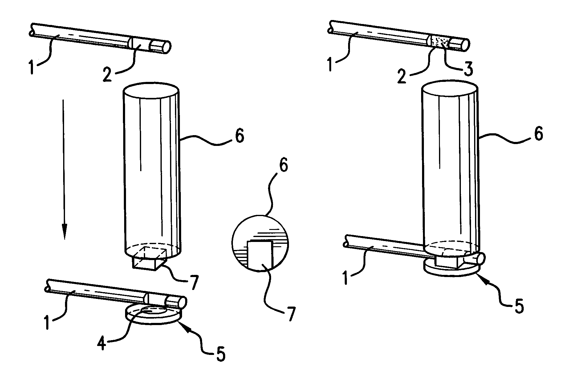
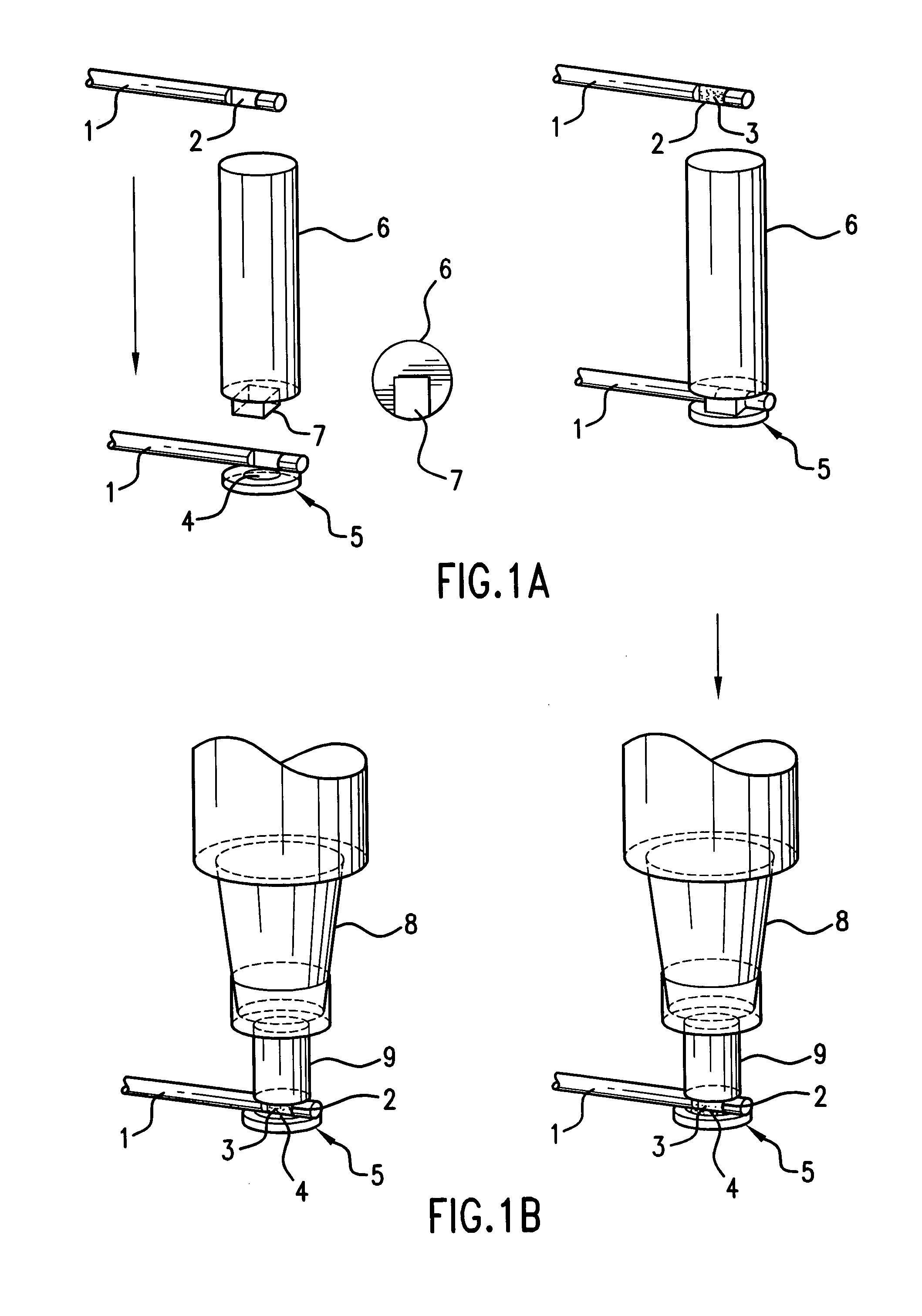
[0022]The apparatus according to the present invention includes biopsy needles, only partially depicted in the FIGS., which have an opening for receiving the biopsy specimen that is substantially reduced in size compared to commercially obtainable products (for example, Pro-Mag™, Manan Medical Products, Inc., Northbrook, Ill. 60062, USA). The opening is, for example, 0.2–0.3 mm deep and only 1.2 mm long; this yields a volume for the resulting biopsies that corresponds to the volume of the specimen well of the preparation plate. The needle is permanently secured in the biopsy gun, so that the inner lance, with the opening for the biopsy material, is immovable with respect to the biopsy gun.

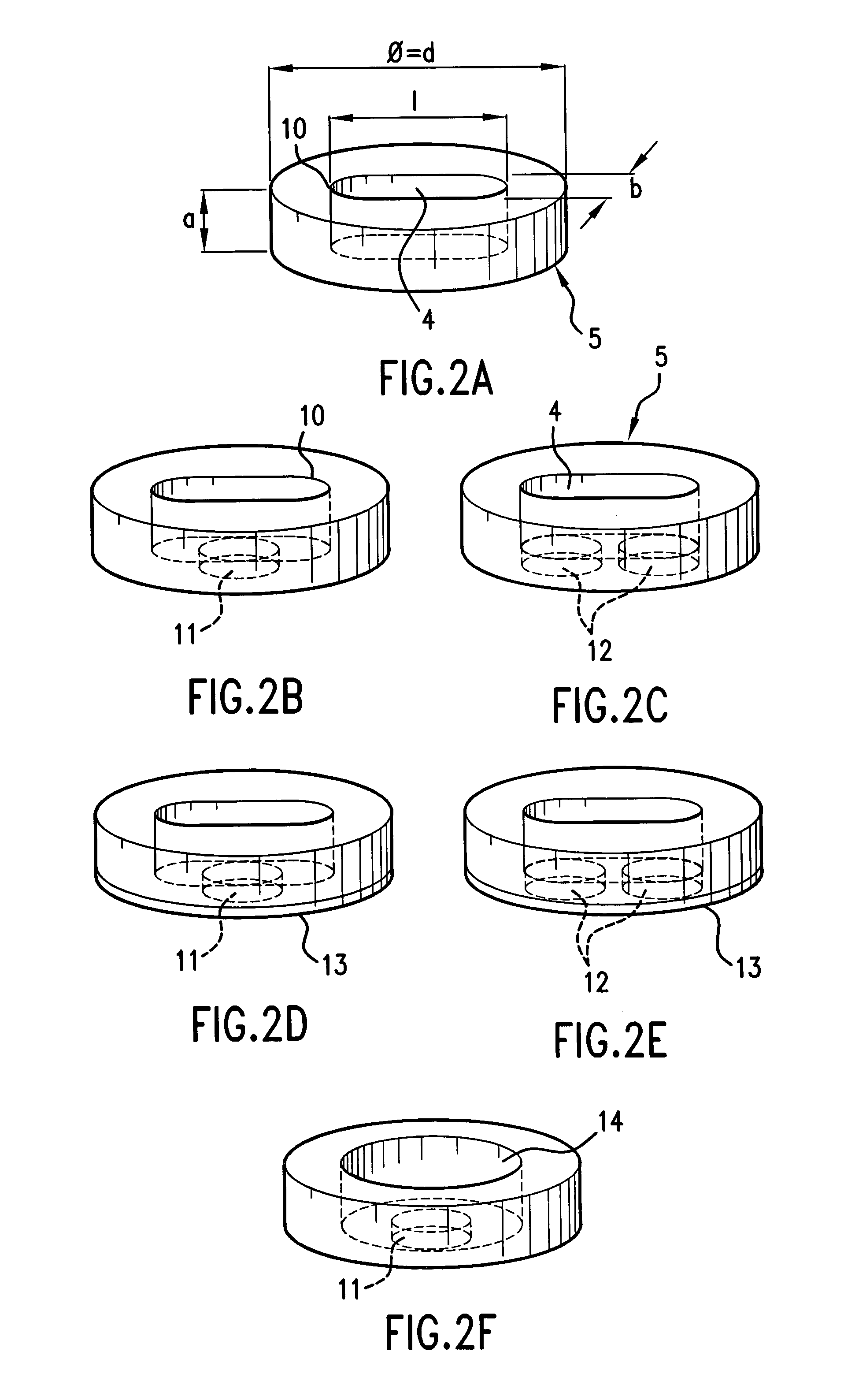
[0023]FIG. 1a shows two embodiments for transferring biopsy specimens using the apparatus according to the present invention. A biopsy specimen 3 is introduced into the empty opening 2 of a biopsy needle 1. The needle filled with this biopsy specimen is centered on specimen well 4 of a preparation ...


PUM

PropertyMeasurementUnit
diameteraaaaaaaaaa
thicknessaaaaaaaaaa
lengthaaaaaaaaaa

Abstract

An apparatus for handling biopsy specimens comprises a transfer part, a slide displaceable in a groove of said transfer part, preparation plates for the reception of biopsy specimens, and a shifter part that serves to introduce the biopsy specimens into the preparation plates. The empty preparation plates are positioned at a predetermined point in the groove of the transfer part, and the shifter part is placed onto the transfer part above the preparation plates. The biopsy specimens are then introduced into the specimen well of a preparation plate by manually actuated instruments or by means of a stream of fluid. The filled preparation plate is displaced back in the groove of the transfer part using the slide, and is transferred into a specimen holder prepared for it.

Description

CROSS REFERENCE TO RELATED APPLICATIONS[0001]This application claims priority of the Swiss patent application 0257 / 02 which is incorporated by reference herein.FIELD OF THE INVENTION[0002]The invention refers to an apparatus for handling biopsy specimens, and to a method for using it.BACKGROUND OF THE INVENTION[0003]Modem electron microscopy of biological specimens is more and more frequently examining specimens that have been immobilized by freezing (cryofixation). Freezing must be performed in such a way that the specimens vitrify, i.e. so that upon cooling, no ice (which would segregate the specimen) can form. “Segregation” is the separation of the specimen into pure water (ice crystals) and concentrated dissolved substances between the branches of the ice crystals. In vitrification, water is immobilized in its liquid configuration. In other words, all the constituents of the biological specimen are not displaced during immobilization, and thus represent a “snapshot” of the livin...

Claims


Application Information

Patent Timeline
Patent Type & Authority Patents(United States)
IPC IPC(8): B01L3/00G01N33/48A61B10/02G01N1/04G01N1/28G01N1/42G02B21/34
CPCG01N1/286G01N1/42G02B21/34Y10T436/11Y10T436/2575G01N2001/2873
Inventor STUDER, DANIEL
Owner LEICA MICROSYSTEMS GMBH
Why Eureka
Try Eureka