
Method and apparatus for dynamically monitoring multiple in vivo tissue chromophores

a tissue chromophores and dynamic monitoring technology, applied in diagnostic recording/measure, instruments, applications, etc., can solve the problems of not reporting absolute biochemical concentrations in tissue, unable to separate light absorption from scattering, and difficult to quantitatively and dynamically monitor biochemical processes

Inactive Publication Date: 2007-07-24
RGT UNIV OF CALIFORNIA
View PDF5 Cites 13 Cited by
  • Summary
  • Abstract
  • Description
  • Claims
  • Application Information

AI Technical Summary

Benefits of technology

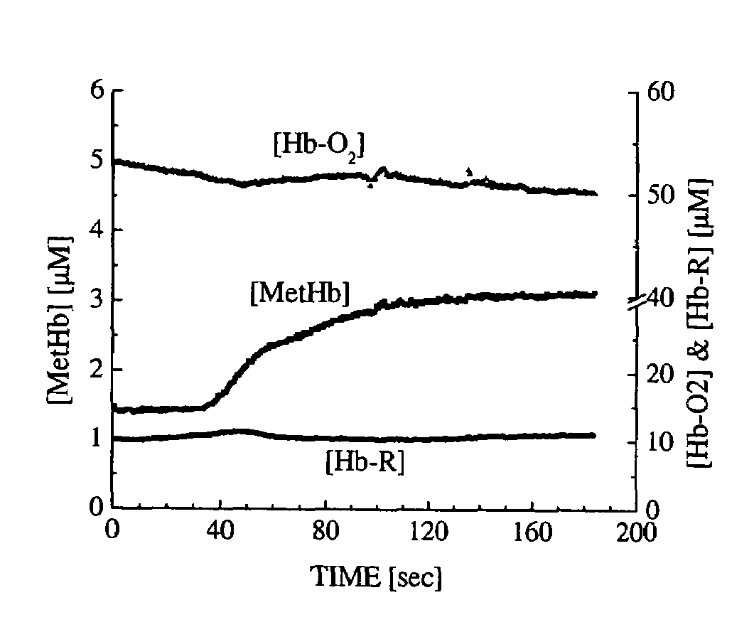
The present invention is a method and device for non-invasively monitoring the concentrations of multiple chromophores in biological tissue. This is done using a broadband diffuse optical spectroscopy device that can dynamically monitor changes in chromophore concentrations in real-time. The device includes a probe with optical fibers that are optically coupled to a detector and a spectrometer. The method involves radiating the tissue with light and detecting the intensity-modulated diffuse reflectance signals using the detector. The device can be used to monitor the therapeutic treatment of methemoglobinemia and cyanide toxicity treatment with methylene blue. The technical effects of this invention include the ability to non-invasively and quantitatively monitor the concentrations of multiple chromophores in real-time, providing a non-invasive method for therapeutic monitoring and assessing the success or failure of therapeutic interventions.

Problems solved by technology

In vivo quantitative non-invasive dynamic monitoring of biochemical processes is one of the most difficult and important challenges in medical diagnostics.
However, conventional methods generally do not separate light absorption from scattering, and as a result, do not report absolute biochemical concentrations in tissue.
However, these methods typically rely on a limited number of optical wavelengths and therefore have poor sensitivity to multiple analytes over a broad range of concentrations.
Pulse oximetry is, however, unreliable in the presence of methemoglobinemia since methemoglobin (MetHb) absorbs light equally well at wavelengths (typically 660 nm and 940 nm) used to determine oxygen saturation.
Arterial blood gas analysis can be also misleading in methemoglobinemia because it will show normal partial pressure of oxygen even in the presence of high MetHb concentration and inaccurate oxygen saturation if values were calculated from the pH and PaO2.
Unfortunately, because co-oximetry relies on the absorption spectra of a few wavelengths for the calculation of MetHb concentration, false positive readings often result from the presence of other pigments such as methylene blue or sulfhemoglobin which have high absorption at the methemoglobin absorption peak at around 630 nm.
However, being an oxidant itself, large doses of methylene blue will overwhelm the reducing effect of leukomethylene blue, and can result in hemolysis and, paradoxically, methemoglobinemia in patients with glucose phosphate dehydrogenase (G6PD) deficiency.
Unfortunately, the three aforementioned existing diagnostic methods (pulse oximetry, arterial blood gas analysis, and co-oximetry) are incapable of simultaneously quantifying in vivo tissue concentrations of MetHb and MB as well as oxyhemoglobin (Hb-O2) and deoxyhemoglobin (Hb-R) to monitor progression and resolution of methemoglobinemia.

Method used


Image

  • Method and apparatus for dynamically monitoring multiple in vivo tissue chromophores
  • Method and apparatus for dynamically monitoring multiple in vivo tissue chromophores
  • Method and apparatus for dynamically monitoring multiple in vivo tissue chromophores

Examples


Embodiment Construction

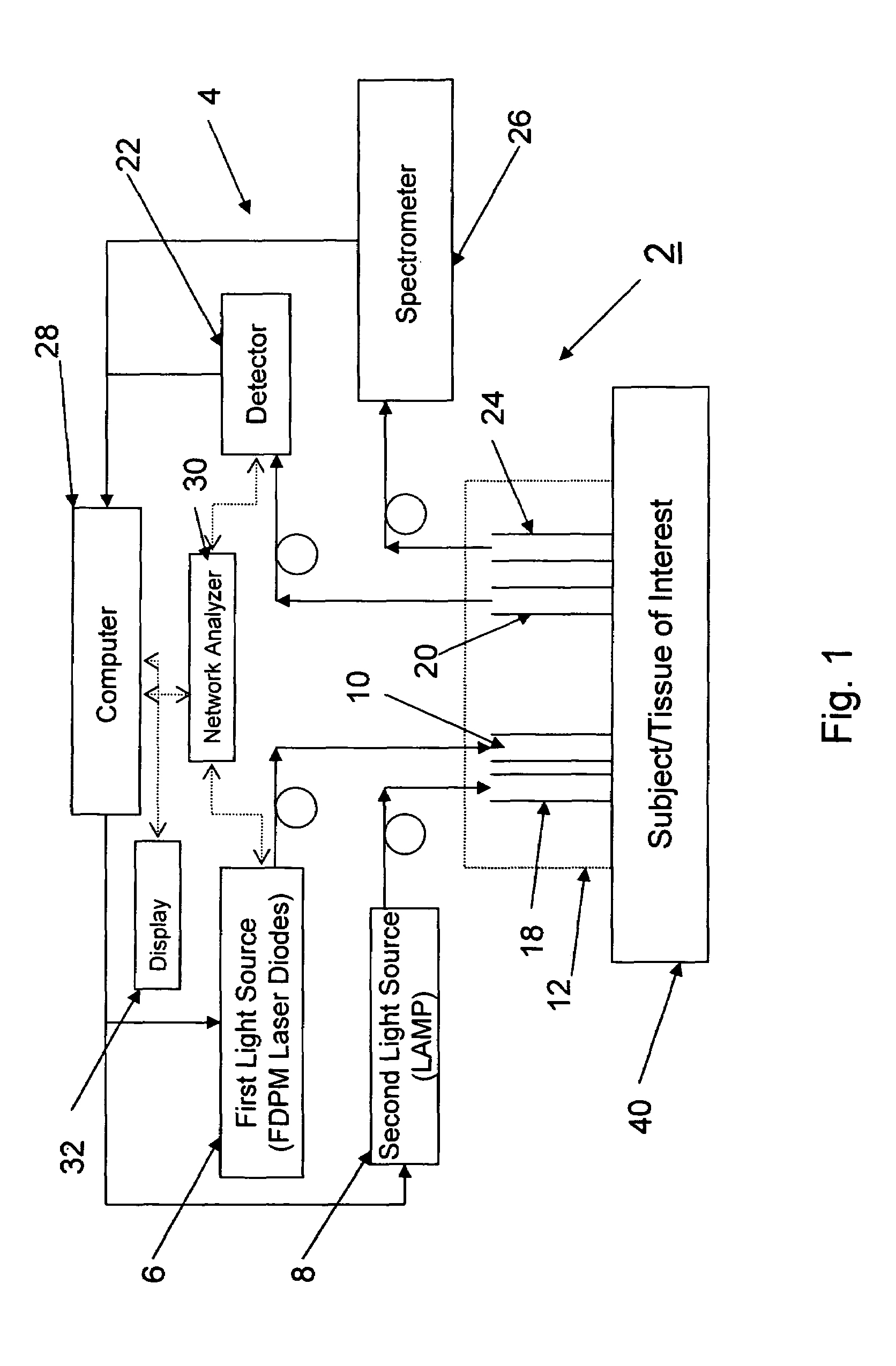
[0031]FIG. 1 illustrates a system 2 according to a preferred aspect of the invention. The system 2 generally includes a diffuse optical spectroscopy (DOS) device 4. The DOS device 4 includes a first light source 6 which preferably emits radiation at multiple wavelengths. For example, the first light source 6 may comprise multiple laser diodes each operating a different wavelengths. One exemplary example uses six laser diodes operating at wavelengths of 661 nm, 681 nm, 783 nm, 823 nm, and 910 nm. Another exemplary example includes six laser diodes operating at wavelengths of 658 nm, 682 nm, 785 nm, 810 nm, 830 nm, and 850 nm. Generally, at least three wavelengths are used to obtain FDPM measurements. In addition, in one aspect of the invention, the wavelengths are chosen within the range of about 600 nm to about 1000 nm. The plurality of laser diodes may be successively switched using a computer-controlled diode controller (ILX Lightwave LDC-3916 Laser Diode Controller) coupled to a ...


PUM

PropertyMeasurementUnit
wavelengthsaaaaaaaaaa
angleaaaaaaaaaa

Abstract

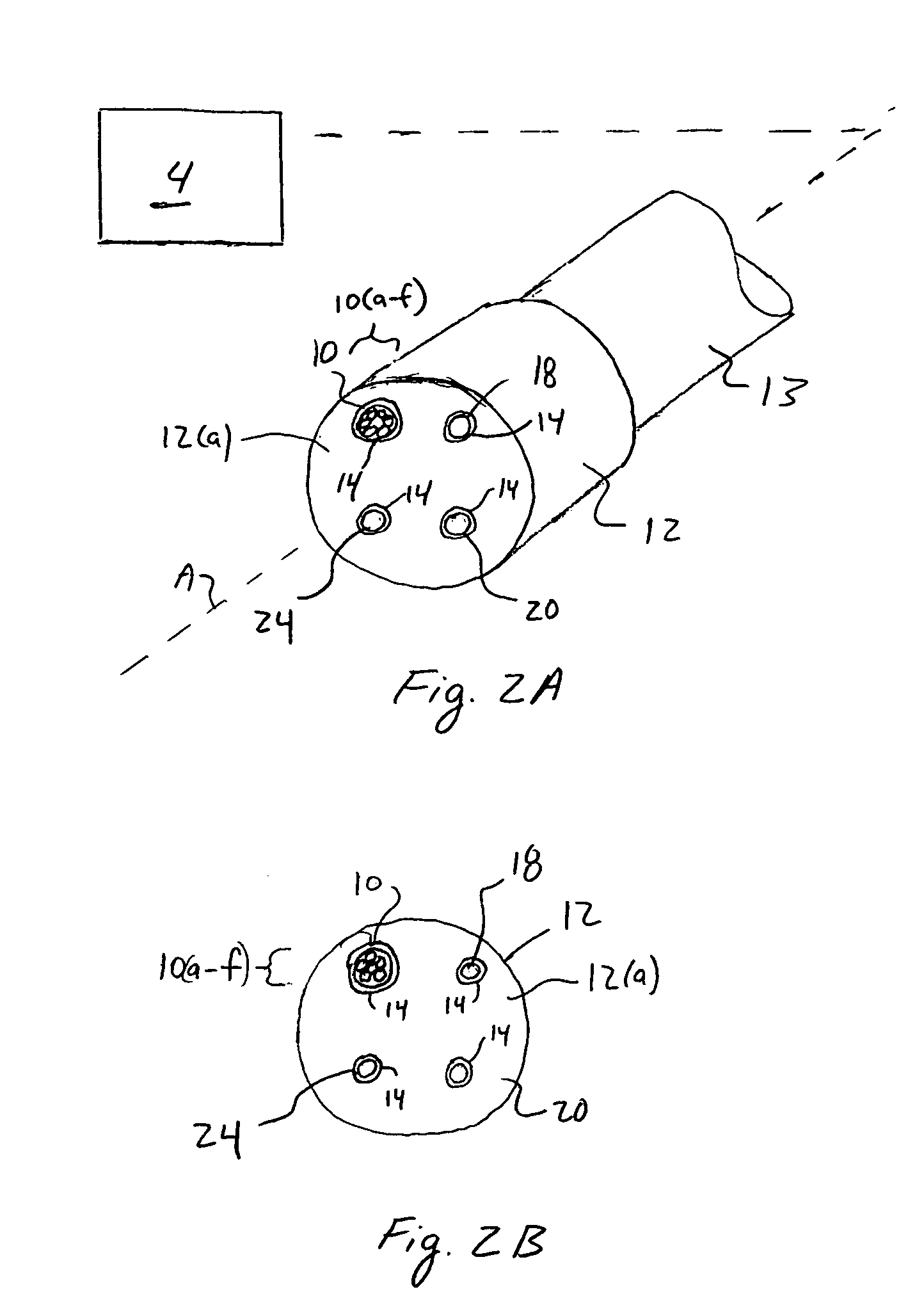
A device and method utilizes a broadband diffuse optical spectroscopy (DOS) system to dynamically calculate the concentrations of multiple chromophores in vivo using a non-invasive probe. The device and method permit dynamic monitoring of multiple in vivo tissue chromophores non-invasively with sensitivities necessary for effective therapeutic monitoring. The device includes a probe containing first and second source optical fibers as well as first and second detector optical fibers. The probe is placed adjacent to a sample of interest and detects reflected light which is passed to a proximally located detector and spectrometer. The concentrations of multiple chromophores are determined in real time. In a preferred embodiment, the multiple tissue chromophores include at least two of methemoglobin (MetHb), deoxyhemoglobin (Hb-R), oxyhemoglobin (Hb-O2), water (H2O), and methylene blue (MB). The device and method can be used quantify and monitor methemoglobin formation in subjects suffering from methemoglobinemia.

Description

REFERENCE TO RELATED APPLICATIONS[0001]This Application claims priority to U.S. Provisional Patent Application No. 60 / 561,839 filed on Apr. 13, 2004. The '839 Application is incorporated by reference as if set forth fully herein.STATEMENT REGARDING FEDERALLY SPONSORED RESEARCH AND DEVELOPMENT[0002]The U.S. Government may have a paid-up license in this invention and the right in limited circumstances to require the patent owner to license others on reasonable terms as provided for by the terms of Grant No. 445574-30133 awarded by the National Institutes of Health (Laser Microbeam and Medical Program: LAMMP) and Department of Defense (Air Force) Contract No. AF49620-00-10371.FIELD OF THE INVENTION[0003]The field of the invention generally relates to methods and devices used in diffuse optical spectroscopy. More specifically, the field of the invention generally relates to broadband diffuse optical spectroscopy methods and devices which are able to dynamically monitor multiple in vivo ...

Claims


Application Information

Patent Timeline
Patent Type & Authority Patents(United States)
IPC IPC(8): A61B5/00G01J3/28G01N21/31G01N21/35G01N21/49
CPCA61B5/14551A61B5/412G01N21/31G01N21/49A61B5/14552G01N2201/08G01N21/359
Inventor LEE, JANGWOENTROMBERG, BRUCE J.CERUSSI, ALBERT E.BRENNER, MATTHEW
Owner RGT UNIV OF CALIFORNIA
Learn More