Dual-band detector system for x-ray imaging of biological samples

a detector system and biological sample technology, applied in imaging devices, instruments, radiography controlled devices, etc., can solve the problems of limiting the image resolution to many tens of micrometers, expensive detectors for extremely fast image readout, artifacts in the reconstructed phase image, etc., to achieve the effect of convenient clinical evaluation

Active Publication Date: 2007-10-23
CARL ZEISS X RAY MICROSCOPY
View PDF29 Cites 55 Cited by
  • Summary
  • Abstract
  • Description
  • Claims
  • Application Information

AI Technical Summary

Benefits of technology

[0008]Dual energy x-ray imaging systems are currently available. They allow for the separation of bone and soft tissues for easier clinical evaluation. These systems accomplish this by acquiring two images; one at a high x-ray energy and one af a low x-ray energy. The x-ray source is typically switched between high energy (100-150 keV) and low energy (60-80 keV) mode while the detector acquires an image at each energy range. These two images are processed using a dual-energy algorithm to extract the soft tissue image and the hard tissue (bone) image.
[0009]The present invention concerns a multi-band or dual-band digital detector that can provide an imaging platform capable of extracting hard and soft tissue images. It can further facilitate the implementation and deployment of recent developments and can permit low cost and practical deployment in clinical applications as well as biomedical research applications where significant improvement in spatial resolution and image contrast is required. These improvements include an increase of feature detection sensitivity by more than an order of magnitude, simultaneous imaging of soft and hard (or mineralized) tissue at extremely high time resolutions that is not limited by detector readout speed, and an increase in the signal to noise ratio of images by reducing the substantial background resulting from Compton scattering.

Problems solved by technology

Motion-induced differences in the subject during the two exposures often cause artifacts in the reconstructed phase image and limit the image resolution to many tens of micrometers.
The imaging tool uses a very specialized x-ray source for fast switching of the x-ray spectra and an expensive detector for extremely fast image readout because each single image contains large amounts of data.

Method used


Image

  • Dual-band detector system for x-ray imaging of biological samples
  • Dual-band detector system for x-ray imaging of biological samples
  • Dual-band detector system for x-ray imaging of biological samples

Examples


Embodiment Construction

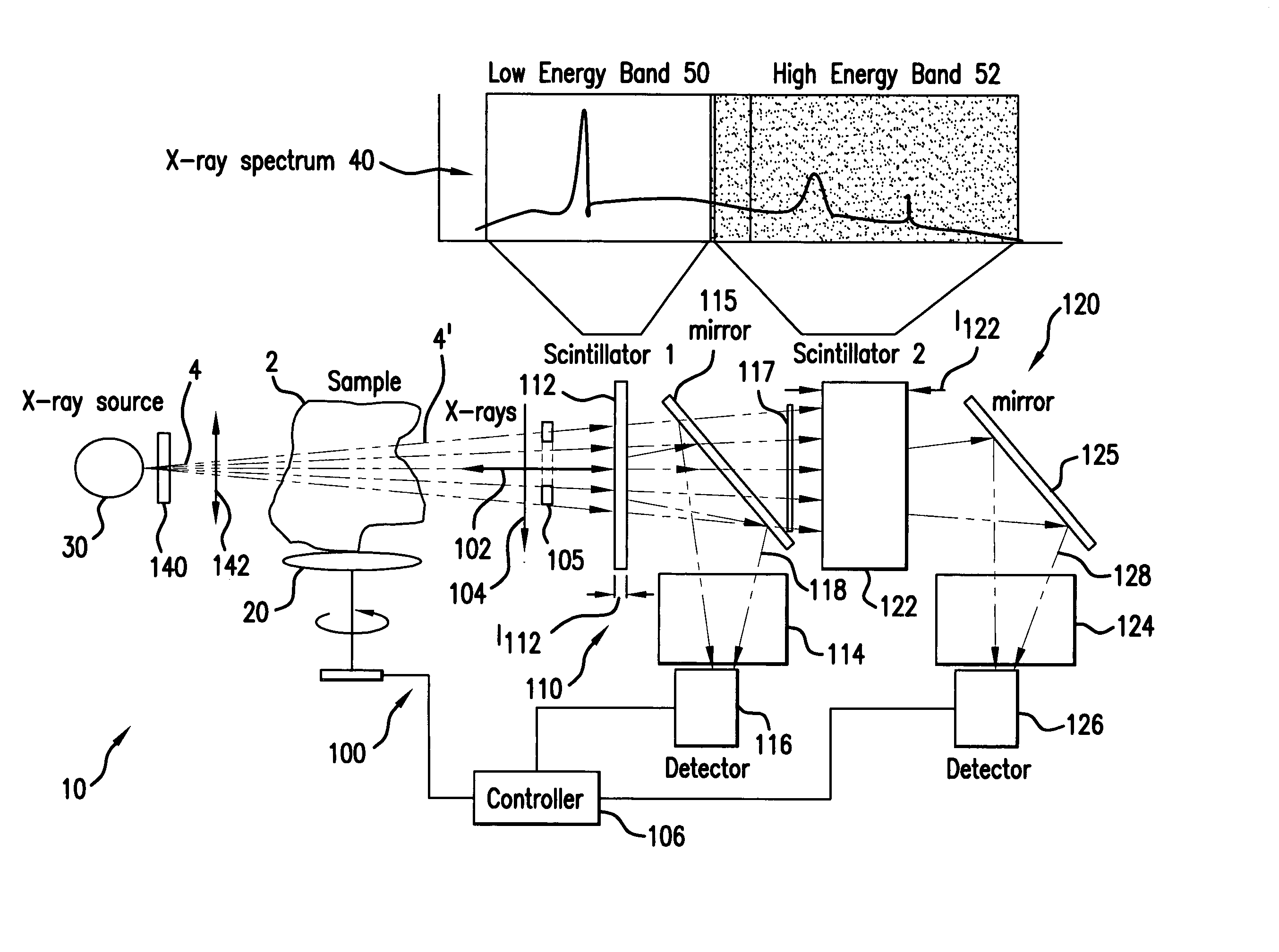
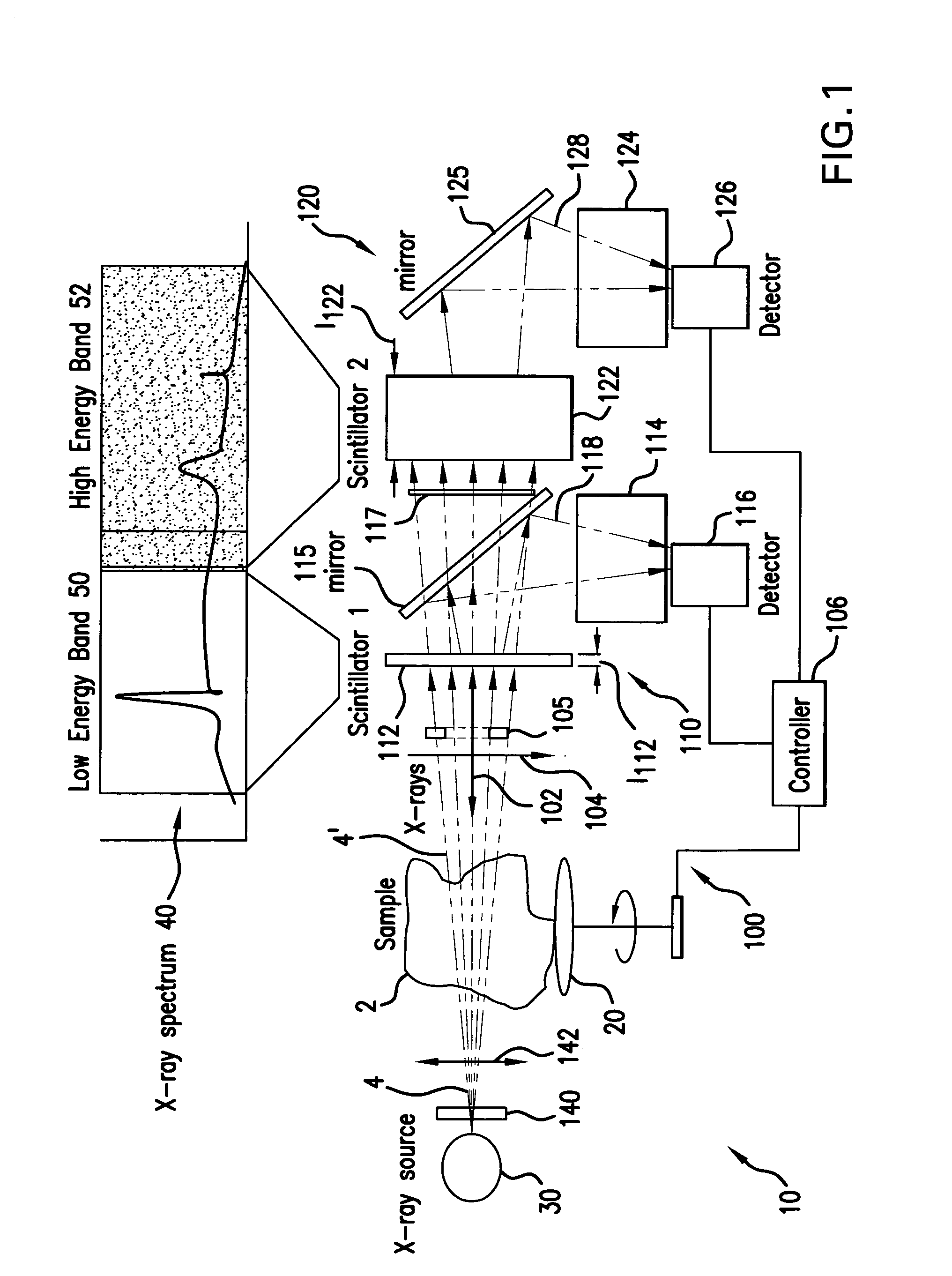
[0026]FIG. 1 shows an x-ray imaging system 10 with an x-ray detector 100 that has been constructed according to the principles of the present invention.

[0027]In general, the detector 100 comprises at least two digital detector systems 110, 120. In the illustrated configuration, the first and second detector systems 110, 120 are arranged in tandem along an optical axis or x-ray path 102.

[0028]The detector systems 110, 120 are configured with the goal of separating the low energy x-rays 50 from the high energy x-rays 52 in the total x-ray spectrum 40 that is generated by the x-ray source 30.

[0029]In more detail, an x-ray beam 4 from preferably a small spot size x-ray source 30 illuminates a sample 2.

[0030]In one example, a synchrotron source is used. In other examples, however, an electron bombardment laboratory X-ray source is used. These systems comprise an electron gun that generates an electron beam that is directed at a target. Typically, the target is selected from the group of:...


PUM

PropertyMeasurementUnit
thicknessaaaaaaaaaa
thicknessaaaaaaaaaa
thicknessaaaaaaaaaa

Abstract

A digital dual-band detector functions as an imaging platform capable of extracting hard and soft tissue images, for example. The detector has a first detector system comprising a first scintillator for converting x-rays from a sample to an first optical signal, and a first detector for detecting the first optical signal in combination with a second detector system comprising a second scintillator for converting x-rays from the sample and passing through the first scintillator to a second optical signal, and a second detector for detecting the second optical signal. The detector can facilitate the implementation and deployment of recent developments and can permit low cost practical deployment in clinical applications as well as biomedical research applications where significant improvement in spatial resolution and image contrast is required.

Description

BACKGROUND OF THE INVENTION[0001]X-ray imaging (radiography) is one of the most important and widely used medical diagnostic methods. It can be traced back to the invention of x-rays more than 100 years ago. Well-known applications include mammography, angiography, x-ray computerized tomography (CT) scans, and dental x-rays. For example, x-ray mammography is currently the primary method for breast cancer screening. Recent randomized clinical trials have shown that screening mammography has reduced mortality from breast cancer by 25 to 30 percent in women between the ages of 50 and 70 and by 18 percent among women between the ages of 40 and 50.[0002]In addition to these essential medical applications, x-ray imaging is finding increasingly important applications in biomedical research laboratories, such as in the study of disease progression or effect of genetic engineering or drug therapy. Often, time-lapsed noninvasive, in vivo images of animal models are used. Often mouse models ar...

Claims


Application Information

Patent Timeline
Patent Type & Authority Patents(United States)
IPC IPC(8): H05G1/64G01T1/20G01J1/42G01N23/04G01T1/00G01T1/202
CPCG01T1/2018G01T1/202G21K2207/005
Inventor YUN, WENBINGWANG, YUXINSCOTT, DAVID DEAN
Owner CARL ZEISS X RAY MICROSCOPY
Why Eureka
Try Eureka