Latent fusion protein

a fusion protein and protein technology, applied in the field of dna constructs, can solve the problems of limiting the use and efficacy of dna/rna, unsatisfactory systemic effects,

Inactive Publication Date: 2007-12-11
STEALTHYX THERAPEUTICS
View PDF17 Cites 2 Cited by
  • Summary
  • Abstract
  • Description
  • Claims
  • Application Information

AI Technical Summary

Benefits of technology

[0018]Amino acid deletions or insertions may also be made relative to the amino acid sequence for the fusion protein referred to above. Thus, for example, amino acids which do not have a substantial effect on the activity of the polypeptide, or at least which do not eliminate such activity, may be deleted. Such deletions can be advantageous since the overall length and the molecular weight of a polypeptide can be reduced whilst still retaining activity. This can enable the amount of polypeptide required for a particular purpose to be reduced—for example, dosage levels can be reduced.
[0030]The LAP may provide a protective “shell” around the pharmaceutically active agent thereby shielding it and hindering, or preventing, its interaction with other molecules in the cell surface or molecules important for its activity.

Problems solved by technology

Pharmacologically active proteins or other medicines based on such agents, which have to be administered at very high concentrations systemically in order to achieve biologically effective concentrations in the tissue being targeted, tend to give rise to undesirable systemic effects, for example toxicity, which limit their use and efficacy.

Method used


Image

  • Latent fusion protein
  • Latent fusion protein
  • Latent fusion protein

Examples


example 1

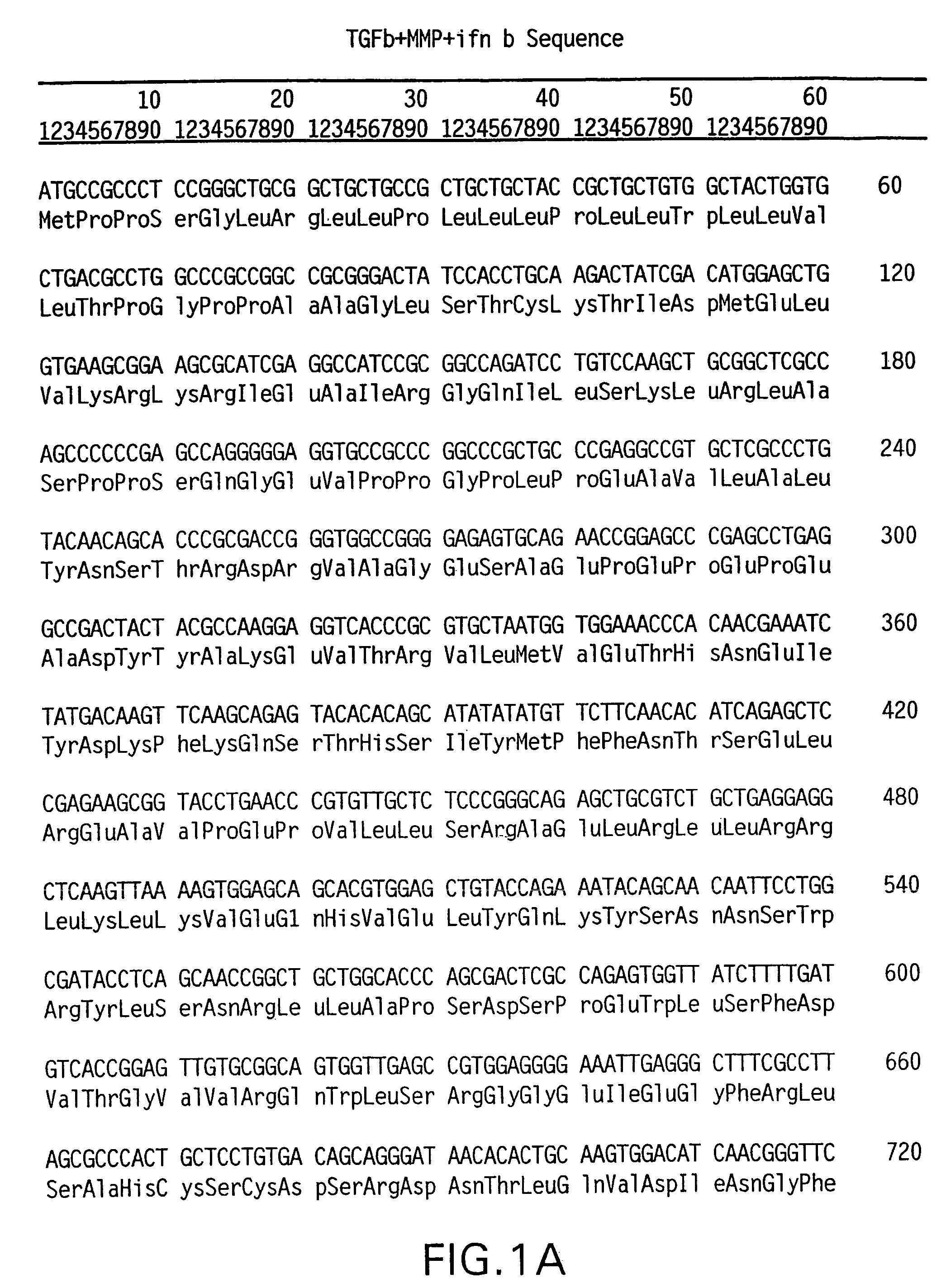
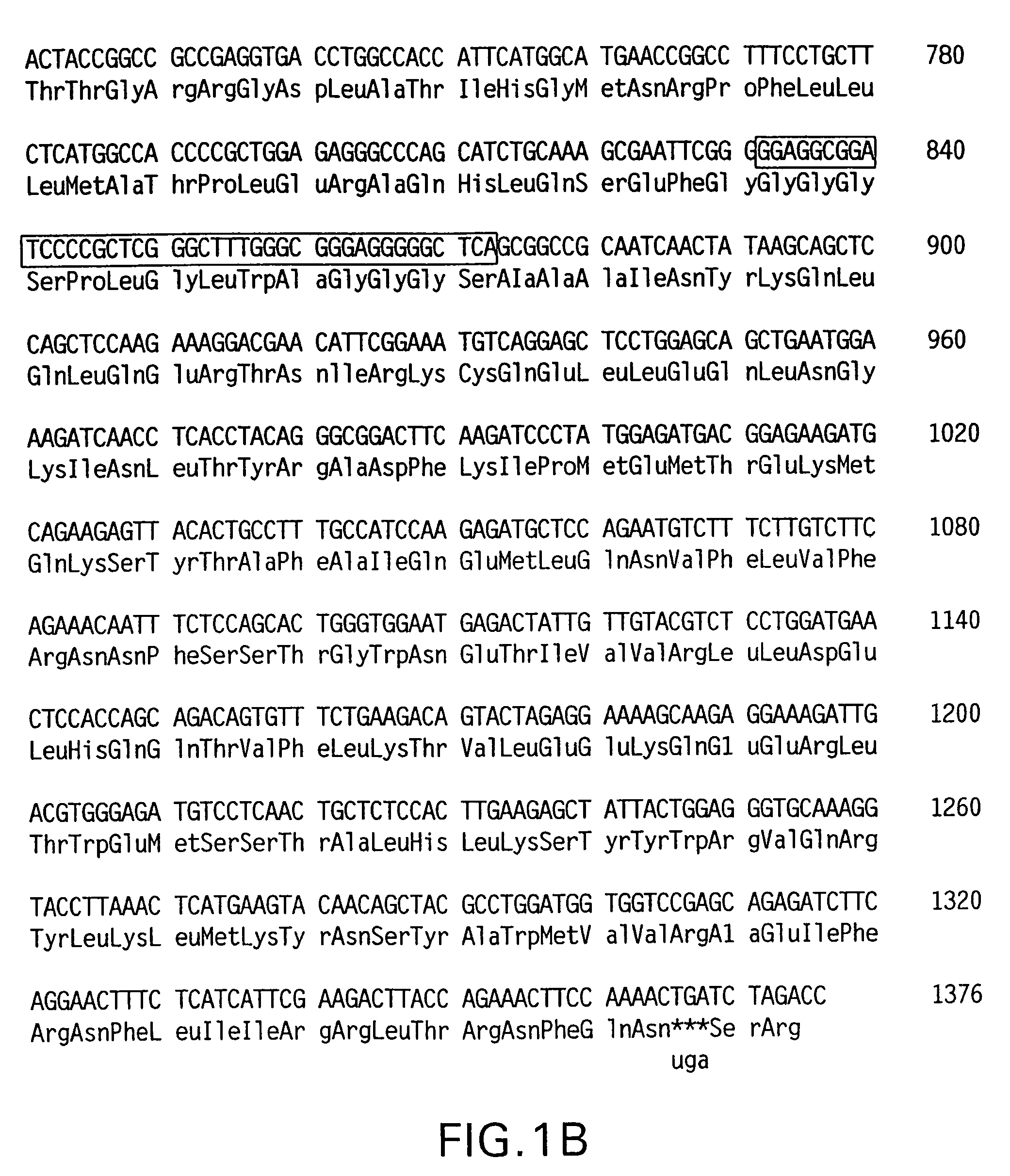
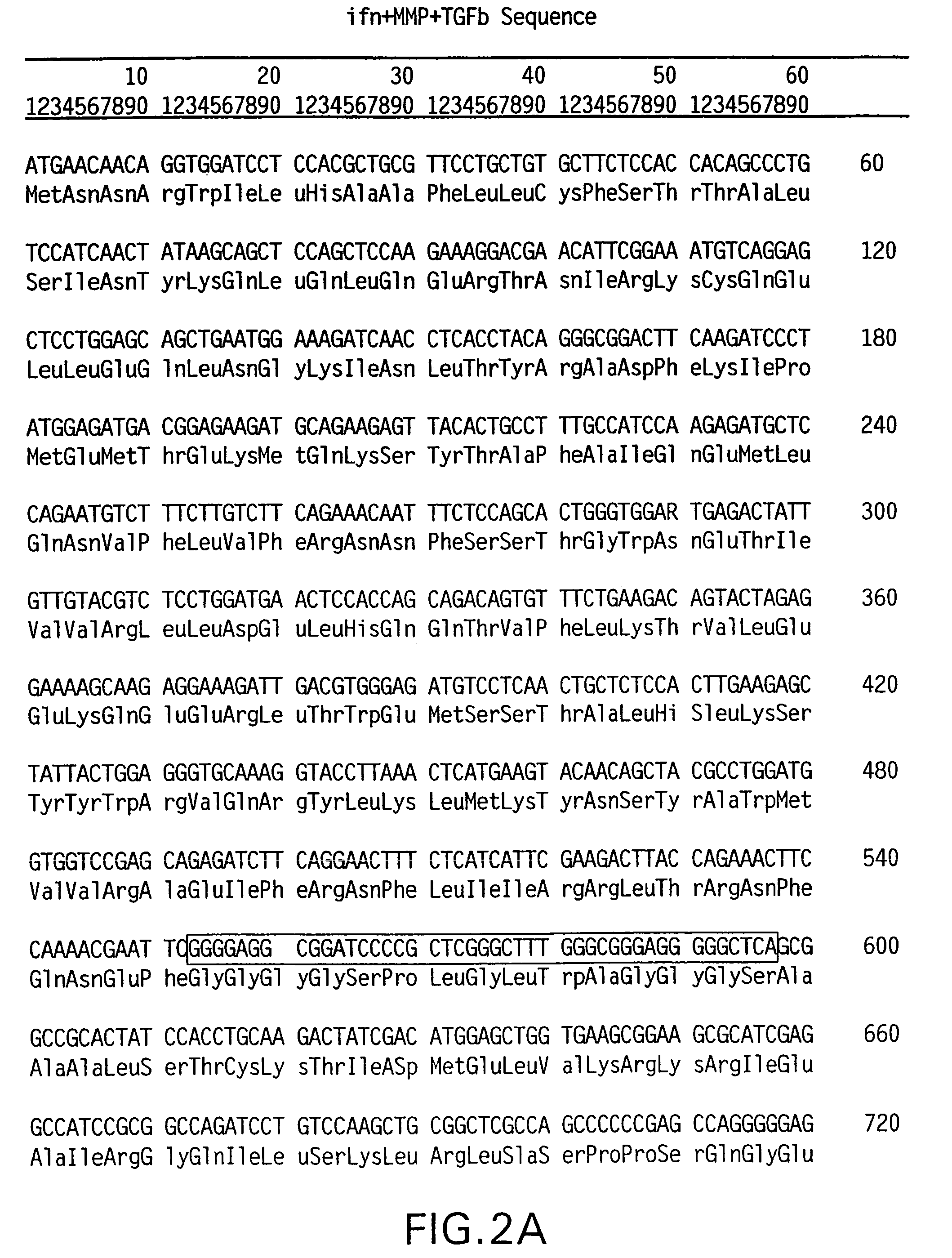
Construction of LAP-mIFNβ and mIFNβ-LAP

Methods

Cloning of GS-MMP-GS Linker into EcoR1-Not1 Sites of pcDNA3

[0108]A vector was constructed by inserting the GS-MMP-GS linker into EcoR1-Not1 cleaved pcDNA3. pcDNA3 is an expression vector (from Invitrogen) which comprises the human cytomegalovirus immediate early promoter and enhancer, together with RNA processing signals allowing transcription.

[0109]Double stranded deoxyoligonucleotide coding for the sequence GLY GLY GLY GLY SER PRO LEU GLY LEU TRP ALA GLY GLY GLY SER (SEQ ID NO: 1), was designed as follows:

[0110]

Sense oligo:(SEQ ID NO:2)5′ AATTCGGGGGAGGCGGATCCCCGCTCGGGCTTTGGGCGGGAGGGGGCTCAGC 3′Antisense oligo:(SEQ ID NO:3)5′ GGCCGCTGAGCCCCCTCCCGCCCAAAGCCCGAGCGGGGATCCGCCT CCCCCG 3′

[0111]Synthetic deoxyoligonucleotides were purchased from Life Technologies Ltd. (Paisley, UK). Annealed deoxyoligonucleotides were cloned into EcoR1-Not1 cleaved pcDNA3 (Invitrogen, Groningen, The Netherlands). The recombinant clone lost its EcoRV site and gai...

example 2

Cell Transfection Studies

Methods

Transfection into DHFR-deficient Chinese Hamster Ovary (CHO) Cells

[0134]Dihydrofolate reductase (DHFR)-deficient CHO cells were maintained in HAM-F12 medium (Life Technologies Ltd., Paisley, UK) with 10% fetal bovine serum (FBS) (Life Technologies Ltd.), penicillin / streptomycin and glutamine.

[0135]pcDNA3 plasmids (20 μg) expressing LAP-mIFNβ or mIFNβ-LAP were each linearized with PvuI and ligated separately with PvuI cut pSV2DHFR (1 μg) (Chemajovsky et al., DNA, 3, 297-308 (1984)). After phenol extraction, the plasmids were ligated in 300 μl with T4 DNA ligase at 16° C. for 3 days. The DNA was precipitated in 0.4 M NH4 acetate and resuspended in water to be added as 1 ml calcium phosphate co-precipitate on 0.5×10⁶ CHO cells on 9 cm plates seeded 24 hrs earlier. 4 hrs later, the cells were treated with 10% glycerol in HAM-F12 without FBS, washed in FBS-free media and left to recover for 48 hrs. Transfected cells were trypsinized and split into six 9 cm...

example 3

[0169]In order to assess whether the latency detected with LAP-mIFNβ required the formation of a putative closed shell structure bounded by the dimeric disulphide linked LAP, a fusion protein was constructed using the porcine LAP that was mutated in Cys 223 and 225 to Ser.

Methods

Preparation of Construct

[0170]Porcine LAP was cloned by PCR as set out in Example 1. The primers used were as set out in Example (cloning of porcine LAP). The cloned porcine LAP was mutated in Cys 223 and 225 to Ser (Sanderson et al., Proc. Natl. Acad. Science, 92, 2572-2576 (1995)).

Transient Transfection into Monkey COS-7 Cells

[0171]20 μg plasmid DNA, PorcLAP-mIFNβ and mIFNβ-LAP & LAP-mIFNβ controls, were transfected by the calcium phosphate co-precipitation method in duplicates to 0.5×10⁶ COS-7 cells seeded in 9 cm plates as described above. The DNA co-precipitate was left on the cells overnight instead of 4 hrs. COS-7 cells were grown in DMEM with antibiotics and 10% FBS. 48 hrs after glycerol shock the s...


PUM

PropertyMeasurementUnit
molecular weightaaaaaaaaaa

Abstract

The present invention provides a method for providing latency to a pharmaceutically active agent. The method has application in overcoming the toxic effect of systemic administration of potent biological agents. The method comprises associating a fusion protein comprising a latency associated peptide and a proteolytic cleavage site with a pharmaceutically active agent. The fusion protein also has application in providing site specific activation to a latent pharmaceutically active agent.

Description

CROSS REFERENCE TO RELATED APPLICATIONS[0001]This application is a continuation of U.S. patent application Ser. No. 10 / 753,181, filed Jan. 7, 2004, now U.S. Pat. No. 7,141,392 which is a divisional of U.S. patent application Ser. No. 09 / 756,283, filed Jan. 9, 2001, now U.S. Pat. No. 6,942,853, issued on Sep. 13, 2005; both of which are fully incorporated herein by reference.BACKGROUND OF THE INVENTION[0002]The present invention relates to the use of DNA constructs, and proteins encoded by the constructs, in medicine with particular application in gene therapy. The present invention also relates to methods of providing latency to pharmaceutically active agents.[0003]Most cytokines and growth factors are expressed under tight control mechanisms. Their gene expression is regulated by environmental stimuli such as infection, cell-cell interactions, change in extracellular matrix composition and interactions with adhesion molecules or via stimulation with other cytokines.[0004]In additio...

Claims


Application Information

Patent Timeline
Patent Type & Authority Patents(United States)
IPC IPC(8): C07K1/00A01N37/18C07K14/00C07K19/00A61K38/00C07H21/04C07K14/495C07K14/565C12N15/62
CPCC07H21/04C07K14/495C07K14/5428C12N15/62C07K14/565C12N9/6491C07K14/55A61K38/00C07K2319/50C07K2319/75
Inventor CHERNAJOVSKY, YUTIDREJA, HANNA STINAADAMS, GILLIAN
Owner STEALTHYX THERAPEUTICS
Why Eureka
Try Eureka