Treatment of retinal detachment

a retinal detachment and treatment technology, applied in the field of retinal detachment treatment, can solve the problems of retinal detachment, loss of vision, and none of these methods are entirely satisfactory

Active Publication Date: 2008-01-08
PEYMAN GHOLAM A
View PDF4 Cites 60 Cited by
  • Summary
  • Abstract
  • Description
  • Claims
  • Application Information

AI Technical Summary

Problems solved by technology

Symptoms of retinal detachment are painless and sudden segmental or total visual loss in one eye.
With time, retinal detachment also results in loss of vision, due to loss of photoreceptor cells located in the outer part of the retina.
None of these methods is entirely satisfactory.
Disadvantages are that the patient must undergo a surgical procedure and endure a long recovery period, and must tolerate the presence of a buckle around the eye that might interfere with movement of the eye.
However, the force of injection itself can force the subretinal fluid through the tear into the vitreous cavity.
Only superior tears can be treated, because air and gas rise above the vitreous fluid, and multiple tears that are separate from each other are difficult to tamponade.
PVR may further detach the retina and is a serious complication of pneumatic retinopexy.
Injection of gas without drainage of fluid may cause a rapid rise in intraocular pressure, thus the patient must be monitored postoperatively for several hours.
Possible side effects, however, are closure of the central retinal artery which could result in diminished blood flow to the retina.
Additionally, in older individuals, the strict head positioning required to effect treatment may be very difficult to achieve and maintain.
Still further, the results are unpredictable and a 24 hour waiting period is required to determine if the subretinal fluid has resorbed.
Disadvantages of vitrectomy are that the patient must undergo a surgical procedure with the possibility of injury to the lens or retina during surgery, and has a 70% chance of postoperative cataract formation due to the vitrectomy and the injection of gas into the eye.

Method used


Image

  • Treatment of retinal detachment
  • Treatment of retinal detachment
  • Treatment of retinal detachment

Examples


Embodiment Construction

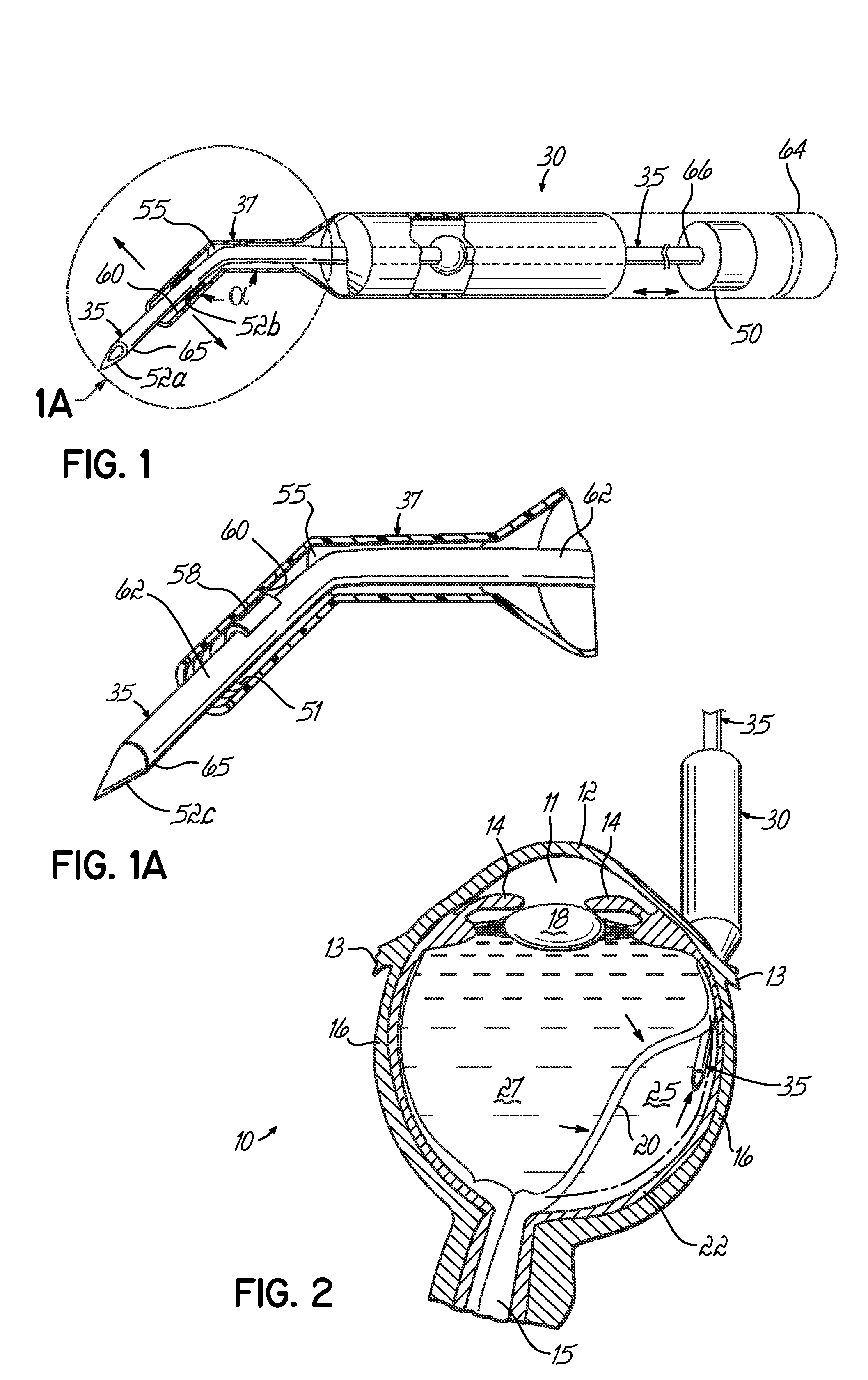
[0027]One embodiment of the invention is an apparatus for repairing a detached retina in an eye 10, shown in FIG. 2. The locations of the anterior chamber 11, cornea 12, conjunctiva 13, iris 14, optic nerve 15, sclera 16, lens 18, retina 20 and choroid 22 are illustrated.

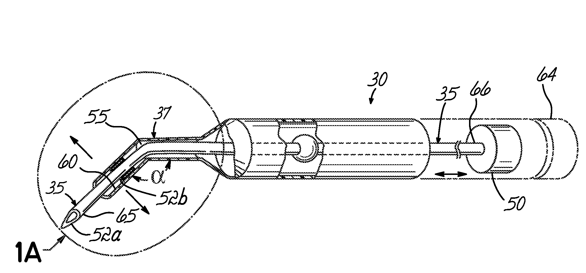
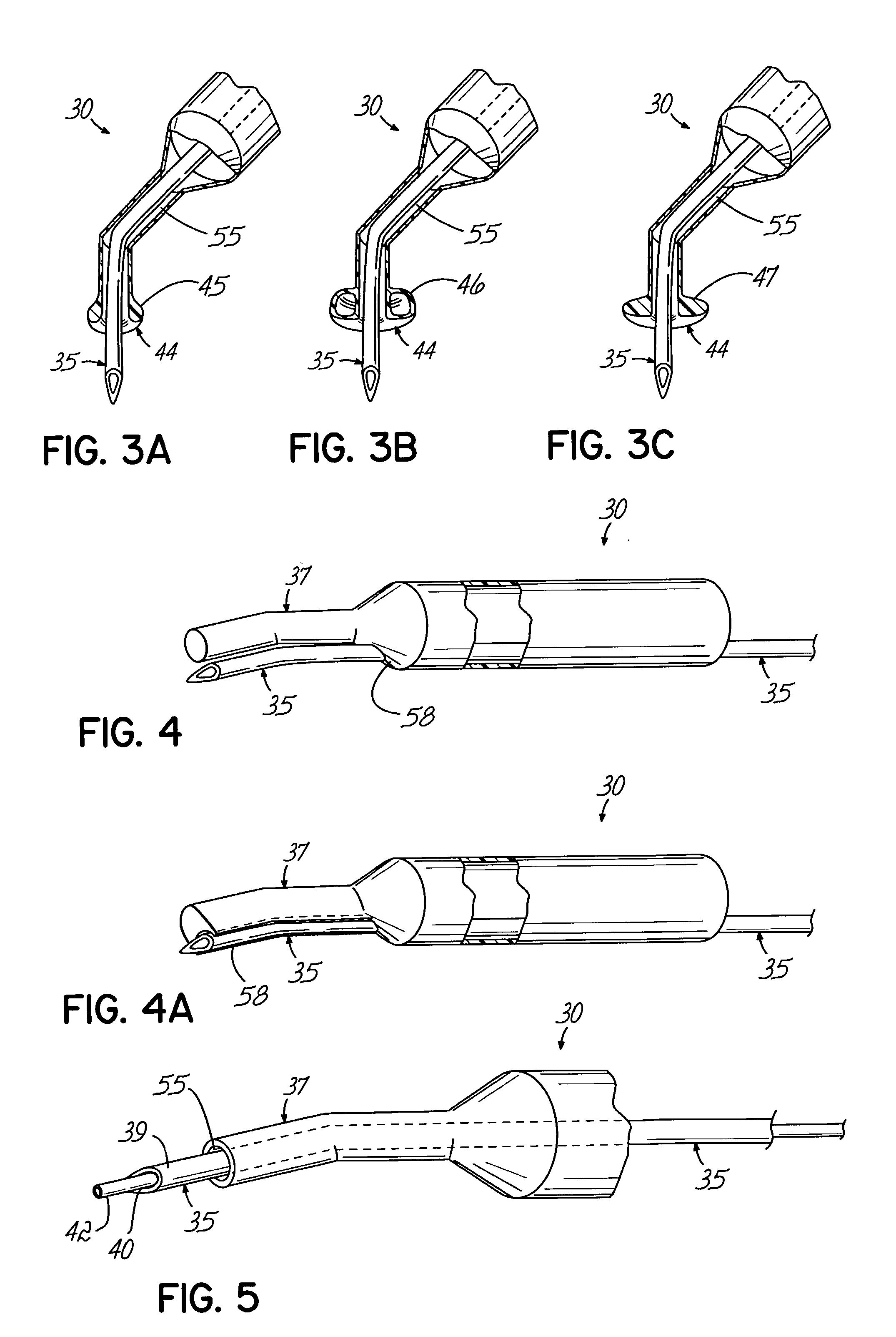
[0028]With reference to FIGS. 1, 1A, 4, 4A, and 5, the apparatus 30 may be a fluid withdrawal device 35, such as a needle or hollow tubing, through which fluid may flow, and a guide tube37 or probe for directing accurate advancement, placement, alignment, and / or positioning of the device 35. In one embodiment, the device 35 is a hollow needle for insertion into a subretinal space 25 of an eye 10, and withdrawal of fluid from the subretinal space 25 through the device 35. The needle gauge may range from 21 gauge to 41 gauge. In another embodiment, shown in FIG. 1A, the device 35 is a solid pin and fluid is removed from the subretinal space 25 by flowing adjacent the device 35, for example, by draining under the conju...


PUM


Abstract

An apparatus and minimally invasive method for removing fluid from a subretinal space to allow a detached retina to flatten. An apparatus, comprising a fluid withdrawal device and a guide for advancing and placing the device, is positioned on the exterior eye surface at the detachment site. Various embodiments of the apparatus are disclosed. Using the guide, the surgeon advances the device into the fluid-filled space and drains fluid, allowing the retina to flatten. Additional injection of saline or gas into the vitreous cavity normalizes intraocular pressure, and the patient is ambulatory immediately afterward. Unlike other retinal attachment techniques, in the inventive procedure the patient receives only a local anesthetic, and has no restraint on head movement.

Description

FIELD OF THE INVENTION[0001]The invention relates to a method and apparatus for treatment of retinal detachment that permits a patient to be ambulatory immediately after treatment.BACKGROUND[0002]Retinal detachment occurs when the sensory layers of the retina become separated from their underlying supporting tissue of retinal pigment epithelium and the choroid. Generally, retinal detachment is caused by a retinal tear or the presence of vitreous traction, either of which may occur spontaneously or may be due to trauma. Retinal detachment may also result from pathology, such as retinopathy of prematurity in premature infants or diabetic retinopathy in diabetic individuals. Symptoms of retinal detachment are painless and sudden segmental or total visual loss in one eye.[0003]When there is a tear, or when there is traction causing separation of the retina from its underlying structures, the liquid vitreous passes through the opening and into the subretinal space, inducing further exuda...

Claims


Application Information

Patent Timeline
Patent Type & Authority Patents(United States)
IPC IPC(8): A61M31/00A61F9/007
CPCA61F9/00727
Inventor PEYMAN, GHOLAM A.LIVIR-RALLATOS, CHARALAMPOS
Owner PEYMAN GHOLAM A
Why Eureka
Try Eureka