Micro-pattern embedded plastic optical film device for cell-based assays

a cell-based assay and micro-pattern technology, applied in the field of biological research and drug discovery, can solve the problems of high cost, high and high cost of this method to make, and achieve the effect of reducing the cost of engraved slides and slips and reducing the cost of production

Active Publication Date: 2008-02-12
NEXCELOM BIOSCIENCE LLC
View PDF13 Cites 41 Cited by
  • Summary
  • Abstract
  • Description
  • Claims
  • Application Information

AI Technical Summary

Benefits of technology

[0017]The micro-pattern embedded optical film may be incorporated into tissue culture vessels by combining the micro-pattern embedded film with supporting components. The supporting components provide mechanical strength for handling, both manually and robotically. They also provide liquid containment for chemical reagents necessary for the cell-based assays.

Problems solved by technology

The throughput of this method is extremely low.
Instruments of this type are typically complex and expensive.
A limitation of this technique is that engraved slides and slips are expensive to make with this method.
In addition, the glass surface also limits the kind of cells that can be grown, since some cells do not grow on glass.

Method used


Image

  • Micro-pattern embedded plastic optical film device for cell-based assays
  • Micro-pattern embedded plastic optical film device for cell-based assays
  • Micro-pattern embedded plastic optical film device for cell-based assays

Examples


example 1

Making Micro-Pattern Embedded Optical Film

[0052]The micro-pattern embedded plastic optical film consists of two layers. The base substrate is a 10-mil (namely, 0.010 inch thick) polycarbonate film: DE 1-1D Makrofo polycarbonate Gloss / Gloss with liners (Tekra Co., New Berlin, Wis.). The second layer is a clear plastic with micro-pattern formed on its surface.

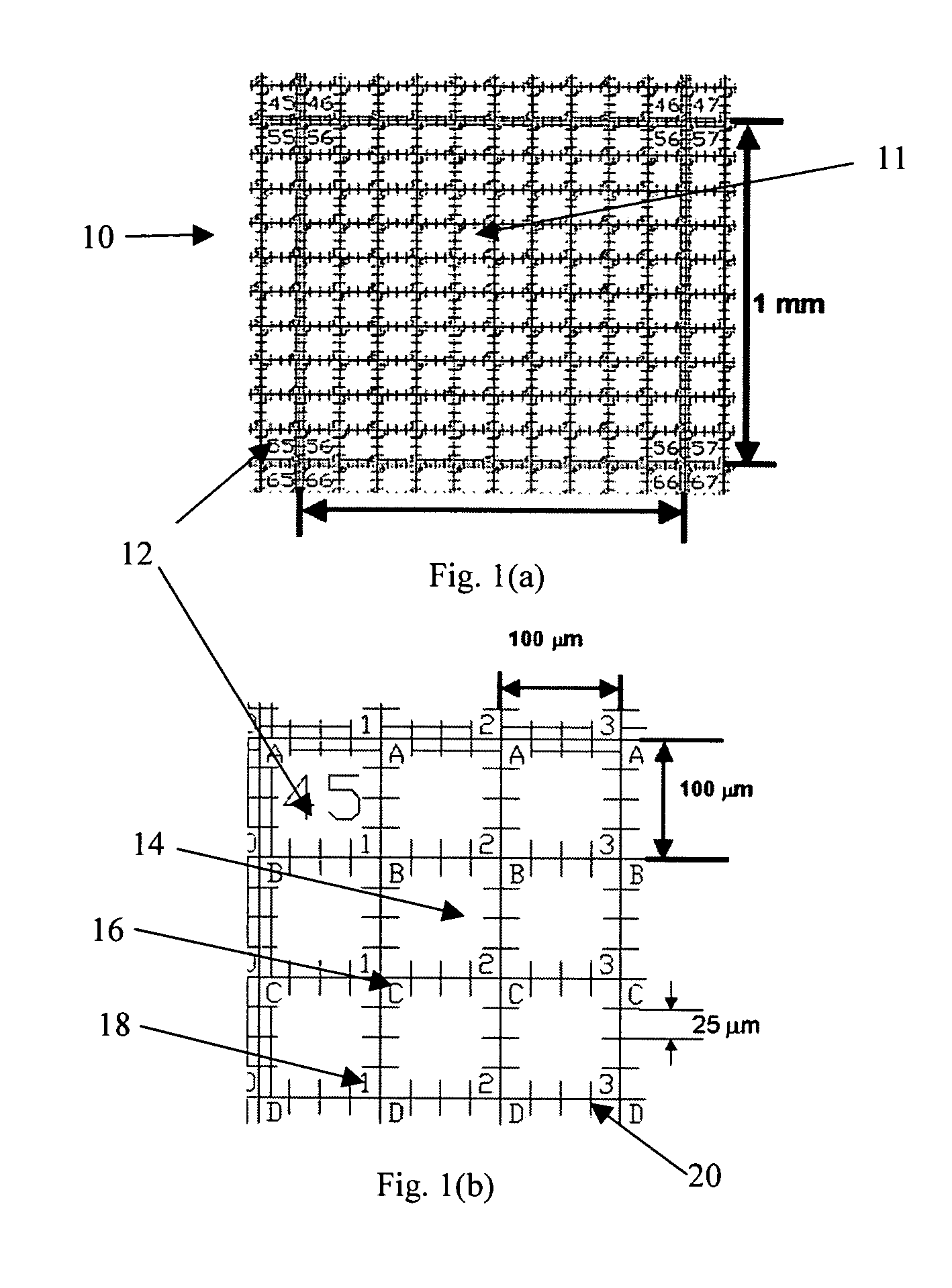
[0053]The process of fabricating the micro-pattern embedded film consists of the following steps.[0054]1. Make a master tool that has a negative image of FIG. 1.[0055]2. Prepare a UV polymerizable solution. (Ex. Lens bond, SK-9 from Summers Optical, Hatfield, Pa.)[0056]3. Cast the UV polymerizable solution between the base substrate and the master tool.[0057]4. Expose the assembly made in step 3 with UV light, resulting in a film with micro-pattern embedded on its surface.[0058]5. Remove the micro-pattern embedded plastic optical film from the master tool.

[0059]Steps 3 to 5 may be repeated to produce multiple micro-pattern embedd...

example 2

Cell Culture on Micro-Pattern Embedded Optical Film

[0061]The micro-pattern embedded film was placed in a cell culture dish and illuminated by UV light over night in a tissue culture hood for sterilization. Neuroblastoma (CCL-131 from American Type Culture Collection, Manassas, Va.) was cultured following standard protocols. FIG. 7 is a phase contrast image of a cluster of Neuroblastoma cells 102 cultured on a micro-patterned optical film 100. Cell growth 104 is observed. Shown in FIG. 7 are a column identification number 106 and a row identification letter 108. Tick marks 110 are also visible.

Cell-Based Assay Device with Micro-Pattern Embedded Optical Film

[0062]One embodiment of the present invention is a cell assay device 120 with micro-pattern embedded plastic optical film 121, as shown in FIG. 8a. The micro-pattern embedded plastic optical film 121 provides a surface for identification and measurement of cells, while a supporting component 122 provides mechanical strength for han...

example 3

Cell Culture on a 24-Well Micro-Pattern Embedded Assay Device

[0079]A 24-well micro-pattern embedded cell assay plate 180 was placed in a tissue culture hood, illuminated by UV light over night for sterilization. Neuroblastoma cells (CCL-131 from American Type Culture Collection, Manassas, Va.) were cultured following standard protocols. FIG. 13 is a phase contrast image of the Neuronblastoma cells on one of the 24 wells. The cell body 190 and axon 192 are clearly visible in the image. Tick marks 194 and 195 are 25 μm apart. The tick marks are used to directly estimate the length of the axon.

Method of Making a Micro-Pattern Embedded Assay Device

[0080]The micro-pattern embedded plastic optical film can be attached to the liquid handling component by various methods. The requirements on the attachment material are to provide sufficient mechanical strength to hold the parts together, and not to release materials into the cell culture media during experiments.

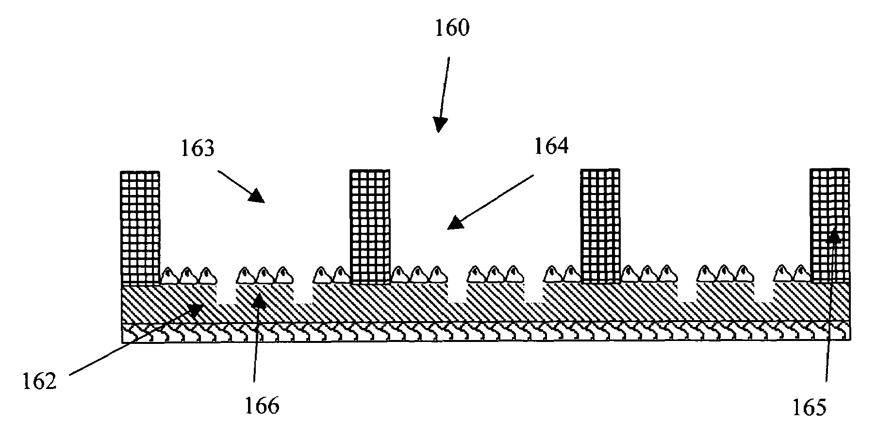
[0081]FIG. 14 illustrates a ...


PUM

PropertyMeasurementUnit
heightaaaaaaaaaa
heightaaaaaaaaaa
sizeaaaaaaaaaa

Abstract

In the present invention, a micro-pattern embedded optical film for cell-based assays is described. The optical film contains micro-patterns in the form of geometric shapes, such as lines and curves, and numbers and letters. Furthermore, the optical film contains a coordinate system that allows identification of each location on the optical film for cell-based assays, including cell growth, identification, and measurements under an optical microscope. Furthermore, an apparatus with the micro-pattern embedded optical film and a supporting part is described. Methods for making the optical film and apparatus are disclosed. A method for performing cell-based assays using the optical film or apparatus is shown.

Description

CROSS-REFERENCE TO RELATED APPLICATIONS[0001]This application claims priority to U.S. Provisional Patent Application Ser. No. 60 / 463,262, filed Apr. 17, 2003. The application is related to commonly owned application Ser. No. 10 / 760,000, filed Jan. 16, 2004, pending.OTHER REFERENCES[0002]Bradke, F., et al., “Differentiated neurons retain the capacity to generate axons from dendrites”, Current Biology, Vol. 10, pp. 1467-1470, November 2000.BACKGROUND OF THE INVENTION[0003]This invention is in the field of biological research and drug discovery, specifically in cell-based assays.[0004]Tissue culture of plant and animal cells in vitro allows researchers to identify critical chemical factors affecting the functions of cells, such as spreading, morphology, motility, and differentiation. Currently, majority of tissue culture experiments are performed on flat surfaces of plastic or glass vessels. The measurements are obtained from large groups of cells without the ability to identify, to mo...

Claims


Application Information

Patent Timeline
Patent Type & Authority Patents(United States)
IPC IPC(8): C12M1/34B01L3/00C12Q1/00
CPCB01L3/5085B01L9/50C12M25/02C12M41/36B01L2200/12B01L2300/0819B01L2300/163B01L9/56
Inventor QIU, JEAN
Owner NEXCELOM BIOSCIENCE LLC
Why Eureka
Try Eureka