Tissue scanning apparatus and method

a tissue scanning and tissue technology, applied in the field of imaging systems, can solve the problems of inability to meet the requirements inability to adjust the time requirement of volume holographic methods, and inability to meet the requirements of tuning time for volume holographic methods

Active Publication Date: 2008-04-29
ARGO AI LLC
View PDF17 Cites 34 Cited by
  • Summary
  • Abstract
  • Description
  • Claims
  • Application Information

AI Technical Summary

Problems solved by technology

However, each of these methods is limited in terms of temporal response time.
Integration times often necessitate much longer acquisition times, often as long as several seconds, which are too long for live biological samples that move on shorter time scales.
The volume holographic method has no tuning time requirement, but is also limited by the CCD read-out rates and integration times. The AOTF imaging method represents the state of art in this field but faces limited throughput and spatial mis-registrations of one color with respect to another.
However, the above-described features and characteristics are not available in one cost-effective instrument.

Method used


Image

  • Tissue scanning apparatus and method
  • Tissue scanning apparatus and method
  • Tissue scanning apparatus and method

Examples


Embodiment Construction

[0021]It is to be understood that the figures and descriptions of the present invention have been simplified to illustrate elements that are relevant for a clear understanding of the present invention, while eliminating, for purposes of clarity, many other elements found in typical optical methods and systems. However, because such elements are well known in the art, and because they do not facilitate a better understanding of the present invention, a discussion of such elements is not provided herein. The disclosure herein is directed to all such variations and modifications known to those skilled in the art.

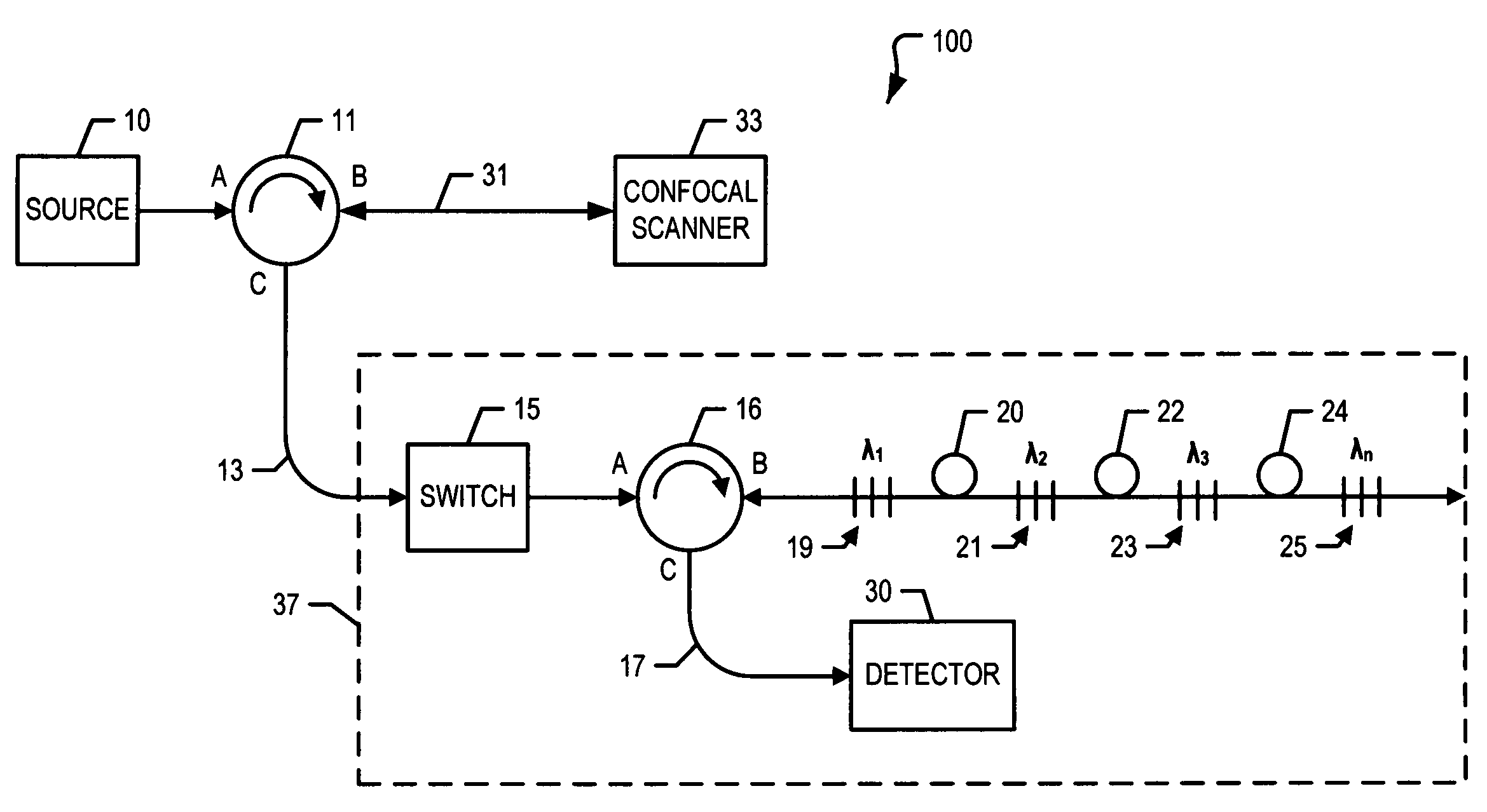
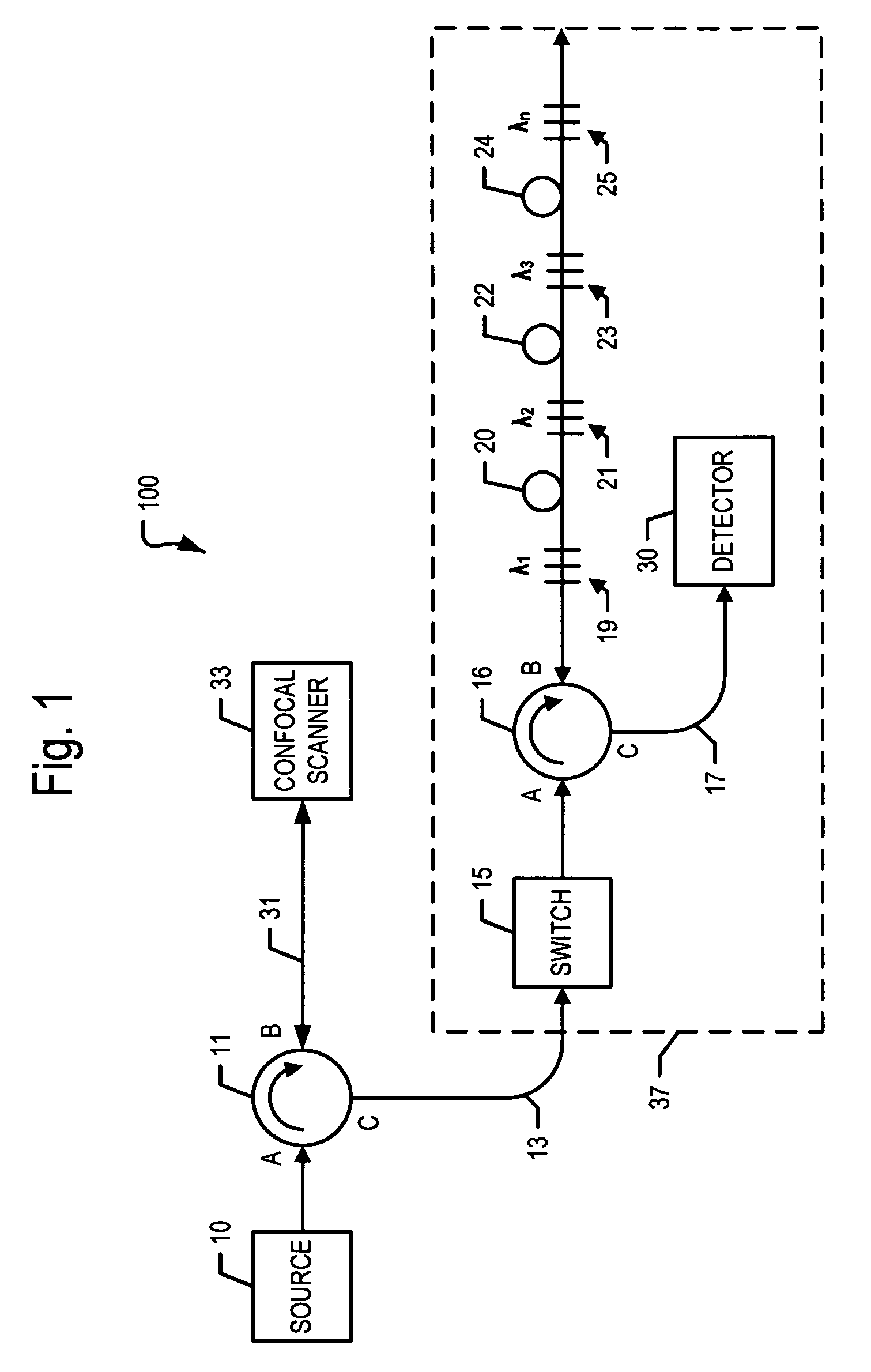
[0022]In general, a confocal spatial scan is employed in conjunction with a fast fiber grating spectrometer. The combination maps wavelengths into time slots and is fiber based. The fiber that connects the fast optical spectrum analyzer to the scanner functions as a pinhole in a confocal microscope. In this manner, the cleaved end-face of a fiber provides the confocal pinhole. ...


PUM


Abstract

A system for multispectral confocal mapping of a tissue, comprising: a light source; an optical fiber having an aperture providing a confocal pinhole, a scanner being free-space coupled to said aperture and operative to scan the tissue and provide intensity signals for each of a plurality of points therein; a switch or modulator operative to convert input signals to optical pulses; a reflector that receives the optical pulses and provides a temporal sequence of wavelength selective reflections; and, a detector optically coupled to receive the temporal sequence of wavelength selective reflections.

Description

RELATED APPLICATION[0001]This application claims the benefit of U.S. Provisional Patent Application No. 60 / 739,863, filed Nov. 23, 2005, entitled “Fast Spectrometer Apparatus” which application is incorporated herein by reference in its entirety.FIELD OF THE INVENTION[0002]The present invention relates generally to imaging systems, and more particularly to a high speed spectrometer for multispectral confocal mapping.BACKGROUND OF THE INVENTION[0003]Various imaging systems and techniques have been used for biomedical applications, such as tissue mapping. One approach to tissue mapping involves multispectral imaging, where the tissue to be mapped is conjugate with a detector array. The array may be a charge coupled device (CCD) or a photo-diode array. In the array, the spatial data is directly obtained in the image. A tunable filter can be placed before the detector array, and images collected for each color setting of the filter. Liquid crystal tunable filters (LCTF) and acousto-opti...

Claims


Application Information

Patent Timeline
Patent Type & Authority Patents(United States)
IPC IPC(8): G02B6/00G01J3/28G02B26/08
CPCA61B5/0068A61B5/0075A61B5/0084G01N21/31A61B5/0071
Inventor CARVER, GARY
Owner ARGO AI LLC
Why Eureka
Try Eureka