

Antibodies specific for phosphorylation sites and screening methods using the same antibodies
- Summary
- Abstract
- Description
- Claims
- Application Information
AI Technical Summary
Benefits of technology
Problems solved by technology
Method used
Image

Examples
example 1
Preparation of Antibodies Specific for Linker Region-Phosphorylated Smads and Ascertainment of Specificity
(1) Outline of Results
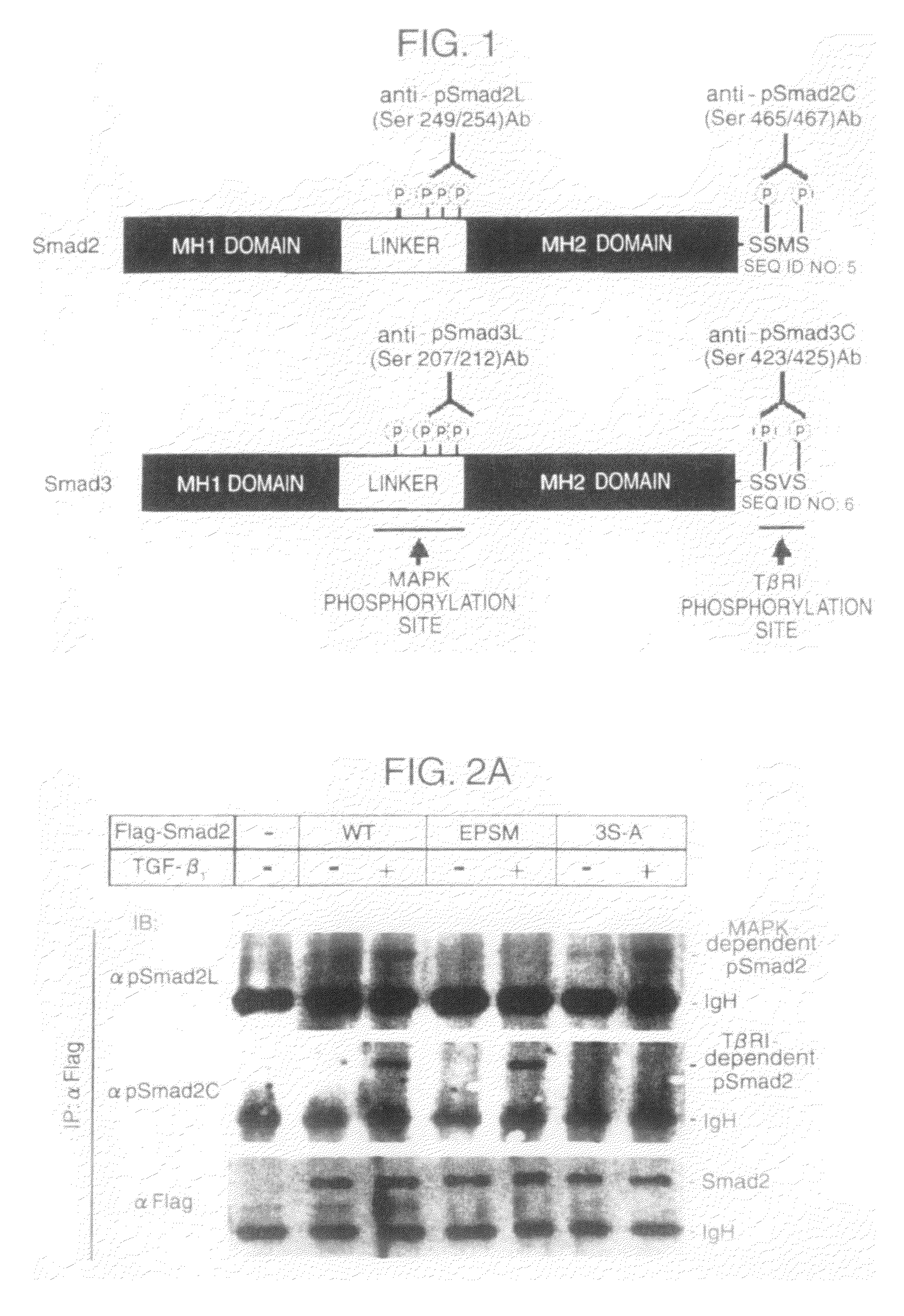
[0122]Phosphorylated peptides specific for the linker regions and the C-terminal regions in Smad2 and Smad3:[0123]Pro Ala Glu Leu p-Ser Pro Thr Thr Leu p-Ser Pro Val Asn His Ser (SEQ ID NO: 1)[0124]Ala Gly Ser Pro Asn Leu p-Ser Pro Asn Pro Met p-Ser Pro Ala (SEQ ID NO: 2)[0125]Pro Ser Val Arg Cys Ser p-Ser Met p-Ser (SEQ ID NO: 3)[0126]Pro Ser Ile Arg Cys Ser p-Ser Val p-Ser (SEQ ID NO: 4)
wherein p-Ser represents phosphorylated serine were prepared, each of the peptides was conjugated with bovine thyroglobulin used as carrier protein, and the mixture of each of the conjugates and an adjuvant was prepared. Three to five rabbits were immunized with the mixture, and then booster was given 4 to 7 times every 2 weeks in the same manner as above. Finally, blood samples were collected from the rabbits to obtain antisera. Then, affinity columns (Activated Thiol Sep...
example 2
Example of Detecting Phosphorylation-Inhibiting Compounds in Test System using the Present Antibodies / Experimental Example Using Known p38 (Kinase) Inhibitor
(1) Outline of Results
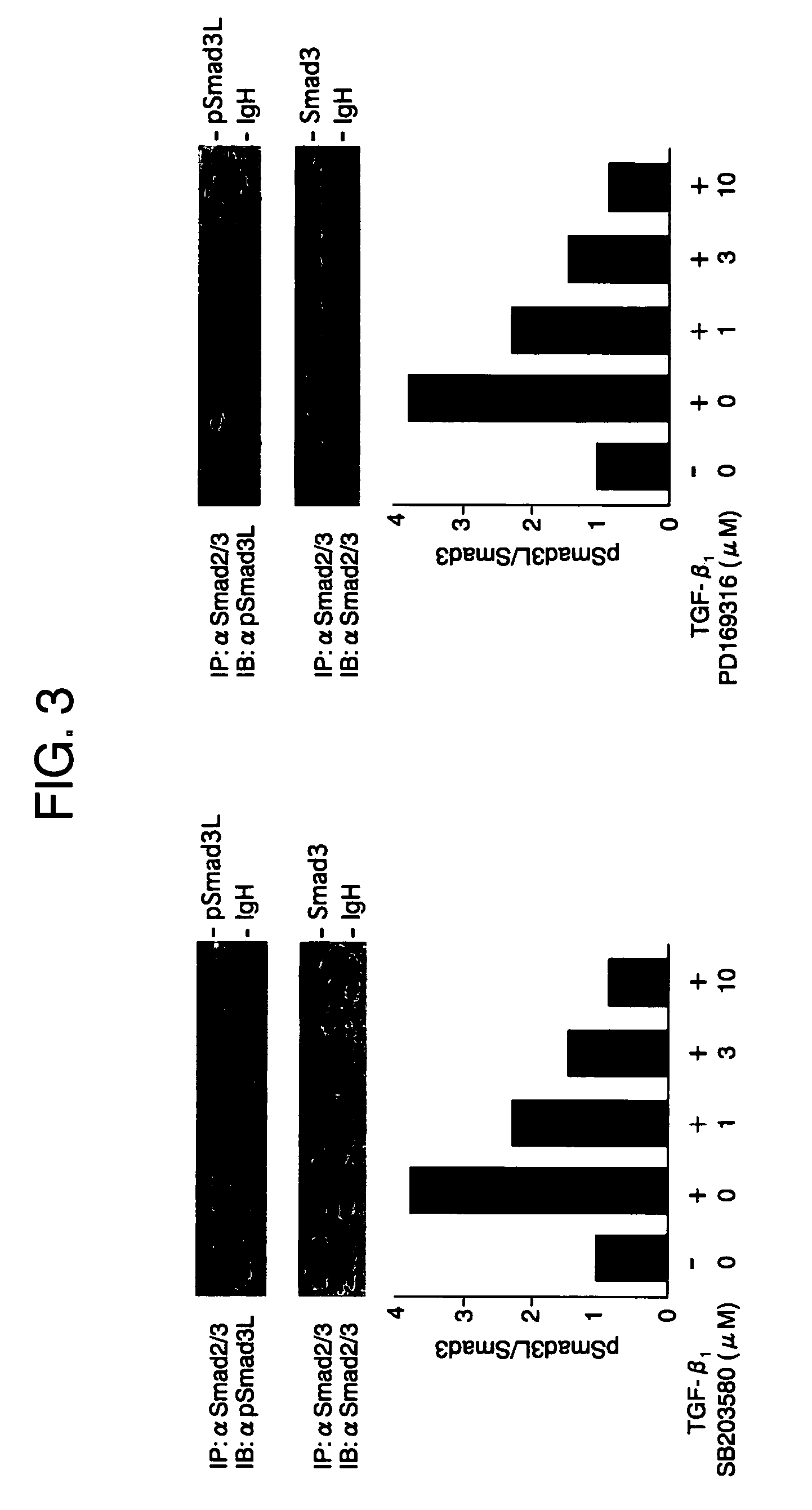
[0132]Compounds that inhibit phosphorylation of Smad proteins can be screened by treating the compounds with a known p38 inhibitor before treatment with TGF-β. In this test system, p38 (kinase) inhibitors such as SB203580, SB210190 and PD169316 (all from Calbiochem) inhibited phosphorylation in the linker regions of Smad proteins (FIG. 3).
[0133]There has been already reported (F. Furukawa, K. Matsuzaki et. al., Hepatology (2003) 38, 879-889) that in cells in which cDNAs with the phosphorylation sites of their linker regions mutated are expressed, the TGF-β-dependent transcription activity is suppressed. Treatment with p38 inhibitors, which inhibited phosphorylation in the linker regions of Smad proteins, also suppressed the TGF-β-dependent transcription activity (FIG. 4).
[0134]As described above, inhibiting...
example 3
Detection of Localization of Smad3 Phosphorylated in the Linker Region using Antibody
(1) Outline of Results
[0137]The cells expressing Smad3 were stimulated with TGF-β, and the phophorylation of the linker region was immunohistochemically detected with the specific antibody. The use of the antibody confirmed that Smad3 phosphorylated in the linker region is localized in nuclei. (FIG. 5A). On the other hand, when the cells were treated with p38 inhibitor and then with TGF-β, the phosphorylation of Smad3 in the linker region was inhibited and the accumulation of Smad3 in nuclei was suppressed (FIG. 5B).
[0138]Immunohistochemical examination using the antibodies was performed in liver tissue sections from CCl4-treated rat with hepatic fibrosis. By the examination, Smad3 phosphorylated in the linker region was detected specifically in the nuclei of α-SMA positive cells as the disease progress (FIG. 6). This indicates that the use of the antibody enables the confirmation of Smad3 phosphory...
PUM
| Property | Measurement | Unit |
|---|---|---|
| Affinity | aaaaa | aaaaa |
Abstract
Description
Claims
Application Information