Single trace multi-channel low coherence interferometric sensor

a single-channel, low-coherence technology, applied in the field of medicine and engineering, can solve the problems of insufficient biomedical diagnostics, inability to perform conventional excisional biopsy, invasive and time-consuming and costly, etc., to achieve rapid identification of radial positions, reduce data collection and interpretation time, and low coherence

Inactive Publication Date: 2008-08-26
MEDEIKON
View PDF56 Cites 84 Cited by
  • Summary
  • Abstract
  • Description
  • Claims
  • Application Information

AI Technical Summary

Problems solved by technology

However, there are many situations where existing biomedical diagnostics are not adequate.
While such examinations are among the most powerful medical diagnostic techniques, they are invasive and can be time consuming and costly.
Furthermore, in many situations conventional excisional biopsy is not possible.
There are many other examples where biopsy can not be performed or conventional imaging techniques lack the sensitivity and resolution for definitive diagnosis.
However, in many applications, for example arteries, there is insufficient room in the cavity or conduit for articulation of the scope tip.
This procedure is not only invasive, time-consuming and expensive but often is not capable of rapid and reliable screening of a large surface such as the colon, esophagus, or stomach.
One drawback of the OCT imaging is the time required to obtain images over a sufficient area.
However, as a typical optical fiber can only emit light and gather back-scattered light along its axial centerline, it is limited to viewing straight ahead.
A view transverse to the axial centerline of the fiber can be obtained by turning or bending the head of the fiber perpendicular to its axial centerline, and this is often very difficult or even impossible in the close confines typically encountered during surgical procedures, or in examining the sides of an artery or vein.
However, these apparatus add bulk to the head of the optical fiber.
The use of either of the aforementioned optical devices thus renders some locations inaccessible and makes the optical fiber more difficult to maneuver.
In addition, extremely small GRIN lenses and mirrored corner cubes are quite expensive, and very fragile.
Their use thus adds to the cost of the probe, and renders it prone to malfunction.

Method used


Image

  • Single trace multi-channel low coherence interferometric sensor
  • Single trace multi-channel low coherence interferometric sensor
  • Single trace multi-channel low coherence interferometric sensor

Examples


example 1

[0135]This prophetic example illustrates how an version of the present invention can be used for detecting plaque within the coronary or other patient vasculature. The apparatus and techniques could also be applied to characterizing lesions in other body lumens which are associated with various disease conditions. The methods and apparatus can be implemented within the body lumen to identify diseased tissue or monitor the course of treatment for a particular condition. The apparatus is able to interrogate the body lumen over a relatively long distance to characterize the tissue in an efficient fashion by providing interferometric information on the tissue from multiple probes in a single sweep of a variable delay of the apparatus.

[0136]Coronary artery disease resulting from the build-up of atherosclerotic plaque in the coronary arteries is a leading cause of death. The build-up of plaque causes a narrowing of the, artery, commonly referred to as a lesion, which reduces blood flow to...

example 2

[0142]This prophetic example illustrate how a sensor of the present invention may be used in photodynamic therapy to treat and monitor a lesion.

[0143]Photodynamic therapy (PDT) is effective in destroying diseased tissue and tumors using light that is absorbed by a photoreactive agent administered to the patient. The photoreactive agent is selectively preferentially absorbed by or linked to the abnormal or diseased tissue and has a characteristic absorption wavelength or absorption band that activates the agent. When activated by the light, the photoreactive agent can produce compounds that destroy the abnormal tissue. The depth of light penetration, and consequently of the tissue necrosis, is a function of light wavelength, and is typically less than one centimeter.

[0144]PDT may be administered locally to a site in or on a patient using light from an external light source such as a laser that is coupled to a plurality of optical fibers or waveguides. The waveguides that propagate th...

example 3

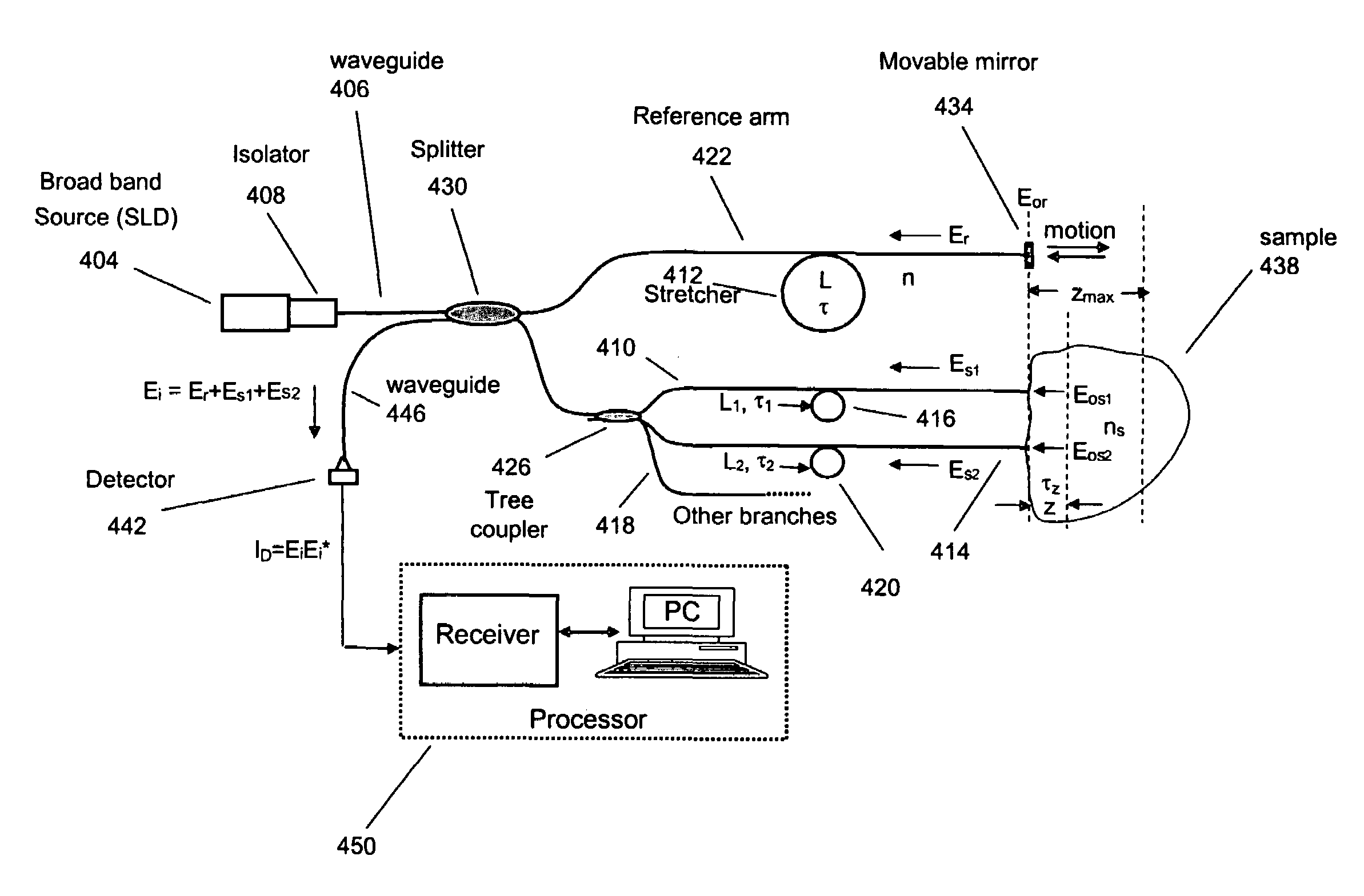
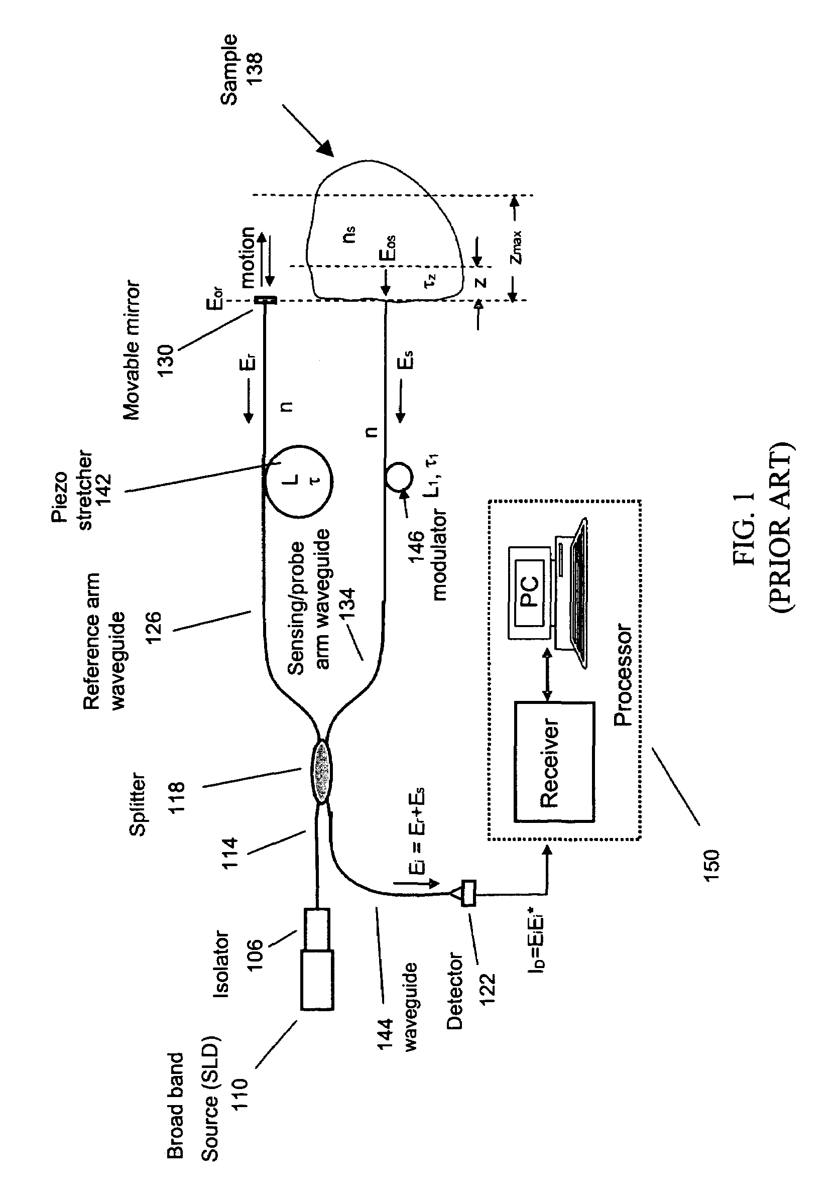
[0149]This example illustrates a method for characterizing a material sample that includes contacting the material with a sensor having two or more probes or sample arms and a delay compensator or a reference section respectively. Each probe arm can include an adjustable delay, the probe arms and reference section configured such that an interference between backscattered source light from the sample for two or more probe arms and reference light occurs and is detected during a single trace of the delay compensator or reference section.

[0150]Preferably the interference signal for the two or more probe or sample arms are at least partially separated from each other. The material can be a tissue where characterization includes the detection of a disease state of the tissue. Preferably the disease state being characterized is a vulnerable plaque.


PUM


Abstract

Interferometers and autocorrelator based sensors are disclosed that are configured to have multiple sample arms which can be scanned and the backscattered low coherence source light from a sample resolved in a single sweep of one or more variable delays of the sensor. Borescopes and catheters capable of scanning multiple sections or areas of materials and tissues using these sensors are described.

Description

BACKGROUND AND SUMMARY[0001]In various fields of medicine and engineering it is often necessary to inspect surfaces that are difficult to reach. For example engine cylinders, compressors for jet aircraft engines, heat exchangers, internal organs, cavities, and arterial passageways in a patient. Biomedical imaging technology, for example, magnetic resonance imaging, X-ray computed tomography, ultrasound, and confocal microscopy can be used to inspect and characterize a variety of tissues and organs. However, there are many situations where existing biomedical diagnostics are not adequate. This is particularly true where high resolution, about 1 micron, imaging is required. Resolution at this level often requires biopsy and histopathologic examination. While such examinations are among the most powerful medical diagnostic techniques, they are invasive and can be time consuming and costly. Furthermore, in many situations conventional excisional biopsy is not possible. Coronary artery d...

Claims


Application Information

Patent Timeline
Patent Type & Authority Patents(United States)
IPC IPC(8): G01B9/02G01B11/02
CPCA61B5/0066A61B5/0084A61B5/0086G01B9/02082G01B9/02007G01B9/0205G01B9/0209G01B9/02028
Inventor ALPHONSE, GERARD A.CARLIN, DONALD B.RAPPAPORT, FRED
Owner MEDEIKON
Why Eureka
Try Eureka