

Method and quantification assay for determining c-kit/SCF/pAKT status
a technology of c-kit and scf, applied in the field of method and quantification assay for determining ckit/scf/pakt status, can solve the problems of affecting normal cells, causing deleterious effects on normal and tumor cells, and preventing growth arrest rather than cell death
- Summary
- Abstract
- Description
- Claims
- Application Information
AI Technical Summary
Benefits of technology
Problems solved by technology
Method used
Image

Examples
example 1
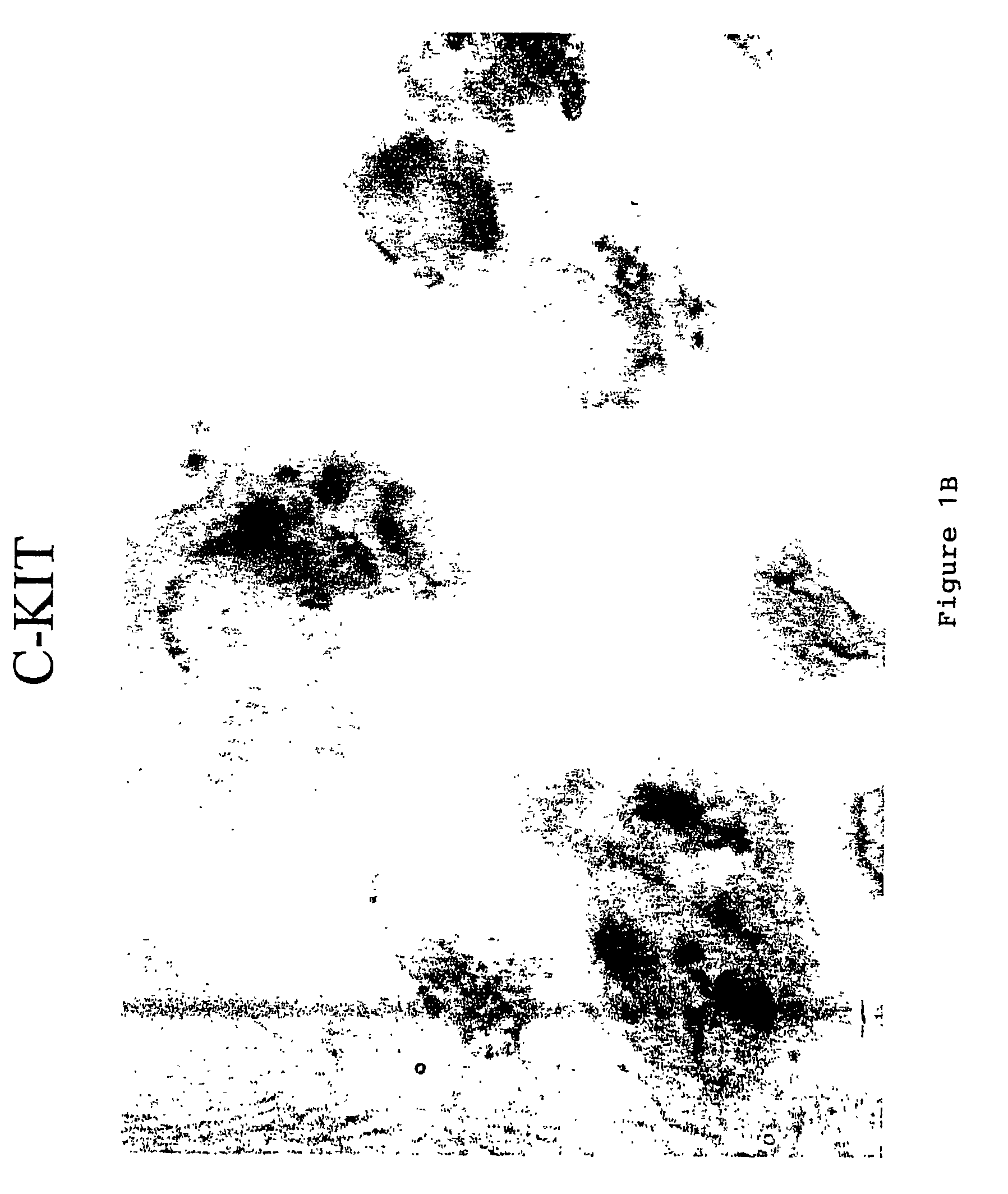
Staining Procedure for c-Kit
[0084]Staining for c-kit was performed on a Benchmark™ automated staining module (Ventana Medical Systems, Inc., Tucson, Ariz.) using a polyclonal antibody to c-kit (NeoMarkers, Fremont, Calif.). 10% Neutral Buffered Formalin fixed 4 micron paraffin sections of the sample were place onto coated slides. The Benchmark bar-code specific for the c-kit staining protocol was placed onto the slide and the slide placed onto the Benchmark. All subsequent steps, drying, deparaffinization antigen retrieval (Cell Conditioning 1 mM EDTA), and detection were performed automatically by the Benchmark. All test reagents are available from Ventana Medical Systems, including the I-View DAB Detection Kit, unless otherwise noted. The protocol implemented in using the automated system is described in Table 1. Where indicated, one drop is one reagent dispense. The sample was then manually counterstained with 4% Ethyl Green 0.1 M pH 4 acetate buffer for 10 minutes. The results o...
example 2
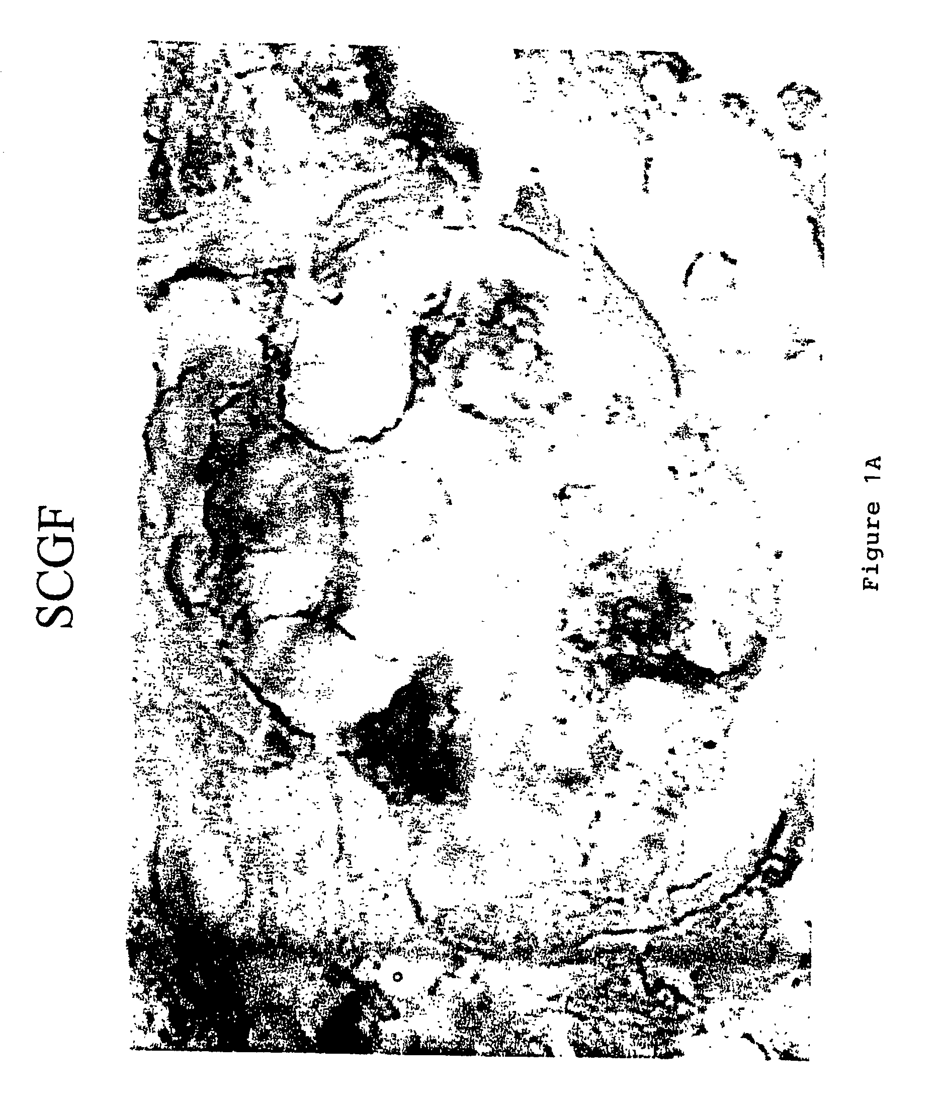
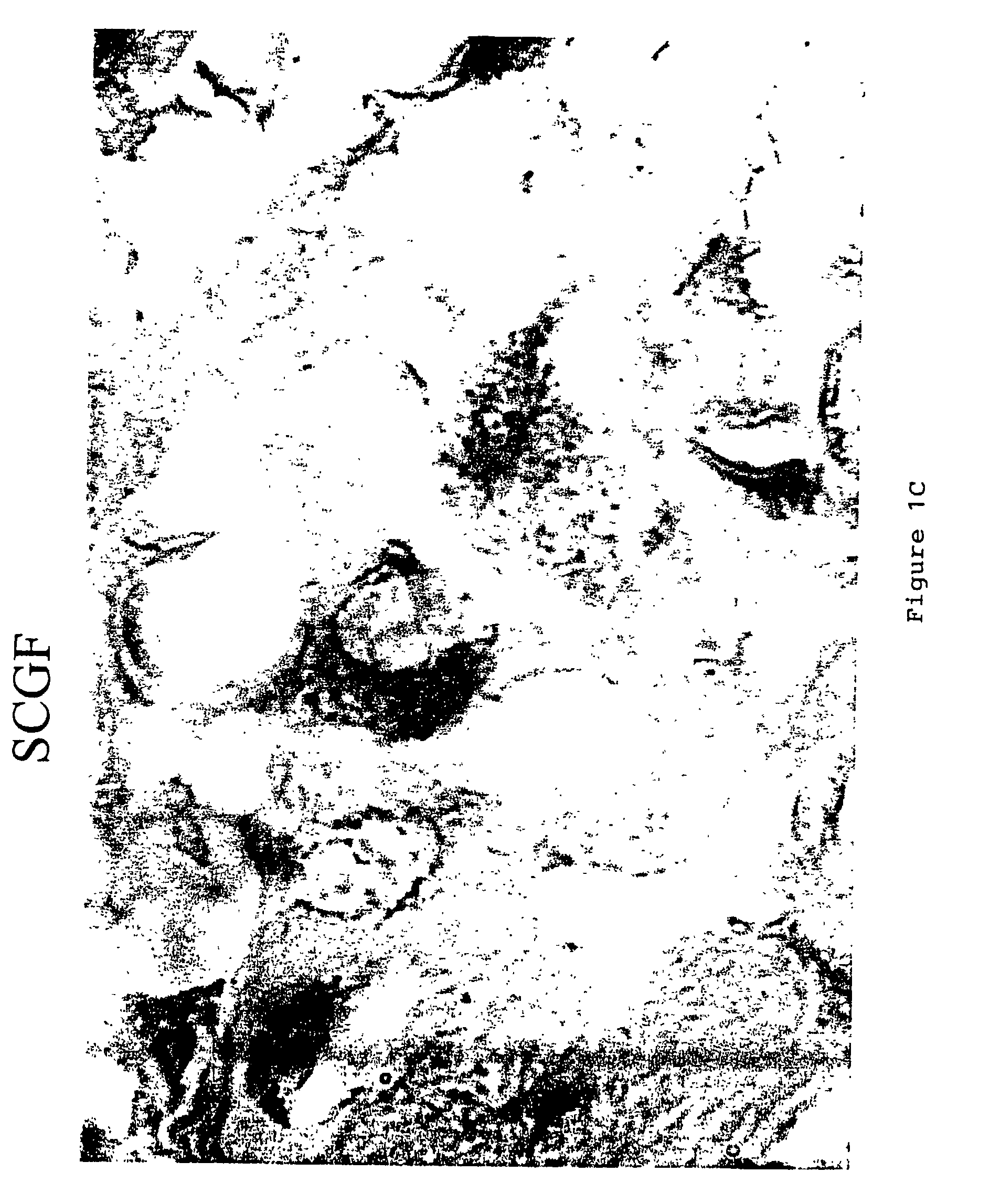
Staining Procedure for SCF
[0086]SCF staining was performed using a polyclonal antibody to SCF (Santa Cruz Biotechnology, Santa Cruz, Calif.). A biotinylated anti-rabbit secondary antibody (Jackson Research Labs, West Lake, Pa.). the “StreptABComplex-HRP” (Streptavidin / Biotin Complex with a Horseradish Peroxidase) label kit (DAKO Corporation, Carpinteria, Calif.), and the DAB chromagen (Dako) were used as the detection system for SCF. 10% Neutral Buffered Formalin fixed 4 micron paraffin sections of the sample were place onto coated slides. To prepare the sample for staining, the sample was first deparaffinized and hydrated to water. Antigen retrieval was preformed enzymatically with Digest-All 1 (Zymed Labs), a Ficin digestion solution. 1-2 drops of the manufacture's ready-to-use solution was added to the sections and incubated for 20 minutes at 37° C. The sections were then washed well with deionized water and rinsed with TBS. Endogenous peroxidase was blocked by incubating the sec...
example 3
Staining Procedure for Phospho-AKT
[0088]p-AKT staining was performed using a Dako Autostainer with the LSAB2 kit (described above). 10% Neutral Buffered Formalin fixed 4 micron paraffin sections of the sample were place onto coated slides. To prepare the sample for staining, the sample was first deparaffinized and hydrated to water. Antigen retrieval was preformed with 0.1 M citrate retrieval buffer, pH 6.0 in a Decloaker Chamber (Biocare Medical, Walnut Grove, Calif.) following the manufacturer's instructions. The sections were allowed to cool for 15 minutes, and then washed well with deionized water. Endogenous peroxidase was blocked by incubating the sections with a 3% hydrogen peroxide / Methanol solution for 10 minutes, followed by a deionized water wash. The sections were then blocked using 10% blocking goat serum in 0.1% BSA / 0.1% Triton X for 10 minutes. The goat serum was then shaken off.
[0089]The sections were then incubated with a 1:75 dilution of p-AKT primary antibody (Cel...
PUM
| Property | Measurement | Unit |
|---|---|---|
| optical density | aaaaa | aaaaa |
| affinity | aaaaa | aaaaa |
| drug resistance | aaaaa | aaaaa |
Abstract
Description
Claims
Application Information