DNA dependent protein kinase catalytic subunit phosphorylation sites and antibodies thereto

a kinase and catalytic subunit technology, applied in the field of cancer treatment, therapeutics and diagnostics, can solve the problems of inability to detect, lack of progress in studying physiological functions, and inability to see any discernible foci, so as to improve cell survival

Inactive Publication Date: 2009-02-17
RGT UNIV OF CALIFORNIA
View PDF1 Cites 0 Cited by
  • Summary
  • Abstract
  • Description
  • Claims
  • Application Information

AI Technical Summary

Benefits of technology

[0014]The present invention is directed to the identification and use of two major DNA-PKcs autophosphorylation sites, Threonine (T) 2609 and Serine (S) 2056, including antibodies specific for phosphorylated T2609 and S2056. It is demonstrated that phosphorylation of these sites, carried out ill vivo by the DNA-PKcs itself (i.e. autophosphorylation), is required for DNA-PK activity and, furthermore, that such activity repairs double strand DNA breaks (DSBs) and improves cell survival to ionizing radiation (IR). For example, it is demonstrated a point mutation at position 2056 from serine to alanine and position 2609 from threonine to alanine results in cells that are radiosensitive.
[0015]The present invention further provides phosphospecific antibodies that recognize these specific sites of phosphorylation in DNA-PKcs. The antibodies do not bind to the unphosphorylated DNA-PKcs protein or peptide. This provides diagnostic tools based on the ability to identify the phosphorylation status of the DNA-PKcs autophosphorylation sites. One can monitor the effectiveness of treatments which target the DNA repair pathway of cancer cells, such as radiation treatment and inhibitor drugs. Also, the ability to intervene in autophosphorylation of T2609 or S2056, either through application of a drug or an antibody, would increase the radiation-induced killing of cancer cells.

Problems solved by technology

This lack of progress in studying the physiological functions of DNA-PK is in part due to the unavailability of the right tools or assays to evaluate DNA-PK in vivo activity.
It is currently not possible to detect DNA-PK foci with the antibodies available because DNA-PK is quite abundant in the nucleus, thus when one performs immunofluorescence with any of the available antibodies, the entire nucleus will produce a signal, making it impossible to see any discernable foci.
Cloning of the DNA-PKcs cDNA is difficult, since the cDNA exceeds 13 kb.

Method used


Image

  • DNA dependent protein kinase catalytic subunit phosphorylation sites and antibodies thereto
  • DNA dependent protein kinase catalytic subunit phosphorylation sites and antibodies thereto
  • DNA dependent protein kinase catalytic subunit phosphorylation sites and antibodies thereto

Examples


example 1

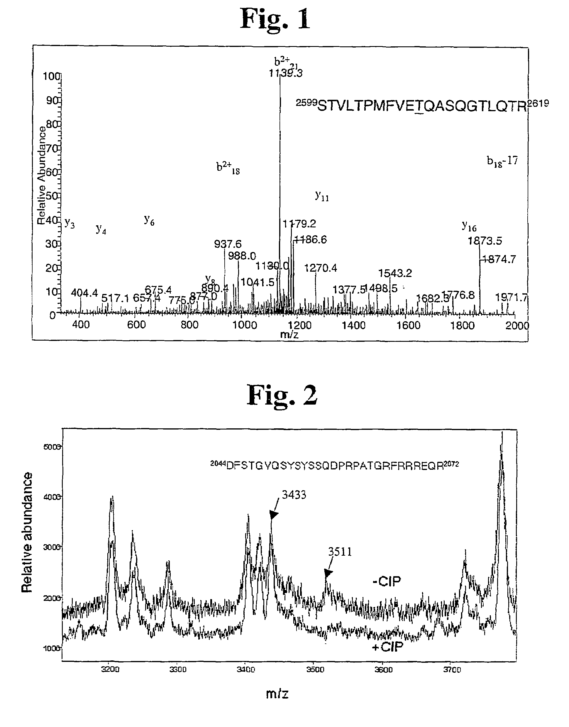
[0081]Determining T2609 and S2056 Sites of Autophosphorylation in DNA-PKcs by Mass Spectrometry

[0082]First, purified human DNA-PKcs and Ku were autophosphorylated as previously described (Chan and Lees-Miler, J Biol Chem 271: 8936-8941, 1996), and hereby incorporated by reference, with the following change: 50 μM ATP was used instead of 250 μM to allow phosphorylation of the most preferential site. Purified DNA-PKcs and Ku proteins were preincubated at 30° C. Reactions contained 25 mM Hepes, pH 7.5. 75 mM KCl, 10 mM MgCl, 1 mM dithiothreitol, 0.2 mM EDTA, 0.1 mM EDTA plus 10 μg / ml sonicated calf thymus DNA, and 0.25 mM ATP containing stabilized [-P]ATP (Sigma Chemicals, St. Louis, Mo.) (specific activity. 500-1000 dpm / pmol) and were started by the addition of purified DNA-PK proteins (usually 0.05-0.1 μg as indicated). Reactions were at 30° C. for 5-10 min and DNA-PK activity was calculated as nmol of phosphate incorporated into the peptide substrate per minute per milligram of prot...

example 2

[0090]DNA-PKcs Fragments and GST Fusion Proteins Containing Autophosphorylated Sites

[0091]20 bp oligomer primers were designed and ordered from Operon (Alameda, Calif.) using SEQ ID NO: 4 (the nucleotide sequence of DNA-PKcs, GenBank Accession Number: P78527) to created primers to amplify cDNA sequence that encodes the phosphorylation sites, T2609 and S2056. Designed DNA-PKcs cDNA fragments that cover the phosphorylation sites in DNA-PKcs found by mass spectrometer were PCR amplified from the full-length DNA-PKcs cDNA (isolated and described by several of the inventors in Kurimasa et al., Mol Cell Biol 19:3877-3884, 1999) using the custom designed PCR primers under normal PCR thermal cycling conditions. The reactions were carried out using pfu DNA polymerase (Stratagene, La Jolla, Calif.) and GeneAmp® 9600 thermocycler (Perkin Elmer). The amplified cDNA fragments were cloned in frame into GEX-KG vector (Guan & Dixon 1991 Analytical Biochem. 192:262-67) for fusion between domains of ...

example 3

[0095]Preparation of Cellular Nuclear Extracts from Cells

[0096]The preparation of nuclear extract from HeLa cells for the Examples that follow were made as generally described by Lees-Miller et al., Mol Cell Biol 10: 6472-6481, 1990 and is herein described. The cells were washed twice with cold PBS, collected, and spun at 2000 g for 5 min. The cell pellet is washed once with 5 ml LSB and spun again. The pellet is resuspended in 1 ml LSB and transfer to a centrifuge tube. (LSB (low salt): 10 mM Hepes pH7.5, 25 mM KCl, 10 mM NaCl. 1 mM MgCl2, 0.1 mM EDTA).

[0097]After spinning down again, the volume of the cell pellet is estimated, then resuspended in 1× Pack cell volume (PCV) of LSB (with 50 mM NaF, 1 mM DTT. 0.5 mM PMSF, and other protease inhibitors), set in ice 5 min, and freezed in liquid N2. Thaw, and spin immediately at 10,000 g for 10 min. Dispose of Supernatant (S10, cytosol fraction.

[0098]The pellet is again resuspended in 1× pack nuclear volume (PNV) of LSB with 0.5M NaCl an...


PUM

PropertyMeasurementUnit
molar ratiosaaaaaaaaaa
massaaaaaaaaaa
pHaaaaaaaaaa

Abstract

The identification and use of two major DNA-PKcs autophosphorylation sites. Threonine (T) 2609 and Serine (S) 2056, including antibodies specific for phosphorylated T2609 and 52056. Peptides and polynucleotides encoding same, that feature these two sites of phosphorylation. The antibodies do not bind to the unphosphorylated DNA-PKcs protein or peptide, thus providing diagnostic tools to monitor the effectiveness of treatments which target the DNA repair pathway of cancer cells, and the ability to intervene or inhibit in phosphorylation of T2609 or 52056, either through application of a drug or an antibody, to increase the radiosensitivity of cancer cells.

Description

CROSS-REFERENCE TO RELATED APPLICATIONS[0001]This application claims priority to U.S. Provisional Patent Application No. 60 / 375,094, which was filed on Apr. 22, 2002, which is incorporated by reference in its entirety.STATEMENT REGARDING SEQUENCE LISTING[0002]Applicants assert that the attached paper copy of the Sequence Listing for the utility application, “DNA Dependent Protein Kinase Catalytic Subunit Phosphorylation Sites and Antibodies Thereto,” claiming priority to U.S. Provisional Patent Application No. 60 / 375,094, filed on Apr. 21, 2003, is identical to the Sequence Listing in computer readable form found on the accompanying computer disk, as required by 37 CFR 1.821(c) and is hereby incorporated by reference in its entirety.BACKGROUND OF THE INVENTION[0003]1. Field of the Invention[0004]This invention generally relates to the field of cancer treatment, therapeutics and diagnostics. More specifically, the invention describes antibodies and a method useful for increasing the ...

Claims


Application Information

Patent Timeline
Patent Type & Authority Patents(United States)
IPC IPC(8): C07K16/40C12N9/12G01N33/50G01N33/573
CPCC07K16/40C07K16/44C12N9/1205C12Y207/01037G01N33/5014G01N33/573G01N2333/9121G01N2800/52
Inventor CHEN, DAVID J.CHEN, PING-CHI BENJAMINCHAN, DOUG W.
Owner RGT UNIV OF CALIFORNIA
Why Eureka
Try Eureka