Cryo-charging specimen holder for electron microscope

a specimen holder and electron microscope technology, applied in the direction of material analysis using wave/particle radiation, instruments, nuclear engineering, etc., can solve the problems of 99.5% of proteins that are difficult to be formed as crystals, conventional nmr technique is only suitable for observation of small molecules, and process may somehow damage the native form of proteins, etc., to reduce radiation damage of biological samples, raise the resolution of the electron microscope, and save costs

Inactive Publication Date: 2010-02-09
CHAO CHIH YU
View PDF3 Cites 4 Cited by
  • Summary
  • Abstract
  • Description
  • Claims
  • Application Information

AI Technical Summary

Benefits of technology

[0014]The invention can greatly reduce the radiation damage of biological samples during the electron beam irradiation. Thus, the native form of biological materials can be observed clearly.
[0015]The invention can break through the key technology of the electron microscope, so that the bio-molecular microscope with atomic-level resolution can be developed successfully.
[0016]The invention can raise the resolution of the electron microscope by the way of saving cost. And only the key technology has to be developed, then the resolution of any electron microscope can be raised.

Problems solved by technology

Crystallization of protein remains a major hurdle, and to date 99.5% of proteins are difficult to be formed as crystal.
Moreover, the crystallization for every kind of protein is a highly difficult process, and process may somehow damage the native form of protein.
On the other hand, the conventional NMR technique is only suitable for the observation of small molecules, not generally applicable to all macromolecular complexes and proteins.
However, the cryo-electron microscopy is not able to provide the atomic resolution.
It means excess electron dosage will damage the biological materials.
The movement of damaged molecular fragments caused by electron radiation can blur the image anisotropically in a relatively unpredictable manner, preventing the atomic resolution from being obtained.
Therefore, the cryo-electron microscopy is not well practicable.

Method used


Image

  • Cryo-charging specimen holder for electron microscope
  • Cryo-charging specimen holder for electron microscope
  • Cryo-charging specimen holder for electron microscope

Examples


Embodiment Construction

[0028]The invention will be understood sufficiently through the description of the following embodiments, and those skilled in the art can complete it accordingly. However, the implementation will not be limited by the following embodiments.

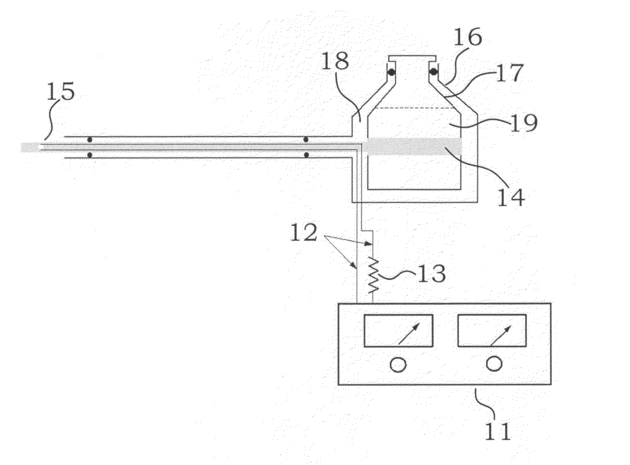
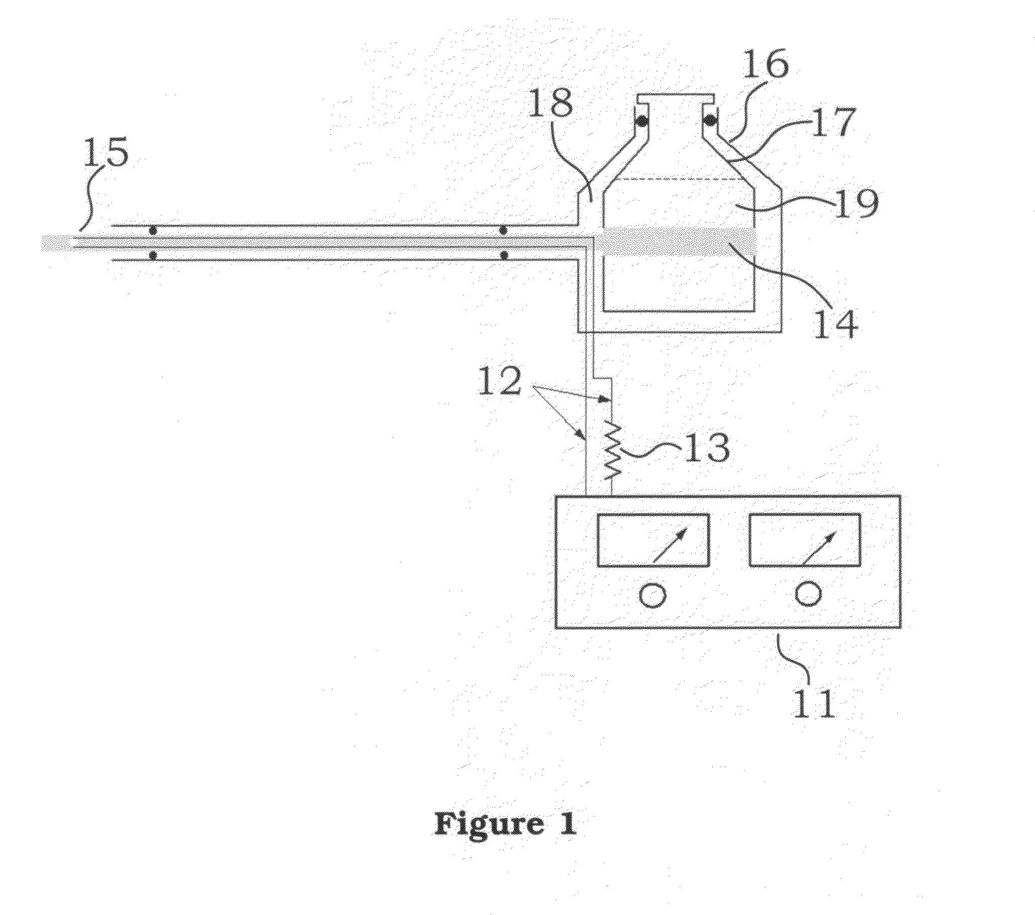
[0029]The present invention relates to a cryo-charging specimen holder for the electron microscope, particularly to a cryo-charging specimen holder for the electron microscope to hold various biological materials, which is suitable to be used by various electron microscopes directly. After the cryo-charging specimen holder for the electron microscope of this invention is installed, the electron microscope can become the bio-molecular microscope with atomic resolution directly without any modification.

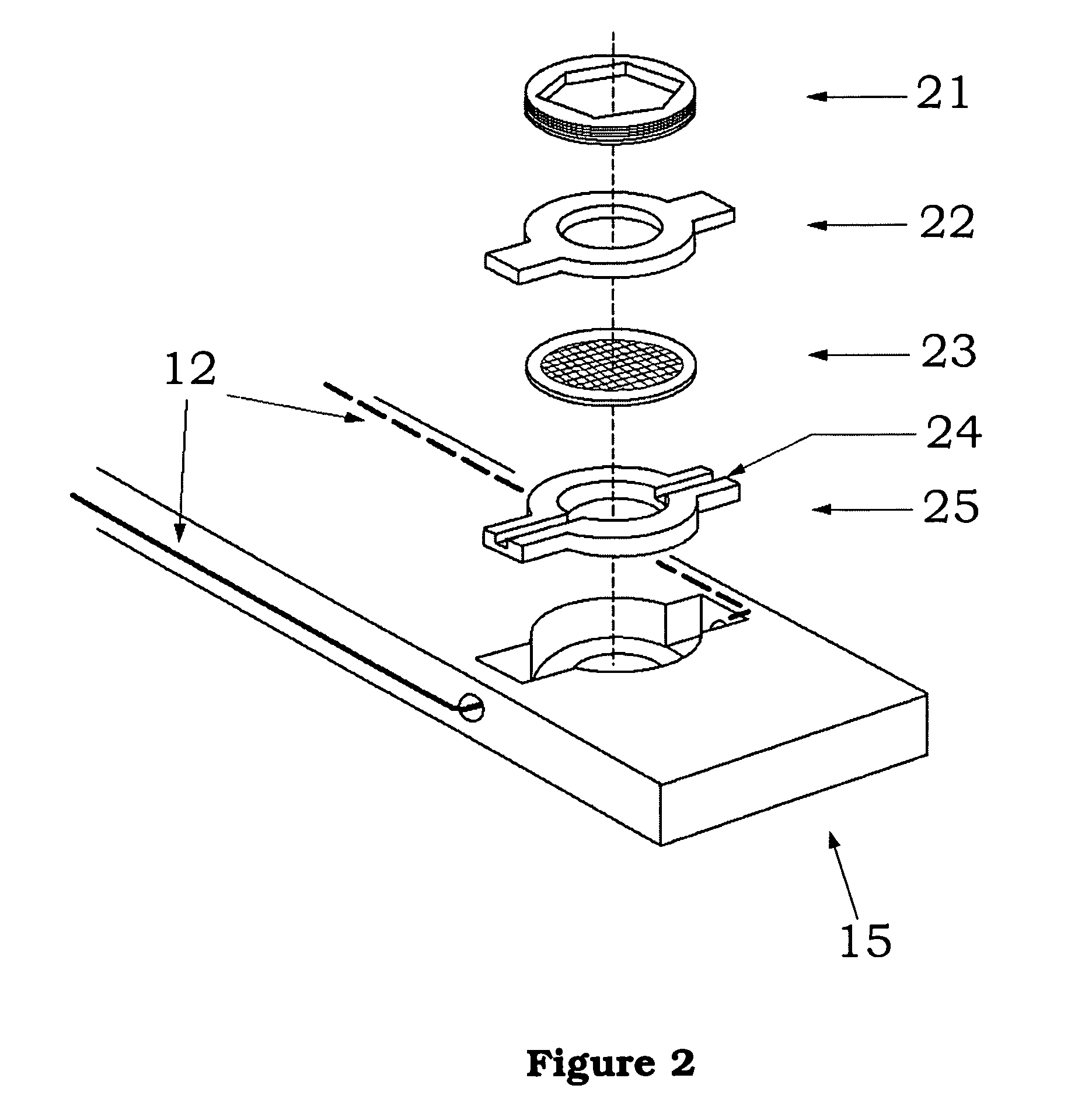
[0030]Please refer to FIG. 1, which shows the schematic of cryo-charging specimen holder of electron microscope of this invention and comprises the following elements:

[0031]Thus, in the embodiment shown in FIG. 1, the elements of cryo-charging specim...


PUM


Abstract

The present invention relates to a cryo-charging specimen holder for the electron microscope, particularly to a cryo-charging specimen holder for the electron microscope to hold various biological materials. The major feature of the invention is to charge the biological specimen and freeze the specimen at low temperature. The ice around the biological sample is also doped, so that after charging the doped ice surrounding the sample has a conductivity level comparable to that of conductor. Therefore, the sample can be embedded by the doped and charged ice obtaining the property of conductor, in order to be observed by the electron microscope.

Description

BACKGROUND OF THE INVENTION[0001]1. Field of the Invention[0002]The present invention relates to a cryo-charging specimen holder for the electron microscope, and more particularly, to a cryo-charging specimen holder for the electron microscope to hold various biological materials.[0003]2. Description of the Prior Art[0004]At present, the knowledge of the mankind have already tried to enter the post gene era, but unfortunately, only can understand less than 0.5% of the protein structure. For the analysis of protein structure, especially the analysis for the nano-scale biological molecular structure is very important on the disease prevention, gene replication and renovation etc.[0005]In the prior art, the primary tools for determining protein structure are the x-ray diffraction (XRD) and the Nuclear Magnetic Resonance (NMR) techniques. Nevertheless, the structures of only a small number of macromolecular complexes and proteins have been successfully determined by using XRD, comprisin...

Claims


Application Information

Patent Timeline
Patent Type & Authority Patents(United States)
IPC IPC(8): H01J49/44
CPCG01N1/42H01J37/20H01J2237/2001H01J2237/2002H01J2237/2003H01J2237/2004H01J2237/26
Inventor CHAO, CHIH-YU
Owner CHAO CHIH YU
Why Eureka
Try Eureka